 Now in my search for a solution to our height increase problem, I have looked through at least 30 different ideas people have thought up as a possible solution. Some of them make no sense, some of them are scams just trying to take your money, other methods are created with good intentions but just can’t work, and some have a chance to work but there has only been circumstantial evidence and testimonials which are hard to believe. Most of us want to see it with our own eyes to believe in something.
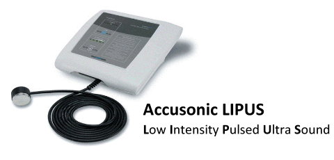
As for the latest technologies that might be coming along that can solve our problems, the 3 big ones are stem cells (probably through implants), gene therapy which is what I believe will ultimately happen if we can figure out how to integrate foreign DNA into our cells and mutate our bodies in a safe way for desired phenotypical traits, and the the joint loading modality (LSJL) which only a few people even know exists. There is some claims over hypnosis and mystic practices working too but that is vert hard to prove. Recently this new technology has come out that is really causing a lot of noise for height increase seekers, but also the dental community as well. It is the method called “Low Intensity Pulsed Ultrasound” or LIPUS for short.
Apparently I have been doing height increase research for so long and even I hadn’t heard about it until Harald of the Biomedical Growth Research Initiative told me that the two major technological developments that currently have the most potential were the LIPUS method and tissue engineering. I knew what tissue engineering did but I had not heard of LIPUS so I did some research. The stuff I found was very interesting. Then Harald explained to me that one of the ideas for his organization was to use LIPUS in some way to increase height, which made the method even more relevant.
Afterwards, I found information on Tyler’s HeightQuest.Com blog about LIPUS so I realized that the guys who are all at the cutting edge of height increase technology and innovation was suggesting that LIPUS could be very big deal. When I was doing research on the Lateral Synovial Joint Loading method on the HeightQuest.Com website, Tyler stated a lot about the potential of the LIPUS in being able to increase our height. That is why that I felt that I should devote quite a bit of time and effort into doing some extensive and serious research on the method and see just how it works, and how effective it will be.
So the first question would be “What is this technology called Low Intensity Pulsed Ultrasound?” . Wikipedia seems to have a clear and short answer to this question so I will paste their answer below (found HERE)
Low-intensity pulsed ultrasound (LIPUS) is a medical technology, generally using 1.5 MHz frequency pulses, with a pulse width of 200 μs, repeated at 1 kHz, at an intensity of 30 mW/cm2, 20 minutes/day.
Applications of LIPUS include:
- Promoting bone-fracture healing.
- Treating orthodontically induced root resorption.
- Regrow missing teeth.[citation needed]
- Enhancing mandibular growth in children with hemifacial microsomia.
- Promoting healing in various soft tissues such as cartilage, inter vertebral disc.
- Improving muscle healing after laceration injury.
Researchers at the University of Alberta have used LIPUS to gently massage teeth roots and jawbones to cause growth or regrowth, and have grown new teeth in rabbits after lower jaw surgical lengthening (Distraction osteogenesis) (American Journal of Orthodontics, 2002). As of June 2006, a larger device has been licensed by the Food and Drug Administration (FDA) and Health Canada for use by orthopedic surgeons. A smaller device that fits on braces has also been developed but is still in the investigational stage and is not available to the public.
It has not yet been approved by either Canadian or American regulatory bodies and a market-ready model is currently being prepared. LIPUS is expected to be commercially available before the end of 2012. The LIPUS foundation website currently announces that Lipus-Plasma application units are available for rental in the USA.
According to Dr. Chen from the University of Alberta, LIPUS may also have medical/cosmetic benefits in allowing people to grow taller by stimulating bone growth.
LIPUS has also been found to stimulate the proliferaton of chondrocytes.
Obviously the last two phrases on the wikipedia article are the most interesting since they state directly that the technology might have the application for stimulating bone growth by the promoting the proliferation of chondrocytes.
Not only this, this is incredible news for people who suffer from dental problems stemming from tooth decay. If this technology is as effective as advertized, it will change the dental industry. However, let’s learn more about what LIPUS does and how it does it.
From the UK website for National Institute For Health And Clinical Excellence located HERE the description for the device states…
Description – Low-intensity pulsed ultrasound aims to speed up fracture healing in broken bones by stimulating bone cells to grow and repair. This involves a short daily treatment using an ultrasound probe that is placed on the skin at the site of the fracture.
For the outline of the prodecure found in this sub-webpage HERE
2.2 Outline of the procedure
2.2.1 The aim of low-intensity pulsed ultrasound is to reduce fracture healing time and avoid non-union by delivering micro-mechanical stress to the bone to stimulate bone healing. This procedure is used to treat fresh fractures, fractures that are slower to heal than expected (delayed healing) and those that have failed to unite (non-union).
2.2.2 This is a non-invasive procedure. The ultrasound probe is positioned on the skin over the fracture and patients self-administer low-intensity pulsed ultrasound daily, usually for 20 minutes. If a patient’s limb is immobilised in a cast, then a hole is cut into the cast for the ultrasound probe. Coupling gel is used on the skin to aid conduction of the ultrasound signal. The operating frequency, pulse width, repetition rate and temporal average power of the ultrasound delivered can be varied.
In the Efficacy section, the experimental and testing results are provided…
2.3 Efficacy (RTC stands for randomized controlled trials)
2.3.1 A meta-analysis of 13 randomised controlled trials (RCTs) including a total of 563 patients (with a mixture of fresh conservatively or operatively managed and non-united fractures) treated by the procedure (n = 280) or a sham procedure (n = 283) reported a 34% (95% confidence interval [CI]: 21 to 44, 6 studies) overall mean reduction in healing time (confirmed by imaging) in patients treated by the procedure (follow-up not stated).
2.3.2 An RCT of 67 patients with closed or open grade I fractures of the tibial shaft (33 low-intensity pulsed ultrasound vs 34 sham) reported a mean healing time (defined as evidence of clinical and radiographic bridging of 3 cortices) of 96 days in the ultrasound group compared with 154 days in the sham group (p < 0.0001).
2.3.3 An RCT of 32 patients with fresh closed or open grade I fractures of the tibial shaft fixed with an intramedullary rod treated by the procedure (n = 15) or a sham procedure (n = 17) reported an average healing time (defined as radiographic bridging of 3 cortices assessed by a radiologist) of 155 days and 125 days respectively (p = 0.76).
2.3.4 An RCT of 21 patients with non-united fractures of the scaphoid treated by pedicle bone graft reported an average healing time (defined as clinical healing [solid and not causing tenderness or pain] and radiographic healing [complete bridging cortices]) of 56 days in patients who also received low-intensity pulsed ultrasound (n = 10) and 94 days in those who received sham therapy (n = 11) (p < 0.001).
2.3.5 An RCT of 30 patients with open tibial fractures or high-energy-induced complex tibial fractures treated by the procedure (n = 16) or a sham procedure (n = 14) reported an average time to full weight bearing of 9.3 weeks and 15.5 weeks respectively (p < 0.05).
Me: I managed to find a great scientific article that really goes into the explanation of the technology (located HERE). Let me take the most important parts of the article.
“”….Low-intensity ultrasound is a biophysical form of intervention in the fracture-repair process, which through several mechanisms accelerates healing of fresh fractures and enhances callus formation in delayed unions and nonunions…Low-intensity pulsed ultrasound is currently applied transcutaneoulsy, although recent experimental studies have proven the efficacy of a trans-osseous application for both enhancement and monitoring of the bone healing process with modern smart implant technologies…”” (published by Elsevier Ltd. 2006)
Key Point: However, one of the fundamental concepts in orthopaedics is the understanding that the mechanical environment at the site of a fracture influences the pattern of fracture repair.
“….The healing of a fractured bone involves the spatial and temporal coordinated action of several different cell types, proteins and the expression of hundreds of genes working towards restoring its structural integrity….”
Basic science
In vitro studies using cell cultures and research on experimental fractures in animal models have demonstrated a stimulatory biologic effect of low intensity ultrasonic energy on the intracellular activity, cytokine release and the bone healing process. In animal models, ultrasound appears to alter the time course or the sequence of gene expression of several genes, notably aggrecan, which is a proteoglycan involved in enchondral osteogenesis. Low-intensity ultrasound elevates intracellular calcium in cultured chondrocytes and stimulates endochondral bone formation in vitro. It also has direct effects on cell physiology by increasing the incorporation of calcium ions in cartilage and bone cell cultures and by stimulating the expression of numerous genes involved in the healing process. It alters potassium flux across the cell membrane in cultured thymocytes, and it modulates adenyle cyclase activity and TGF-b synthesis in osteoblastic cell
lines. In addition to modulating gene expression, ultrasound may enhance angiogenesis and increase blood flow around the fracture. Despite these well documented studies, the mechanism through which LIUS interacts with living tissue and stimulates bone healing remains unclear.
In addition to the above-mentioned molecular interactions, the acoustic pressure waves at the fracture site, facilitate fluid flow which, in turn, increases nutrient delivery and waste removal (acoustic streaming phenomenon), thus stimulating proliferation and differentiation of the fibroblasts, chondroblasts and osteoblasts.
In addition, the acoustic pressure waves produce micro-stress fields resulting in a mechanical response of the bone, analogous to the phenomena described by Wolf’s law. Small temperature fluctuations (<1 8C) appear at the fracture site as a result of the conversion of ultrasound energy to heat. Some enzymes, such as collagenase, are exquisitely sensitive to these small temperature variations, thus, ultrasound may also facilitate some enzymatic processes.
Trans-cutaneous application of ultrasound in the management of fresh fractures
Ultrasound increases soft callus formation and results in the earlier onset of endochondral ossification, suggesting that the most prominent effect is on the chondrocyte population….In a placebo-controlled study of bilateral mid-shaft fibular osteotomies in rabbits, Pilla et al. found that low-intensity pulsed ultrasound applied for 20 min/day significantly accelerated the recovery of torsional strength and stiffness.
The effect of ultrasound in distraction osteogenesis
Callus distraction is currently an established treatment for the management of defects larger than 3—4 cm in the long bones. However this technique carries the problem of the long time for healing and maturation of the newly formed bone and the burden to wear the external fixator for a very long time. The ossification process in distraction and maturation involves intramembranous bone as the dominant type of tissue formation while endochondral ossification normally is of minor importance.
The effects of low-intensity pulsed ultrasound on maturation of the distracted callus have been investigated in several animal studies, with controversial results. In a rabbit study, Shimazaki et al. found that bone mineral density, hard callus area, and mechanical test scores were greater in distraction callus treated with low-intensity pulsed ultrasound than in the control group. In a study of rats, Eberson et al. found that radiographicaly assessed healing occurred earlier in ultrasound treated bones than in control bones and that bone volume fraction and trabecular bone pattern, were higher in the ultrasound-treated bones. In a study of rabbits, Tis et al. found a greater hard callus area and less fibrous tissue in bones treated with low-intensity pulsed ultrasound than in control bones. Neither Eberson et al.
nor Tis et al. found a difference in bone mineral density or mechanical strength of distraction callus between ultrasound-treated bones and controls, although Eberson et al. observed a trend toward greater mechanical strength in ultrasound-treated bones. Uglow et al. found no substantial difference in bone mineral content, crosssectional area, or strength of distraction callus between ultrasound-treated bones and control bones of rabbits.
In a sheep metatarsal bone transfer model for the study of distraction osteogenesis, pulsed low-intensity ultrasound were applied transcutaneously after the distraction was complete and only throughout the maturation phase. Histologic analysis of the cortical defect zone showed approximately 32% more bone in the group stimulated by ultrasound. Although it presented seven times more intramembranous bone formation compared to endochondral in the control group, which is in accordance with results of another study, there was a three times higher rate of endochondral ossification in the specimens treated with ultrasound. Biomechanical tests showed significantly higher axial compression stiffness (1.4—2.7 times the control values) and significantly higher indentation stiffness of callus tissue in the healing zone of the treated bones. In all of the animal studies mentioned above, osteotomy and distraction were performed at the diaphysis, which consists of thick cortical bone. A recent investigation on rabbits showed that low-intensity pulsed ultrasound stimulates bone formation most effectively during the distraction phase.
In a randomised study (block randomisation) in humans with internal controls, the low-intensity pulsed ultrasound applied only during the consolidation phase (after distraction had ceased) on hemicallotasis after high tibial ostetomy, significantly enhanced the mineralisation of the callus. The bone mineral density in the metaphyseal segment adjacent to the distraction callus, in the previous study and also in animal studies collectively suggest that metaphyseal trabecular bone might be more susceptible than diaphyseal cortical bone to the mechanicalultrasonic stimuli.
Future clinical studies should address the question of whether additional low-intensity pulsed ultrasound treatment during the distraction phase can further shorten the period necessary for callus maturation. The distraction osteogenesis-specific mechanism that translates mechanical forces due to low-intensity pulsed ultrasound into bone formation need further clarification.
Me: Overall the paper was extremely insightful on the utility of the LIPUS technology. All I can say is that after reading the paper I can say with a 97% confidence that this technology is very amazing at what it can do. It really does have the potential to help people heal bones faster and to induce chondrocyte proliferation and bone formation.
Note: Since this topic is a very large topic, I will stop right here and continue on this study on the device and technology in a later post. You can get to Part II by clicking HERE.

Pingback: Complete List Of Posts - |
Where do you use the Lipus machine on your body for increased height? How many weeks or months do you need to use it for this?
How many weeks for months is recommended to regrow teeth using Lipus?
Thank you
Debra
I am from india.I wanted to know if this technique is available for public use and does it bring about increase in height.can u pls forward the details on my email Id.i need it for cosmetic reasons for a 20 year old male patient.
Thank you,
And if u are posting the text then please keep my name anonymous.
Thank you.
How effective is this device ?what is the price?