Monthly Archives: August 2012

Gene Shortage Might Lead To Shorter Height

We continue to look at the possible causes of short stature. This article talked about an idea on something that I never thought was possible. Again I will highlight the parts of the article which I found the most interesting and informative. A full analysis, and review on the information will be done to connect it to how the technology can be used towards height increase.

I found the article in USA Today. You can get to the original article by clicking HERE.


Gene shortage might lead to shorter height, study says

By Maureen Salamon, HealthDay

Updated 11/27/2011 6:04 PM

Tall or short, it’s long been known that genes account for much of a person’s height. Now, scientists have found that short people actually might be missing copies of certain genes, which can leave them significantly smaller than average.

Studying DNA from more than 11,000 children and adults, an international team of researchers learned that those of short stature — defined roughly as falling into the shortest 2.5 percent of their peer group — had an excess number of rare deletions, or missing copies, of specific genes. Thus far, most research into genes and height has centered on identifying variations in common genes instead of an absence of others, study author Dr. Joel Hirschhorn said.

“We were a little bit surprised, since we didn’t really know what we would find going in [to the study] and whether we would see enough of an effect,” said Hirschhorn, a professor of genetics at Children’s Hospital Boston. “We were trying to figure out what’s the underlying genetics of height and things like it, and this is a class of variation less well studied.”

The study is published in the December issue of the American Journal of Human Genetics.

Common gene variants linked to height explain only about 10 percent of the variation in adult height, Hirschhorn said, but perhaps half of such variation might eventually be explained by some of the differences his team studied.

First analyzing the DNA of more than 4,400 children whose genetic material was collected for other purposes, the researchers observed that many more CNVs or “copy-number variants” — in this case, fewer copies of a gene — were present in those of short stature.

Extending the findings to a larger, population-based group of nearly 6,900 African Americans, the scientists again found that shorter participants had an excess of such missing gene copies. These deletions would typically be inherited from one’s parents, but not always, Hirschhorn said.

“Usually [researchers] look at variants one at a time, but this is a cumulative-effect type of variation,” said Hirschhorn, also a senior associate at the Broad Institute, a biomedical research organization in Cambridge, Mass.

Several limitations might affect the validity of the study results, the authors acknowledged. One is the fact that children whose DNA was evaluated had initially undergone genetic analysis for other reasons such as developmental delays, autism spectrum disorders and multiple birth defects. So it’s possible that those with many missing gene copies are likelier to have conditions leading to poor growth, the study said, but the replication of results in a more representative population suggests the findings can be generalized to others.

SOURCES: Joel N. Hirschhorn, M.D., Ph.D., professor of genetics, Children’s Hospital Boston, senior associate, Broad Institute, Cambridge, Mass.; December 2011,American Journal of Human Genetics


Me: One of the theories I have been proposed with was that the growth plates also go through the process of senescence so the idea is that taller people have growth plates that go through senescence slower than people who have growth plates that senescence affects more pronouncely. If the senescence of  growth plates is effected in the same way that the process of senescence affects the entire human body, we can say that the telomere lengths thus determines how long the growth plates stay active. Since telomere shortening is believed to be the main cause of aging (senescence) it would make sense that a a telomere with extra genes of any kind would lengthen at a shorter rate. So from that logic, we can make the logical claim that people with less genes means shorter telomeres so they would result in being shorter than average. 

The Short Stature Homeobox SHOX Gene Effect On Overall Height

We continue in our quest to learn more about the disorders, illnesses, and possible ways a person can develop short stature. Today I learned that people who have the Short Stature Homeobox SHOX Gene can more easily develop into short stature. Let’s first figure out what exactly is this SHOX gene and what does it do.

From the National Institute of Health (NIH) website found HERE


What is the official name of the SHOX gene?

The official name of this gene is “short stature homeobox.” SHOX is the gene’s official symbol. The SHOX gene is also known by other names, listed below.

What is the normal function of the SHOX gene?

The SHOX gene provides instructions for making a protein that regulates the activity of other genes. On the basis of this role, the SHOX protein is called a transcription factor. The SHOX gene is part of a large family of homeobox genes, which act during early embryonic development to control the formation of many body structures. Specifically, the SHOX gene is essential for the development of the skeleton. It plays a particularly important role in the growth and maturation of bones in the arms and legs.

One copy of the SHOX gene is located on each of the sex chromosomes (the X and Y chromosomes) in an area called the pseudoautosomal region. Although many genes are unique to either the X or Y chromosome, genes in the pseudoautosomal region are present on both chromosomes. As a result, both females (who have two X chromosomes) and males (who have one X and one Y chromosome) have two functional copies of the SHOX gene in each cell.

Does the SHOX gene share characteristics with other genes?

The SHOX gene belongs to a family of genes called homeobox (homeoboxes). It also belongs to a family of genes called PAR (pseudoautosomal regions).

A gene family is a group of genes that share important characteristics. Classifying individual genes into families helps researchers describe how genes are related to each other.

How are changes in the SHOX gene related to health conditions?

Langer mesomelic dysplasia – caused by mutations in the SHOX gene
Langer mesomelic dysplasia results from genetic changes involving both copies of the SHOXgene in each cell. Deletions of this gene are the most common change responsible for this condition. Mutations in the SHOX gene can also cause the condition, as can deletions of nearby genetic material that normally helps regulate the gene’s activity. These changes greatly reduce or eliminate the amount of SHOX protein that is produced. A lack of this protein disrupts normal bone development and growth starting before birth. The resulting skeletal abnormalities include very short stature, extreme shortening of the long bones in the arms and legs (mesomelia), and an abnormality of the wrist and forearm bones known as Madelung deformity.
Léri-Weill dyschondrosteosis – caused by mutations in the SHOX gene
Léri-Weill dyschondrosteosis results from genetic changes involving one copy of the SHOX gene in each cell. Most commonly, this skeletal disorder is caused by a deletion of the SHOX gene. Other genetic changes that can cause the disorder include mutations in the SHOX gene or deletions of nearby genetic material that normally helps regulate the gene’s activity. These changes reduce the amount of SHOX protein that is produced. A shortage of this protein disrupts normal bone development and growth starting before birth. The resulting skeletal abnormalities are similar to those of Langer mesomelic dysplasia, although they tend to be less severe.
Turner syndrome – associated with the SHOX gene
Turner syndrome occurs when one normal X chromosome is present in a female’s cells and the other sex chromosome is missing or structurally altered. Because the SHOX gene is located on the sex chromosomes, most women with Turner syndrome have only one copy of the gene in each cell instead of the usual two copies. Loss of one copy of this gene reduces the amount of SHOX protein that is produced. A shortage of this protein likely contributes to the short stature and skeletal abnormalities (such as unusual rotation of the wrist and elbow joints) often seen in females with this condition.
other disorders – caused by mutations in the SHOX gene
    Deletions of the entire SHOX gene or mutations within or near the gene have been identified in some people with short stature. This short stature is usually described as idiopathic, which means it is not associated with the characteristic features of a disease or syndrome. However, some people with short stature and changes in the SHOX gene have been found to have subtle skeletal abnormalities.

Where is the SHOX gene located?

Cytogenetic Location: Xp22.33;Yp11.3

Molecular Location on the X chromosome and the Y chromosome: base pairs 535,078 to 570,145

The SHOX gene is located on the short (p) arm of the X chromosome at position 22.33 ; on the short (p) arm of the Y chromosome at position 11.3.

The SHOX gene is located on the short (p) arm of the X chromosome at position 22.33 ; on the short (p) arm of the Y chromosome at position 11.3.

More precisely, the SHOX gene is located from base pair 535,078 to base pair 570,145 on the X chromosome and the Y chromosome.

What other names do people use for the SHOX gene or gene products?

  • GCFX
  • growth control factor, X-linked
  • PHOG
  • pseudoautosomal homeobox-containing osteogenic gene
  • SHOX_HUMAN
  • SS

From the website Science 2.0 (Resource) I wanted to post another article on what the functions of the gene are. As always, I will highlight the parts which are the most important and applicable to our height increase endeavors

Not Just The Gene – Abnormal SHOX Regulation Implicated In Short Stature And Leri-Weill Syndrome

By News Staff | August 25th 2009 12:00 AM |
The so-called SHOX gene (short stature homeobox gene) is responsible for the normal growth of bones and is often mutated in short-stature patients. Short stature is considered when final height of an individual is no more than 160 cm (men) or 150 cm (women).

Researchers in Heidelberg have now discovered that sequences of genetic material on the X and Y chromosome that regulate this gene are also crucial for growth in children.

These gene regulators determine how frequently a gene is copied, thus how effective it is. In many cases, the mutation of one regulatory sequence of the homeobox transcription factor gene SHOX is sufficient to give rise to the full-blown syndrome. Professor Gudrun Rappold, Director of the Department of Human Molecular Genetics at Heidelberg University Hospital and her team of researchers have published their results in the Journal of Medical Genetics, available as an open access article at the link below.

These results could open up new possibilities for diagnosing the cause of short stature and initiating treatment before it is too late.

There are many causes of short stature, e.g. hormone disorders, malnutrition, chronic disease, or a genetic disorder. If, in addition to short stature, other symptoms such as short forearms and lower legs or other bone malformations also occur, it is considered a syndrome. However, often no exact cause can be determined and other typical features are lacking – this is then known as idiopathic short stature.

SHOX gene mutation is frequently the cause of short stature 

Professor Rappold’s team discovered back in 2007 that in over 4 percent of children with idiopathic short stature, the trigger for the disorder was a mutation in the SHOX gene. This gene lies on the X chromosome and is responsible for growth in the epiphyseal plate, where the long bones of the arms and legs grow in length. After puberty, epiphyseal fusion takes place. When there is a mutation of the SHOX gene, patients reach a height up to 20 cm less than expected. Up to about 15 cm can be regained if the disease is diagnosed early enough and treated with growth hormones. The SHOX gene is involved in various other syndromes with growth disorders (Léri-Weill, Langer, Ullrich-Turner syndrome).

The researchers’ latest studies show that not only the gene itself, but its regulators as well can be crucial for developing the disease. Regulatory sequences ensure that the respective gene is copied more or less frequently and thus is more or less effective.

The researchers in Heidelberg examined the genetic material from a total of 893 subjects. About 5 percent of the patients with idiopathic short stature and 80 percent of the patients with Léri-Weill syndrome had mutations in the segment either including or around the SHOX gene. Some patients had an intact SHOX gene but an unexpectedly high number of mutations in its enhancer sequences: for 26 percent of patients with SHOX deficiency and idiopathic short stature and for 45 percent of patients with SHOX deficiency and Léri-Weill syndrome, the disease could be attributed solely to a genetic mutation of the enhancer sequence. “The astounding thing is that this enhancer mutation is quite far away from the affected gene and yet it still leads to the exact same clinical symptoms as a mutation in the gene itself,” says Professor Rappold.

Genes that are responsible for growth and development are needed more or less frequently in different phases of growth. In these genes in particular, mutations in the regulatory sequences can be the decisive factor for illness. The researchers hope that their results will give them a better understanding of the causes of the disease and allow them to optimize the diagnostic possibilities for patients with SHOX gene mutations.

“Patients who suffer from their short stature often have a great need to be able to name the cause. Even if it is not possible to treat the cause, patients with mutations of the SHOX gene can benefit from a treatment of the symptoms with growth hormones,” explains Professor Rappold.

Article: Jianjun Chen, Gabriele Wildhardt, Zilin Zhong, Ralph Roeth, Birgit Weiss, Daniela Steinberger, Jochen Decker, Werner F Blum and Gudrun A Rappold, ‘Enhancer mutations of the SHOX gene as a frequent cause of short stature – the essential role of a 250 kb downstream regulatory domain’, J Med Genet. Published Online First: 2 July 2009. OPEN ACCESS.   doi:10.1136/jmg.2009.067785


Me: So clearly this is a big major breakthrough in understanding at least which genes hav a profound affect on our over height. What amazed me was that if there is a mutation of the SHOX gene, the patients can be up to 8 inches shorter than expected. However, if one can realize and diagnose the problem early enough, growth hormone treatments can help get back around 6 inches of height. Now, this is great news for people , especially children, who have been on the lower percentile in the height and growth rate range while they are still developing.  

{Tyler-

Turner syndrome presented with tall stature due to overdosage of the SHOX gene.

“Turner syndrome is one of the most common chromosomal disorders. It is caused by numerical or structural abnormalities of the X chromosome and results in short stature and gonadal dysgenesis. The short stature arises from haploinsufficiency of the SHOX gene, whereas overdosage contributes to tall stature. This report describes the first Korean case of Turner syndrome with tall stature caused by SHOX overdosage. The patient presented with primary amenorrhea and hypergonadotropic hypogonadism at the age of 17 years. Estrogen replacement therapy was initiated at that time. She displayed tall stature from childhood, with normal growth velocity, and reached a final height of 190 cm (standard deviation score, 4.3) at the age of 30 years. Her karyotype was 46,X, psu idic(X)(q21.2), representing partial monosomy of Xq and partial trisomy of Xp. Analysis by multiplex ligation-dependent probe amplification detected a duplication at Xp22.3-Xp22.2, encompassing the PPP2R3 gene near the 5′-end of the SHOX gene through the FANCD gene at Xp22.2.”

” The SHOX gene is expressed in limbs, pharyngeal arches, osteogenic cells, and bone marrow fibroblasts, and is involved in skeletal growth and development. The loss of the SHOX gene therefore leads to short stature and various skeletal abnormalities, such as short metacarpals, high-arched palate, cubitus valgus, Madelung deformity, and mesomelia. The SHOX gene is expressed on both the inactive X chromosome and the active X or Y chromosome, thereby escaping from X chromosome inactivation. According to the altered SHOX dosage, haploinsufficiency causes short stature, while overdosage contributes to tall stature.”

“She did not menstruate until she was 30 years old and still presented with breast and pubic hair at Tanner stage 3.”

“SHOX overdosage and gonadal dysgenesis[ progressive loss of germ cells on the developing gonads of an embryo] contribute to sustained growth, with constant height velocity from infancy to adolescence”

“This patient continued to grow after 18 years of age, despite estrogen replacement therapy lasting 10 years, suggesting that SHOX overdosage surpasses the skeletal maturing effect of estrogen. However, other deleted genes on Xq or duplicated genes on Xp could have contributed to the tall stature of our patient.”

Public Apology To The Biomedical Growth Research Initiative

This post is a public apology to the people and the organization Biomedical Growth Research Initiative. I have previously written a post stating at the end that I was suspicious of its intentions and believed it was a scam. Since then, I have been in contact with its main representative Harald Oberlaender. After exchanging upwards of a dozen emails and messages back and forth I have changed my opinion of the organization and retract my old judgement that it was a scam. I will be editing and changing the old post and leave an updated message saying that the organization is legitimate. It seems that the orgnization has been around at least sincce 2007 and for the last 5 years it has been difficult for Harald and others who represent the organization to get the amount of funding they are looking for.

I signed a Non-Disclosure Agreement and was able to obtain 5 short .doc files which contained only a part of one of the proposal ideas. In his emails to me, the 4 ideas were stated in very general terms so no details were told.

– The first research proposal is about a tissue engineered growth plate implant.
– The second research proposal is about creating a multidisciplinary research institute based on bone and cartilage tissue enginnering principles.
– The third research proposal is about using LIPUS to stimulate new bone growth.
– The fourth research idea is about using genes and growth factors to stimulate new bone growth (but no official research proposal for this idea yet).

I have promised that if I can get that interview with Dave Asprey, not only will I try to get his opinions on our endeavors to find  way to increase our height and other great biohacks and post the interview on the website, I will also try to mention the organization to him and see if he or anyone he know would be interested in assisting and/or funding the organization’s proposal ideas. I will try to get him in contact with Harald.

If you personally wanted to get in contact with Harald to see the 4 biomedical proposals and ideas , email him at harald_oberlaender@hotmail.com

Thank you.

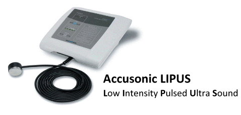
Teeth Regrowth Using Low Intensity Pulsed Ultrasound, LIPUS

This article has nothing really to do with how to increase our height but it does have another very important function and utility. We will focus back on the Low Intensity Pulsed Ultrasound technology to see how it will work to regrow our teeth and maybe ever regenerate lost teeth.

However, I have found a few links (like HERE) which claim that all you really need is a low frequency ultrasound source. You can’t get a high frequency ultrasound device legally unless you are a doctor. There is supposed to be a Novasonic device that can generate a sound vibration of 20,000 Hertz. If you touch the device to your skin, you can fell it working on the inside of the skin. There is also supposed to be a second type of device you can find on Ebay if you type in “ultrasound massager”. The link above says that they cost about $100-150. They are much more powerful and can generate sound vibrations of up to 3-5 mHz frequency. You put these device close to your teeth and gums every day and they massage your gums and teeth to help them become stronger and regenerate.

It is a little bad and the resource seems a little non-credible but from a badly designed Rex Research website found HERE, I copy and pasted the articles that have talked about the LIPUS technology for the use of teeth regeneration.

http://www.breitbart.com/news/2006/06/28/060628204537.2422eofv.html
http://news.yahoo.com/s/afp/20060628/wl_canada_afp/canadaussciencehealth&printer=1;_ylt=Ai0n21.IDNpv5KbEzjuV2Pz6OrgF;_ylu=X3oDMTA3MXN1bHE0BHNlYwN0bWE-
Wed Jun 28, 4:47 PM ET

Smile! A New Canadian Tool Can Regrow Teeth Say Inventors
Snaggle-toothed hockey players and sugar lovers may soon rejoice as Canadian scientists said they have created the first device able to re-grow teeth and bones.

The researchers at the University of Alberta in Edmonton filed patents earlier this month in the United States for the tool based on low-intensity pulsed ultrasound technology after testing it on a dozen dental patients in Canada.

“Right now, we plan to use it to fix fractured or diseased teeth, as well as asymmetric jawbones, but it may also help hockey players or children who had their tooth knocked out,” Jie Chen, an engineering professor and nano-circuit design expert, told AFP.

Chen helped create the tiny ultrasound machine that gently massages gums and stimulates tooth growth from the root once inserted into a person’s mouth, mounted on braces or a removable plastic crown.

The wireless device, smaller than a pea, must be activated for 20 minutes each day for four months to stimulate growth, he said.

It can also stimulate jawbone growth to fix a person’s crooked smile and may eventually allow people to grow taller by stimulating bone growth, Chen said.

Tarek El-Bialy, a new member of the university’s dentistry faculty, first tested the low-intensity pulsed ultrasound treatment to repair dental tissue in rabbits in the late 1990s.

His research was published in the American Journal of Orthodontics and Dentofacial Orthopedics and later presented at the World Federation of Orthodontics in Paris in September 2005.

With the help of Chen and Ying Tsui, another engineering professor, the initial massive handheld device was shrunk to fit inside a person’s mouth.

It is still at the prototype stage, but the trio expects to commercialize it within two years, Chen said.

The bigger version has already received approvals from American and Canadian regulatory bodies, he noted.

Copyright © 2006 Agence France Presse. All rights reserved. The information contained in the AFP News report may not be published, broadcast, rewritten or redistributed without the prior written authority of Agence France Presse. // Copyright © 2006 Yahoo! Inc. All rights reserved.


http://myprofile.cos.com/telbialy

Tarek El-Baily
University of Alberta
Faculty of Medicine and Dentistry
Dentistry
Orthodontics
Associate Professor Appointed: 2005

Mailing Address:

University of Alberta, Graduate Orthodontic Program
Faculty of Medicine and Dentistry
4051 Dent/Pharm Bldg.
Edmonton, Alberta T6G 2N8
Canada

Contact Information

Phone: (780) 492-2751
Fax: (780) 492-1624
telbialy@ualberta.ca
http://www.uofaweb.ualberta.ca/ortho/nav02.cfm?nav02=10606&nav01=1

Profile Details: Last Updated: 6/20/2006 — COS Expertise ID #896844 — Reference this profile directly: http://myprofile.cos.com/telbialy


http://www.dent.ualberta.ca/nav02.cfm?nav02=47501&nav01=44192

Dr. Tarek El-Bialy, Dr. Jie Chen & Dr. Ying Tsui Awarded Grant
Congratulations to Dr. Tarek El-Bialy and his team Dr. Jie Chen and Dr. Ying Tsui (from the Electrical Engineering department) who has recently been awarded with the NSERC (121) [Idea to Innovation] grant for “Intraoral Wireless Device for Dental Tissue Formation and Tooth-Root Healing”.

Moreover, Dr. Tarek El-Bialy has discovered that this ultrasound can stimulate lower jaw growth especially in patients with craniofacial problems like Hemifacial Microsomia. Usually these patients have to undergo many surgeries during their lives. This new device will be expected to improve many unsolved problems in Dentistry and Craniofacial areas. A provisional patent has been filed based on this research as well as the awarded grant.  More details about this research can be found at the following website http://myprofile.cos.com/telbialy

For the second time in UA history, our research team has awarded an NSERC (I2I)[Idea to Innovation] grant to miniaturize a small ultrasound device for stimulating teeth healing and dental tissue formation. This team includes in addition to Dr. Tarek El-Bialy  (in the Orthodontic Graduate program and Biomedical Engineering), Drs. Jie Chen and Ying Tsui from the Electrical Engineering department. When it was published by Dr. Tarek El-Bialy at the American Journal of Orthodontics for the first time in History that new dental tissue can be reformed after the teeth are grown, this research team and patent was planned for.


http://www5.eurekalert.org/pub_releases/2006-06/uoa-umh062806.php

Contact: Phoebe Dey
phoebe.dey@ualberta.ca
780-492-0437
University of Alberta

Ultrasound may help regrow teeth
Hockey players, rejoice! A team of University of Alberta researchers has created technology to regrow teeth–the first time scientists have been able to reform human dental tissue.

Using low-intensity pulsed ultrasound (LIPUS), Dr. Tarak El-Bialy from the Faculty of Medicine and Dentistry and Dr. Jie Chen and Dr. Ying Tsui from the Faculty of Engineering have created a miniaturized system-on-a-chip that offers a non-invasive and novel way to stimulate jaw growth and dental tissue healing.

“It’s very exciting because we have shown the results and actually have something you can touch and feel that will impact the health of people in Canada and throughout the world,” said Chen, who works out of the Department of Electrical and Computer Engineering and the National Institute for Nanotechnology.

The wireless design of the ultrasound transducer means the miniscule device will be able to fit comfortably inside a patient’s mouth while packed in biocompatible materials. The unit will be easily mounted on an orthodontic or “braces” bracket or even a plastic removable crown. The team also designed an energy sensor that will ensure the LIPUS power is reaching the target area of the teeth roots within the bone. TEC Edmonton, the U of A’s exclusive tech transfer service provider, filed the first patent recently in the U.S. Currently, the research team is finishing the system-on-a-chip and hopes to complete the miniaturized device by next year.

“If the root is broken, it can now be fixed,” said El-Bialy. “And because we can regrow the teeth root, a patient could have his own tooth rather than foreign objects in his mouth.”

The device is aimed at those experiencing dental root resorption, a common effect of mechanical or chemical injury to dental tissue caused by diseases and endocrine disturbances. Mechanical injury from wearing orthodontic braces causes progressive root resorption, limiting the duration that braces can be worn. This new device will work to counteract the destructive resorptive process while allowing for the continued wearing of corrective braces. With approximately five million people in North America presently wearing orthodontic braces, the market size for the device would be 1.4 million users.

In a true tale of interdisciplinary work, El-Bialy met Chen at the U of A’s new staff orientation. After hearing about Chen’s expertise in nanoscale circuit design and nano-biotechnology, El-Bialy explained his own research and asked if Chen might be able to help produce a tiny ultrasound device to fit in a patient’s mouth. The two collaborated and eventually along with Tsui received a grant from NSERC’s “Idea to Innovation,” program to expand on their prototype.

Dr. El-Bialy first discovered new dental tissue was being formed after using ultrasound on rabbits. In one study, published in the American Journal of Orthodontics and Dentofacial Orthopedics, El Bialy used ultrasound on one rabbit incisor and left the other incisor alone. After seeing the surprising positive results, he moved onto humans and found similar results. He has also shown that LIPUS can improve jaw growth in cases with hemifacial microsomia, a congenital syndrome where one side of the child’s jaw or face is underdeveloped compared to the other, normal, side. These patients usually undergo many surgeries to improve their facial appearance. This work on human patients was presented at the World Federation of Orthodontics in Paris, September 2005.

“After proving it worked, we looked at creating a smaller ultrasound carrier where we can take the patient out as a variable,” said El-Bialy. “Before this, a patient has to hold the ultrasound for 20 minutes a day for a year and that is a lot to ask.”

The researchers are currently working on turning their prototype into a market-ready model and expect the device to be ready for the public within next two years.

For more information, please contact:

Dr. Tarek El-Bialy, Faculty of Medicine and Dentistry
University of Alberta, 780-492-2751

Dr. Jie Chen, Faculty of Engineering
University of Alberta, 780-492-9820

Dr. Ying Tsui, Faculty of Engineering
University of Alberta 780-492-3192

Phoebe Dey, Public Affairs
University of Alberta, 780-492-0437


http://www.cbc.ca/story/science/national/2006/06/28/teeth-grow.html

Dentist, Engineer Team up to Regrow eeth
CBC News

A tiny ultrasound device could help people regrow teeth, researchers at the University of Alberta say.

The prototype device offers a way to reform human dental tissue for the first time, the team said Wednesday.

Everyone from hockey players to children who knock out a tooth could benefit.

The treatment, called low-intensity pulsed ultrasound, massages the gums to stimulate jaws, encourage growth in the roots of teeth and aid healing in dental tissue.

“If the root is broken, it can now be fixed,” said Dr. Tarak El-Bialy of the Faculty of Medicine and Dentistry. “And because we can regrow the teeth root, a patient could have his own tooth rather than foreign objects in his mouth.”

El-Bialy discovered ultrasound could be used to form new dental tissue from his research on rabbit incisors, which was published in the American Journal of Orthodontics and Dentofacial Orthopedics.

He then tested the technique on people who needed to get their teeth pulled.

Participants held the bulky ultrasound device for 20 minutes a day for four weeks against a tooth that had a problem, such as erosion after a root canal.

When El-Bialy looked at the extracted teeth under the microscope, he found new tissue was added to the roots of treated teeth, but not to untreated ones. The therapy regenerates the inner part of the tooth, but not the enamel.

He then teamed up with engineers Jie Chen and Ying Tsui to make the ultrasound device smaller so it could fit comfortably inside a patient’s mouth.

The prototype can be mounted on braces or a plastic removable crown.

The team has filed for a patent on their prototype in the U.S. They expect to have a version that is ready for patients within two years.


Increase Height And Grow Taller Using Low Intensity Pulsed Ultrasound, LIPUS

Now in my search for a solution to our height increase problem, I have looked through at least 30 different ideas people have thought up as a possible solution. Some of them make no sense, some of them are scams just trying to take your money, other methods are created with good intentions but just can’t work, and some have a chance to work but there has only been circumstantial evidence and testimonials which are hard to believe. Most of us want to see it with our own eyes to believe in something.

As for the latest technologies that might be coming along that can solve our problems, the 3 big ones are stem cells (probably through implants), gene therapy which is what I believe will ultimately happen if we can figure out how to integrate foreign DNA into our cells and mutate our bodies in a safe way for desired phenotypical traits, and the the joint loading modality (LSJL) which only a few people even know exists. There is some claims over hypnosis and mystic practices working too but that is vert hard to prove. Recently this new technology has come out that is really causing a lot of noise for height increase seekers, but also the dental community as well. It is the method called “Low Intensity Pulsed Ultrasound” or LIPUS for short.

Apparently I have been doing height increase research for so long and even I hadn’t heard about it until Harald of the Biomedical Growth Research Initiative told me that the two major technological developments that currently have the most potential were the LIPUS method and tissue engineering. I knew what tissue engineering did but I had not heard of LIPUS so I did some research. The stuff I found was very interesting. Then Harald explained to me that one of the ideas for his organization was to use LIPUS in some way to increase height, which made the method even more relevant.

Afterwards, I found information on Tyler’s HeightQuest.Com blog about LIPUS so I realized that the guys who are all at the cutting edge of height increase technology and innovation was suggesting that LIPUS could be very big deal. When I was doing research on the Lateral Synovial Joint Loading method on the HeightQuest.Com website, Tyler stated a lot about the potential of the LIPUS in being able to increase our height. That is why that I felt that I should devote quite a bit of time and effort into doing some extensive and serious research on the method and see just how it works, and how effective it will be.

So the first question would be “What is this technology called Low Intensity Pulsed Ultrasound?” . Wikipedia seems to have a clear and short answer to this question so I will paste their answer below (found HERE)


Low-intensity pulsed ultrasound (LIPUS) is a medical technology, generally using 1.5 MHz frequency pulses, with a pulse width of 200 μs, repeated at 1 kHz, at an intensity of 30 mW/cm2, 20 minutes/day.

Applications of LIPUS include:

  • Promoting bone-fracture healing.
  • Treating orthodontically induced root resorption.
  • Regrow missing teeth.[citation needed]
  • Enhancing mandibular growth in children with hemifacial microsomia.
  • Promoting healing in various soft tissues such as cartilage, inter vertebral disc.
  • Improving muscle healing after laceration injury.

Researchers at the University of Alberta have used LIPUS to gently massage teeth roots and jawbones to cause growth or regrowth, and have grown new teeth in rabbits after lower jaw surgical lengthening (Distraction osteogenesis) (American Journal of Orthodontics, 2002). As of June 2006, a larger device has been licensed by the Food and Drug Administration (FDA) and Health Canada for use by orthopedic surgeons. A smaller device that fits on braces has also been developed but is still in the investigational stage and is not available to the public.

It has not yet been approved by either Canadian or American regulatory bodies and a market-ready model is currently being prepared. LIPUS is expected to be commercially available before the end of 2012. The LIPUS foundation website currently announces that Lipus-Plasma application units are available for rental in the USA.

According to Dr. Chen from the University of Alberta, LIPUS may also have medical/cosmetic benefits in allowing people to grow taller by stimulating bone growth.

LIPUS has also been found to stimulate the proliferaton of chondrocytes.


Obviously the last two phrases on the wikipedia article are the most interesting since they state directly that the technology might have the application for stimulating bone growth by the promoting the proliferation of chondrocytes.

Not only this, this is incredible news for people who suffer from dental problems stemming from tooth decay. If this technology is as effective as advertized, it will change the dental industry. However, let’s learn more about what LIPUS does and how it does it.

From the UK website for National Institute For Health And Clinical Excellence located HERE the description for the device states…

Description – Low-intensity pulsed ultrasound aims to speed up fracture healing in broken bones by stimulating bone cells to grow and repair. This involves a short daily treatment using an ultrasound probe that is placed on the skin at the site of the fracture.

For the outline of the prodecure found in this sub-webpage HERE

2.2   Outline of the procedure

2.2.1  The aim of low-intensity pulsed ultrasound is to reduce fracture healing time and avoid non-union by delivering micro-mechanical stress to the bone to stimulate bone healing. This procedure is used to treat fresh fractures, fractures that are slower to heal than expected (delayed healing) and those that have failed to unite (non-union).

2.2.2  This is a non-invasive procedure. The ultrasound probe is positioned on the skin over the fracture and patients self-administer low-intensity pulsed ultrasound daily, usually for 20 minutes. If a patient’s limb is immobilised in a cast, then a hole is cut into the cast for the ultrasound probe. Coupling gel is used on the skin to aid conduction of the ultrasound signal. The operating frequency, pulse width, repetition rate and temporal average power of the ultrasound delivered can be varied.

In the Efficacy section, the experimental and testing results are provided…

2.3  Efficacy (RTC stands for randomized controlled trials)

2.3.1  A meta-analysis of 13 randomised controlled trials (RCTs) including a total of 563 patients (with a mixture of fresh conservatively or operatively managed and non-united fractures) treated by the procedure (n = 280) or a sham procedure (n = 283) reported a 34% (95% confidence interval [CI]: 21 to 44, 6 studies) overall mean reduction in healing time (confirmed by imaging) in patients treated by the procedure (follow-up not stated).

2.3.2  An RCT of 67 patients with closed or open grade I fractures of the tibial shaft (33 low-intensity pulsed ultrasound vs 34 sham) reported a mean healing time (defined as evidence of clinical and radiographic bridging of 3 cortices) of 96 days in the ultrasound group compared with 154 days in the sham group (p < 0.0001).

2.3.3  An RCT of 32 patients with fresh closed or open grade I fractures of the tibial shaft fixed with an intramedullary rod treated by the procedure (n = 15) or a sham procedure (n = 17) reported an average healing time (defined as radiographic bridging of 3 cortices assessed by a radiologist) of 155 days and 125 days respectively (p = 0.76).

2.3.4  An RCT of 21 patients with non-united fractures of the scaphoid treated by pedicle bone graft reported an average healing time (defined as clinical healing [solid and not causing tenderness or pain] and radiographic healing [complete bridging cortices]) of 56 days in patients who also received low-intensity pulsed ultrasound (n = 10) and 94 days in those who received sham therapy (n = 11) (p < 0.001).

2.3.5  An RCT of 30 patients with open tibial fractures or high-energy-induced complex tibial fractures treated by the procedure (n = 16) or a sham procedure (n = 14) reported an average time to full weight bearing of 9.3 weeks and 15.5 weeks respectively (p < 0.05).


Me: I managed to find a great scientific article that really goes into the explanation of the technology (located HERE). Let me take the most important parts of the article. 

“”….Low-intensity ultrasound is a biophysical form of intervention in the fracture-repair process, which through several mechanisms accelerates healing of fresh fractures and enhances callus formation in delayed unions and nonunions…Low-intensity pulsed ultrasound is currently applied transcutaneoulsy, although recent experimental studies have proven the efficacy of a trans-osseous application for both enhancement and monitoring of the bone healing process with modern smart implant technologies…”” (published by Elsevier Ltd. 2006)

Key Point: However, one of the fundamental concepts in orthopaedics is the understanding that the mechanical environment at the site of a fracture influences the pattern of fracture repair.

“….The healing of a fractured bone involves the spatial and temporal coordinated action of several different cell types, proteins and the expression of hundreds of genes working towards restoring its structural integrity….”

Basic science 

In vitro studies using cell cultures and research on experimental fractures in animal models have demonstrated a stimulatory biologic effect of low intensity ultrasonic energy on the intracellular activity, cytokine release and the bone healing process. In animal models, ultrasound appears to alter the time course or the sequence of gene expression of several genes, notably aggrecan, which is a proteoglycan involved in enchondral osteogenesis. Low-intensity ultrasound elevates intracellular calcium in cultured chondrocytes and stimulates endochondral bone formation in vitro. It also has direct effects on cell physiology by increasing the incorporation of calcium ions in cartilage and bone cell cultures and by stimulating the expression of numerous genes involved in the healing process. It alters potassium flux across the cell membrane in cultured thymocytes, and it modulates adenyle cyclase activity and TGF-b synthesis in osteoblastic cell
lines. In addition to modulating gene expression, ultrasound may enhance angiogenesis and increase blood flow around the fracture. Despite these well documented studies, the mechanism through which LIUS interacts with living tissue and stimulates bone healing remains unclear.

In addition to the above-mentioned molecular interactions, the acoustic pressure waves at the fracture site, facilitate fluid flow which, in turn, increases nutrient delivery and waste removal (acoustic streaming phenomenon), thus stimulating proliferation and differentiation of the fibroblasts, chondroblasts and osteoblasts.
In addition, the acoustic pressure waves produce micro-stress fields resulting in a mechanical response of the bone, analogous to the phenomena described by Wolf’s law. Small temperature fluctuations (<1 8C) appear at the fracture site as a result of the conversion of ultrasound energy to heat. Some enzymes, such as collagenase, are exquisitely sensitive to these small temperature variations, thus, ultrasound may also facilitate some enzymatic processes.

Trans-cutaneous application of ultrasound in the management of fresh fractures

Ultrasound increases soft callus formation and results in the earlier onset of endochondral ossification, suggesting that the most prominent effect is on the chondrocyte population….In a placebo-controlled study of bilateral mid-shaft fibular osteotomies in rabbits, Pilla et al. found that low-intensity pulsed ultrasound applied for 20 min/day significantly accelerated the recovery of torsional strength and stiffness.

The effect of ultrasound in distraction osteogenesis

Callus distraction is currently an established treatment for the management of defects larger than 3—4 cm in the long bones. However this technique carries the problem of the long time for healing and maturation of the newly formed bone and the burden to wear the external fixator for a very long time. The ossification process in distraction and maturation involves intramembranous bone as the dominant type of tissue formation while endochondral ossification normally is of minor importance.

The effects of low-intensity pulsed ultrasound on maturation of the distracted callus have been investigated in several animal studies, with controversial results. In a rabbit study, Shimazaki et al. found that bone mineral density, hard callus area, and mechanical test scores were greater in distraction callus treated with low-intensity pulsed ultrasound than in the control group. In a study of rats, Eberson et al. found that radiographicaly assessed healing occurred earlier in ultrasound treated bones than in control bones and that bone volume fraction and trabecular bone pattern, were higher in the ultrasound-treated bones. In a study of rabbits, Tis et al. found a greater hard callus area and less fibrous tissue in bones treated with low-intensity pulsed ultrasound than in control bones. Neither Eberson et al.
nor Tis et al. found a difference in bone mineral density or mechanical strength of distraction callus between ultrasound-treated bones and controls, although Eberson et al. observed a trend toward greater mechanical strength in ultrasound-treated bones. Uglow et al. found no substantial difference in bone mineral content, crosssectional area, or strength of distraction callus between ultrasound-treated bones and control bones of rabbits.

In a sheep metatarsal bone transfer model for the study of distraction osteogenesis, pulsed low-intensity ultrasound were applied transcutaneously after the distraction was complete and only throughout the maturation phase. Histologic analysis of the cortical defect zone showed approximately 32% more bone in the group stimulated by ultrasound. Although it presented seven times more intramembranous bone formation compared to endochondral in the control group, which is in accordance with results of another study, there was a three times higher rate of endochondral ossification in the specimens treated with ultrasound. Biomechanical tests showed significantly higher axial compression stiffness (1.4—2.7 times the control values) and significantly higher indentation stiffness of callus tissue in the healing zone of the treated bones. In all of the animal studies mentioned above, osteotomy and distraction were performed at the diaphysis, which consists of thick cortical bone. A recent investigation on rabbits showed that low-intensity pulsed ultrasound stimulates bone formation most effectively during the distraction phase.

In a randomised study (block randomisation) in humans with internal controls, the low-intensity pulsed ultrasound applied only during the consolidation phase (after distraction had ceased) on hemicallotasis after high tibial ostetomy, significantly enhanced the mineralisation of the callus. The bone mineral density in the metaphyseal segment adjacent to the distraction callus, in the previous study and also in animal studies collectively suggest that metaphyseal trabecular bone might be more susceptible than diaphyseal cortical bone to the mechanicalultrasonic stimuli.

Future clinical studies should address the question of whether additional low-intensity pulsed ultrasound treatment during the distraction phase can further shorten the period necessary for callus maturation. The distraction osteogenesis-specific mechanism that translates mechanical forces due to low-intensity pulsed ultrasound into bone formation need further clarification.

Me: Overall the paper was extremely insightful on the utility of the LIPUS technology. All I can say is that after reading the paper I can say with a 97% confidence that this technology is very amazing at what it can do. It really does have the potential to help people heal bones faster and to induce chondrocyte proliferation and bone formation. 

Note: Since this topic is a very large topic, I will stop right here and continue on this study on the device and technology in a later post. You can get to Part II by clicking HERE.

Growth Plate Regeneration By Robert Ballock Orthopaedic Surgeon

When I was in contact with Tyler from HeightQuest.Com he gave me two leads or ideas to look into so I could be better acquainted with what options and ideas are current available or in the process of being developed in our search for an increase in height. One of the ideas was the Joint Loading Modality done by Yokota which I wrote a very short review and analysis found HERE. The other idea he wanted me to do research on was the idea of “Growth Plate Regeneration” being developed by Robert Ballock from the Cleveland Clinic.

Let me do a little review and critical analysis on what I managed to find out about this method/technique. The first  thing I found from searching about the type of growth plate regeneration research done by Robert Ballock was either a paper submitted to the National Institute of Health (located HERE) or it was maybe a proposal for a grant or project.

However let’s get a summary of how Dr. Ballock describes his own research from his profile webpage found on the Cleveland Clinic website (located HERE) If you would like to contact him all his contact information is available on his profile webpage which you get get to again by clicking on the link above.

“”….I had the good fortune of being taken in by Drs. Michael Sporn and Anita Roberts in the Laboratory of Chemoprevention at the National Cancer Institute. Their laboratory had recently discovered TGF-beta and they were interested in exploring the role of this new peptide growth factor in regulating growth and differentiation in the skeleton.During these two years, I focused on the role of TGF-beta in the growth plate and developed the three-dimensional pellet culture model of growth plate chondrocyte differentiation that is now used by many laboratories throughout the world.This experience resulted in three publications, including first author papers in Developmental Biology and Journal of Cell Physiology….”

“”….I spent a third year in his laboratory at Johns Hopkins where I formulated the serum-free, chemically defined culture conditions for growth plate chondrocyte pellet cultures that are also now used by many laboratories world-wide.For example, Dr. Brian Johnstone and colleagues at CWRU used our pellet culture model and serum-free, chemically-defined conditions to demonstrate for the first time that mesenchymal stem cells could undergo chondrogenesis in vitro. This year at Johns Hopkins was marked by several other key observations, including the discovery that terminal differentiation of growth plate chondrocytes was a default pathway that could be accelerated by thyroid hormone, and that morphogenesis of columnar cartilage in the growth plate could be recapitulated in our serum-free three-dimensional pellet cultures by addition of thyroid hormone….”

“”….my research program began its focus on the molecular mechanisms of thyroid hormone action in the growth plate that continues to be the major thrust of our laboratory work today.We initially determined that the principal site of thyroid hormone action during skeletal maturation was regulation of the critical transition between cell proliferation and terminal hypertrophic differentiation in the growth plate.In addition to characterizing the expression of thyroid hormone receptors in this tissue, we also explored the interactions between thyroid hormone, vitamin D, and retinoic acid in regulating terminal differentiation of growth plate chondrocytes.Other work established an important link between thyroid hormone-induced terminal differentiation and upregulation of cell cycle proteins p21cip-1, waf-1 and p57 kip-1, indicating that growth arrest at the G1-S restriction point of the cell cycle is an obligatory step in the terminal differentiation process of growth plate chondrocytes….”

“”….we also began to investigate the role of obesity in the dysfunction of the growth plate that results in slipped capital femoral epiphysis, a potentially devastating hip condition in adolescents. These studies have demonstrated that peroxisome proliferator activated receptors (PPARs), which are upregulated in response to a high fat diet, are also expressed in growth plate chondrocytes and interfere with the normal transcriptional activation function of thyroid hormone receptors in these cells, eventually resulting in the inhibition of terminal differentiation and matrix mineralization that allows the subsequent mechanical failure of the growth plate to occur….”

“”….we initiated DNA microarray studies of growth plate chondrocytes in order to identify the direct downstream genetic targets of the thyroid hormone receptor in the growth plate.These studies resulted in the surprising identification of the gene encoding carboxypeptidase Z (CPZ) as a direct target of thyroid hormone action.CPZ is an enzyme that removes C-terminal amino acid residues, particularly arginines, from proteins, and has also been shown to modulate Wnt signaling.Further experiments in our laboratory have demonstrated that Wnt-4, which is the principal Wnt family member expressed in the mammalian growth plate, contains a C-terminal arginine, and that removal of this C-terminal arginine enhances the biological activity of Wnt-4 in inducing terminal differentiation of these cells….”

Me: As for what I can contribute to the effort or knowledge right now, there is little I can do that has not already been covered and researched by Tyler from HeightQuest.Com. He has already looked into the proposal ideas. And yes, it appears the first link was a grant proposal of some kind to get money to fund for a project. The project was supposed to have started in April of 2012 and end in March 2014. They appeared to have gotten some money, around $140,000 which in my opinion is really NOT a lot.  I’ll copy and paste the entire abstract right below here. I’ll highlight the areas which I feel are the most important and applicable to us height increase seekers. Again you can get to the paper submitted by clicking HERE.

Abstract Text:
DESCRIPTION (provided by applicant): The growth plate, also known as the epiphyseal plate or physis, is the area of growing tissue near the end of the long bones in children and adolescents that determines the future length and shape of the mature bone. Fractures through the cartilage growth plates of the long bones of children may result in growth arrest with subsequent leg length inequality and progressive deformity. This growth arrest is due to formation of a bony bar across the traumatic growth plate defect that acts as an tether to resist further longitudinal growth. If the bar is large or is located in the central portion of the growth plate, a complete growth arrest ensues. A bar located in the peripheral portion of the physis tethers growth asymmetrically, producing a progressive angular deformity of the limb. Once a physeal bar forms, surgical excision is technically difficult and resumption of further growth is quite variable. Previous studies of experimental growth plate injury have focused on the histological events in the growth plate defect leading to bar formation. However, our understanding of the factors that regulate the proliferation and differentiation of growth plate chondrocytes, as well as the principles of cartilage tissue engineering, have increased dramatically over the past decade. These advances now provide a unique opportunity to develop strategies for regeneration of normal physeal cartilage following serious growth plate injuries. Successful regeneration of growth plate cartilage architecture in vivo would have a transformational impact on the practice of pediatric orthopaedic surgery, providing for the first time not only the ability to replace growth plates irreversibly damaged by trauma, infection or irradiation, but also the possibility of restoring longitudinal growth in individuals beyond the age of skeletal maturity. Our hypothesis is that co-cultured chondrocytes and osteoblasts implanted into tibial bone defects in vivo will recapitulate the function of the normal growth plate and result in the reformation of columnar physeal architecture and resumption of longitudinal growth. This hypothesis will be tested by using a tissue engineering approach to determine the degree to which this optimized physeal construct replicates the function of the normal growth plate in vivofollowing implantation into a complete growth plate defect.PUBLIC HEALTH RELEVANCE: Our understanding of the factors that regulate the proliferation and differentiation of growth plate chondrocytes, as well as the principles of cartilage tissue engineering, have increased dramatically over the past decade. These advances now provide a unique opportunity to develop strategies for regeneration of normal physeal cartilage following growth plate injury. Successful regeneration of growth plate cartilage architecture in vivo would have a transformational impact on the practice of pediatric orthopaedic surgery, providing for the first time not only the ability to replace growth plates irreversibly damaged by trauma, infection or irradiation, but also the possibility of restoring longitudinal growth in individuals beyond the age of skeletal maturity.

Me: If you took the time to read the abstract, it might still be a little hard to understand what Dr. Ballock is talking about. The basic idea proposed is that for many children who are still growing, when they suffer an injury like a fracture on their growth plates, they can actually stunt their growth or growth irreuglarly afterwards because of how the growth plates cartilage form and heal themselves. To solve this problems, as well as give all height increase seekers around the world hope, is that they want to test this hypothesis and radical on implanting some cultured (made in the lab) chondrocytes (growth plate cells) and osteoblasts (bone cells) into the tibia fractures and defects and see if they can result in the growth plates in regrowing again and providing longitudinal growth (increase in bone length, thus increase in height). This technology is defenitely something every serious height increase seekers should stay up to date with. One might even be able to participate in the study and even get their height increase for free during the experimental trials and be paid some extra money for being a willing trial subject. Just something to think about. 

To be completely honest, it is actually really hard to find anymore information about Dr. Ballock or his research being done since professor doctors who do really advanced scientific research are not as easily found on the internet as a Kim Kardashian sex tape. To find the latest and most advanced innovations and scientific breakthroughs, you really have to dig through the science online databases like Pub Med. The problem is that I don’t have an account with PubMed and I am not sure I want to pay for something like a yearly fee to become some form of member.