Monthly Archives: September 2012

Regeneration Of The Growth Plate

I found this really interesting article entitle” Regeneration of the growth plates” and I wanted to read and review it. Unfortunately it is another scientific article submitted to PubMed that I can’t the full Text File for.

source HERE. The article abstract is found below…


Acta Anat (Basel). 1989;134(2):113-23.

Regeneration of the growth plate.

Langenskiöld A, Heikel HV, Nevalainen T, Osterman K, Videman T.

Source

Orthopaedic Hospital of the Invalid Foundation, Helsinki.

Abstract

The occurrence of growth plate regeneration has been doubted. However, in 5 different series of experiments reported between 1950 and 1986 regeneration of injured parts of growth plates in long bones of rabbits and pigs could be demonstrated. The 1st series implied partial X-ray injury of growth plates in rabbits aged 3-6 weeks. The 2nd series implied autotransplantation of the head of the fibula in rabbits aged 10-21 days. The 3rd, 4th and 5th series implied transplantation of autologous fat grafts into provoked defects of growth plates in rabbits and pigs. The findings show that regeneration of a growth plate occurs when a part of it is injured in such a manner that a bone bridge is not formed between the epiphysis and the metaphysis. Regeneration of a plate is much faster in relation to the growth in length of the bone in the rabbit than in the pig. The 1st and 2nd series suggest that regeneration takes place by interstitial proliferation of cells from the germinal layer of the uninjured parts of the plate. Signs of partial regeneration of growth plates have been seen in radiographs after operation for partial closure of growth plates in children.

PMID: 2718724 [PubMed – indexed for MEDLINE]

**[Me: I really wanted to ask you people how do I get these full doc files? And how much would it cost me to get these files? i don’t think they are free.] 

Tallness And Height Gene Discoveries

From source Med Page Today and American Journal of Human Genetics

So I found this article and I felt that it was a nice addition to the site to show just how many genes and factors have been found so far that seems to influence the human height. It is really overwhelming and at this, point I don’t think geneticists and scientists can do gene therapy to increase height on a potential embryonic specimen.

My suggestion is similar to what the doctors are saying right now. We want to focus on learning about the possible ways height decreased disorders come about and learn their mechanism. If we can disrupt the genetic mechanistic pathway of the process, at least we can prevent and treat people who are suffering from pathologies leading to height deficiencies and prevent people from growing short.

Again, I will highlight the parts which I found the most important.


Tallness Gene Discoveries Could Give Legs to Other Research

By Crystal Phend, Senior Staff Writer, MedPage Today

Published: December 30, 2010

Reviewed by Robert Jasmer, MD; Associate Clinical Professor of Medicine, University of California, San Francisco and Dorothy Caputo, MA, RN, BC-ADM, CDE, Nurse Planner

Action Points

  • Explain that scientists probing the genetics of height have discovered dozens more uncommon variants, adding to the nearly 200 already known.
  • Note that the study was a meta-analysis of studies done using the same genotyping chip with dense coverage of genes for uncommon single nucleotide polymorphisms (SNPs) and detected 30 new signals missed by regular chips in the past.
  • Explain that the researchers expect their process to be helpful in unraveling the genetics of complex diseases such as diabetes and MS.

Using a process that could help unravel the causes of a variety of complex diseases, scientists probing the genetics of height have discovered dozens more uncommon variants, adding to the nearly 200 already known.

Meta-analysis of studies using the same genotyping chip with dense coverage of genes for uncommon single nucleotide polymorphisms (SNPs) turned up 30 new signals missed by regular chips in the past, Hakon Hakonarson, MD, PhD, of Children’s Hospital of Philadelphia, and colleagues reported online in the American Journal of Human Genetics.

Still more variants remain to be found, Hakonarson’s group noted.

One recent analysis suggested that about 300,000 SNPs would be required to explain just 45% of the variance in height across the population, they wrote.

That’s a situation that may be similar to the genetics underlying some common health problems, Hakonarson explained in an interview.

“The ultimate goal is really to look at heart disease, cardiovascular risk factors, and diabetes,” he told MedPage Today.

Executive director or the American College of Medical Genetics, Michael S. Watson, PhD, agreed that the study carries broad implications for genetic research.

This study offers a reality check on how complex it can get with many, many genes involved plus environmental factors, and still only a minority of the heritability of the trait identified,” Watson wrote in an e-mail to MedPage Today and ABC News.

Few diseases are caused by so-called one-hit genetic mutations, noted Jerry W. Shay, PhD, of the University of Texas Southwestern Medical Center in Dallas.

“Most human diseases such as cancer, type 1 diabetes, multiple sclerosis, Crohn’s disease, and psychiatric disorders are much more complicated and may involve interplay between multiple genetic and epigenetic changes and the environment,” he said in an e-mail to MedPage Today and ABC News.

Understanding the genetic basis of something as straightforward as height is just the tip of the iceberg,” he added. “It is proof that our DNA contains valuable information that we are only beginning to dissect.”

Hakonarson’s group used height as a prototype to determine what could be done mining into known genes in greater detail with more density of the SNPs in each gene.

The researchers pooled results from 47 studies that genotyped 114,223 adults across six ethnicities using a gene chip targeted to cardiovascular-related gene loci but still contained many plausible height-related loci.

Comparing height to the results on the nearly 50,000 SNPs across about 2,000 loci, a total of 64 loci contained an SNP that predicted height with array-wide significance (P<2.4 x 10⁻⁶).

Among them, 42 loci exceeded the conventional genome-wide significance threshold of P<5 × 10⁻⁸.

Although 20 of the 64 had previously been described in studies as linked to height, Hakonarson’s group identified associations with several new regions containing genes with “interesting biological roles,” including growth hormone receptor, glucokinase regulatory protein, circadian rhythm, and collagen formation.

They paid special attention to variants of lower minor allele frequency (carried by less than 5% of the population) “that would go undetected in studies relying on imputation or in studies with fewer participants” and found 22 such SNPs in eight loci significantly associated with height.

Such genetic findings could be clinically useful in evaluating children of short stature of unknown cause, commented Ronald Bachman, MD, a geneticist at Kaiser Permanente in Oakland, Calif.

However, there’s limited value for the general public, and height shouldn’t be the subject of consideration for direct-to-consumer genetic testing, he warned.

“The interpretation of the results would probably lead to a great deal of stress and then perhaps unnecessary testing and treatments,” Bachman said in an e-mail to MedPage Today and ABC News.

Psychosocial Short Stature aka Kaspar Hauser Syndrome And The Relationship Between Mental Health And Height

I recently found a Wikipedia article (HERE) on a topic which made me really think about a critical element to what determines our overall height, which is our mental and emotional state.

We all know that genetics, sleep, nutrition, and exercise plays a role in determining our height, but what about our mental health? There are many claims going around the height increase and grow taller niche and one of them is that of using hypnosis. A lot of people state that for you to be taller, you have to really be positive and believe you WILL grow taller and increase your height. So the obvious question is. How much does our mental state affect our height?

From the article on Psychosocial Short Stature, it seems that the effects are really dramatic, at least when it some to how it can limit our grow if we are not in the healthiest of mental states.

From the wiki article…

Psychosocial short stature (PSS) or psychosocial dwarfism, sometimes called psychogenic or stress dwarfism, or Kaspar Hauser Syndrome,[3] is a growth disorder that is observed between the ages of 2 and 15, caused by extreme emotional deprivation or stress.

The symptoms include decreased growth hormone (GH) secretion, very short stature, weight that is inappropriate for the height, and immature skeletal age. This disease is a progressive one, and as long as the child is left in the stressing environment, his or her cognitive abilities continue to degenerate. Though rare in the population at large, it is common in feral children and in children kept in abusive, confined conditions for extended lengths of time. It can cause the body to completely stop growing but is generally considered to be temporary; regular growth will resume when the source of stress is removed.

Etiology

Children with PSS have extremely low levels of growth hormone. These children possibly have a problem with growth hormone inhibiting hormone (GHIH) or growth hormone releasing hormone (GHRH). The children could either be unresponsive to these hormones or too sensitive.

Children who have PSS exhibit signs of failure to thrive. Even though they appear to be receiving adequate nutrition, they do not grow and develop normally compared to other children of their age.

An environment of constant and extreme stress causes PSS. Stress releases hormones in the body such as epinephrine and norepinephrine, engaging what is known as the ‘fight or flight’ response. The heart speeds up and the body diverts resources away from processes that are not immediately important; in PSS, the production of growth hormone (GH) is thus affected. As well as lacking growth hormone, children with PSS exhibit gastrointestinal problems due to the large amounts of epinephrine and norepinephrine, resulting in their bodies lacking proper digestion of nutrients and further affecting development.

While the cure for PSS is questionable, some studies show that placing the child affected with the disease in a foster or group home increases growth rate and socialization skills.

Me: I asked a person I am working with on what she can find on the link between mental health and human growth and she pointed out two articles which I looked over. They are located HERE (resource 1) and HERE (resource 2).

From resource 1, HERE

Other endocrine interactions

Growth, reproductive function, and the thyroid axis are also influenced by stress system activation. In the acute setting of stress, glucocorticoids stimulate the growth hormone gene, leading to enhanced growth hormone secretion.53 However, with more prolonged stress, growth hormone release is suppressed by CRH‐induced elevations in somatostatin levels.54 This results in an inverse relationship between the diurnal concentrations of cortisol and growth hormone. Also, glucocorticoids directly inhibit growth hormone effects at target tissues by inhibiting insulin‐like growth factor‐1 (IGF‐1) and other growth factors.55 The effects of stress on the growth axis may account for the delay in growth often seen in chronic disease and emotional deprivation in childhood.

CRH, β‐endorphin, and glucocorticoids inhibit GnRH secretion from the hypothalamus. Glucocorticoids also suppress pituitary gonadotrophin release and inhibit gonadal tissue.56 Patients with illnesses associated with increased HPA‐axis activity, such as anorexia nervosa, hyperthyroidism, and malnutrition, may experience abnormalities of menstrual and reproductive function.57–,59

From resource 2, HERE

Me: I really coudn’t figure out the link to the study and how human height is effected. The conclusion is that behavior created from stress is from the Corticotropin-releasing hormone receptor, but don’t need the actual hormone. The Abstract below…

Stress-induced behaviors require the corticotropin-releasing hormone (CRH) receptor, but not CRH

+

Author Affiliations

  • *Program in Neuroscience, Howard Hughes Medical Institute, and Division of Endocrinology and Department of Neurology, Children’s Hospital, and Harvard Medical School, Boston, MA 02115; Department of Pharmacology, Louisiana State University Medical Center, Shreveport, LA 71130; Department of Psychology, Tufts University, Medford, MA 02155; and **Department of Psychology, University of Wisconsin, Madison, WI 53706
  • Edited by Bruce S. McEwen, The Rockefeller University, New York, NY, and approved April 30, 1999 (received for review October 6, 1998)


Next Section

Abstract

Corticotropin-releasing hormone (CRH) is a central regulator of the hormonal stress response, causing stimulation of corticotropin and glucocorticoid secretion. CRH is also widely believed to mediate stress-induced behaviors, implying a broader, integrative role for the hormone in the psychological stress response. Mice lacking the CRH gene exhibit normal stress-induced behavior that is specifically blocked by a CRH type 1 receptor antagonist. The other known mammalian ligand for CRH receptors is urocortin. Normal and CRH-deficient mice have an identical distribution of urocortin mRNA, which is confined to the region of the Edinger–Westphal nucleus, and is absent from regions known to mediate stress-related behaviors. Since the Edinger–Westphal nucleus is not known to project to any brain regions believed to play a role in anxiety-like behavior, an entirely different pathway must be postulated for urocortin in the Edinger–Westphal nucleus to mediate these behaviors in CRH-deficient mice. Alternatively, an unidentified CRH-like molecule other than CRH or urocortin, acting through the CRH receptors in brain regions believed to mediate stress-induced behaviors, may mediate the behavioral response to stress, either alone or in concert with CRH.

Stress, defined as the response of the body to any threatening demand (1), can be broadly separated into physiological responses, including the stimulation of adrenal glucocorticoid secretion, and behavioral responses, including anxiety and fearful behavior. Alterations in the stress-response system are believed to underlie many anxiety-related disorders (2–5). Corticotropin-releasing hormone (CRH) has been implicated in both physiological and behavioral stress responses. CRH was identified by its ability to stimulate adrenocorticotropic hormone (ACTH) secretion from anterior pituitary corticotrophs, thus activating the hypothalamic–pituitary–adrenal (HPA) axis (6). In addition, infusion of CRH into the brain was found to cause stress-like behaviors (7), suggesting that CRH integrates physiological and behavioral activities into a generalized stress response. Subsequently, many other pharmacological studies have implicated CRH in the behavioral response to stressors. These studies confirmed that intracerebral infusion of CRH induces stress-like behaviors and additionally that intracerebral infusions of CRH antagonists blunt the behavioral response to a stressor (8–10). Furthermore, transgenic mice that overexpress CRH exhibit increased anxiety-like behavior (11).

The role of the CRH receptor in the behavioral stress response has been further evaluated. The two known CRH receptors, type 1 and type 2 (12), both consist of a 7-transmembrane helix functionally coupled to adenylate cyclase via Gs. For the most part, the anatomical distributions of the two CRH receptors are distinct, with the type 1 receptor expressed in the central nervous system in regions including neo-, olfactory, and hippocampal cortices, subcortical limbic structures in the septal region and amygdala, certain relay nuclei in the brainstem, and the cerebellum (13). One splice variant of the type 2 receptor is expressed in the periphery, whereas another splice variant is expressed in specific subcortical structures, the lateral septal nuclei, and the hypothalamus (14, 15). Two reports of mice lacking the CRH type 1 receptor have confirmed a role for this receptor in anxiety-related behavior (16, 17). CRH type 1 receptor-deficient mice display decreased anxiety-like behavior in the dark–light emergence task and the elevated-plus maze, both behavioral paradigms thought to measure anxiety in rodents. Both studies conclude that CRH mediates the behavioral responses to stressors by means of the CRH type 1 receptor (16, 17). More recently, a CRH type 1 receptor antagonist has been found to block both the acquisition and expression of stress-induced behaviors in rats (18).

Thus, the CRH type 1 receptor clearly mediates behavioral responses to stress, with the evidence to date implicating CRH as its most likely ligand (9, 10, 19). The other known mammalian ligand for CRH receptors is urocortin (20). Urocortin, acting either alone or in concert with CRH, is a potential mediator of in vivo stress responses, because in the studies cited above, infused or transgenically overexpressed CRH could act at receptor sites normally occupied by urocortin, and CRH antagonists could block urocortin actions as well as those of CRH (21). Therefore, we examined the role of CRH in this pathway by characterizing the behavioral responses to stressors of CRH-deficient (knockout, KO) mice (22) and the effects of CRH antagonists on these responses. We also examined the expression of urocortin mRNA in wild-type (WT) and CRH KO mouse brain to assess whether its distribution provides insight into its potential role in mediating stress-induced behaviors.

Me: What I can say is that stress does cause the human brain to release certain hormones that seems to inhibit the human longitudinal growth process. Here is my theory, from using evolutionary biology. The human body can be viewed as a system of work done that needs energy input. For the body/system to become bigger, it needs a lot more energy than before to even get it to grow to that size. 

So, while you are still young, stay happy and dont get too stress out. There is really no good point and you won’t be inhibiting your growth process.

If we view stress, as a form of resistance or internal conflict, we can use the analogy froom physics that stress is like friction, which is lost energy. If we are losing the energy, we don’t have the energy to go through our growth spurts. So stress is taking away the energy our young bodys/minds need to make that quantum energy leap. 

From the researcher, she states…

“cortisol is released during stress, and cortisol inhibits growth,….Yes, being in a happier less stressful situation, a person raising their chances of growing.”


Increase Height By Quad Rod Implants Into The Vertebrate For Scoliosis Patients

Me: I wanted to make this post be about using surgery to correct for spinal deformities. In effect, the rod implanted into the vertebrate to stop the curvature of the spine and relieve much pain will also increase height.

I found this article HEY Clinic For Spine & Surgery at Raleigh, NC where a spine surgeon talks about using a new 4-rod implant to make a patient who already had implants in her back stronger and more durable since her current rods have broken apart..


The Hey Clinic for Scoliosis and Spine Surgery

This is where Dr. Hey shares his thoughts regarding recent patients he has cared for, and shares photos and case summaries to benefit patients, their families and friends, other physicians, medical students, residents, and fellows. Dr. Hey works at the Hey Clinic for Scoliosis and Spine Surgery, Raleigh, NC USA http://www.heyclinic.com.

FRIDAY, SEPTEMBER 14, 2007
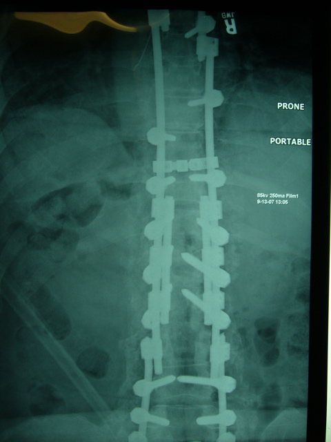
Hey Clinic Sep 13 2007 AM Surgery: New “QUAD ROD” technique for stabilizing thoracic kyphotic fracture with history of rod breakage.

Yesterday morning, September 13th 2007, we helped a Nancy, a 75 yo woman who had a T12 severe wedge fracture above a previous fusion which caused myelopathy and severe pain. This was fixed using a laminectomy, and extension instrumentation and fusion. Postoperatively, she had trouble with rod breakages at that high load area around T12, with 2 rod construct, and then 3 rod construct. She how presented with recurrent rod breakage at that same T12 area, breaking both rods, and breaking conectors for the triple rod, resulting in recurrent kyphosis and pain. Given the amount of wedging at T12, we had discussed possibility of fixing this problem with an anterior/posterior procedure, removing body of T12 and replacing the vertebra with a strut expandable cage with graft, then instrumenting posteriorly. However, that is a very big operation, which involves having to take down diaphragm, etc., and Nancy was not interested in that at all. So, for the past couple of months, I have been working on several different engineering designs to make a stronger construct across this junction, “playing” with my set of Synthes “erector set” tools, and trying to come up with a new design which would be at least ten times stronger than the last one, but also be low profile enough that it would not cause problems with soft tissue prominence.

What seemed to be the best solution is a new surgical technique that I call the “Quad Rod”. Instead of just having 2 rods going across the area of the fracture, there are actually four rods, all closely coupled to each other with very strong rod to rod connecters. The combination of this second rod on each side closely coupled to the main rod means that there is major improvement in bending and twisting strength and rigidity, while still keeping a low profile instrumentation, while leaving the lateral “gutter” over the transverse processes wide open for BMP and bone graft.

Over the past couple months, I have done a bunch of different permutations of the “Quad Rod” on the spine model, and also experimented with other combinations including a 6 rod construct, which proved to be very strong, but very bulky. I also got a chance to try a small version of the Quad Rod unilaterally on a surgery a couple weeks ago which went well, and helped me think through some of the complexities of the rod insertion.

For my resident and fellow and younger spine surgeon readers, let me go over a few basic points.

1. Rods and/or screws are not meant to be able to withstand a lifetime of loading for most normal-sized people: they are meant to hold things in position hopefully long enough for the bone to heal, forming a fusion along the spine. This fusion can be thought of as “cement” that very slowly “hardens” around the metal superstructure, forming a very solid construct. However, the bone can take up to a year or even more to completely heal, especially in older patients who have weaker bone that does not make bone as quickly. If the bone does not heal, it is possible that the rods could break, or that the screws could loosen in the bone, and start to “toggle”. This could cause pain and deformity.

2. Whenever you face a revision instrumentation case, you always want to be thinking about making your new construct much stronger than the old one, while trying to get the bone to heal quicker using biological enhancers like bone morphogenic protein (BMP) and local or autologous bone graft. My military analogy here is this: If you get beaten in battle with 20 guys, don’t go back the next time with 20 or 21 guys —-> bring 100+ guys the next time, maybe with fire power from aircraft and a tank or two!

In this particular case, my first revision instrumentation used a triple rod technique with BMP, which had been my rock-solid revision instrumentation technique for broken rods, which had worked without fail for past 3 years or so without fail… Until Nancy broke the Triple Rod a couple months ago! So this time I am calling out the Marines, Air Force, Army, Navy and the Six Million Dollar Man! Here are the things I planned to do, and did differently with this revision:

1. Replace the screws above and below the unstable fracture with new screws, at least 1mm in diameter larger than the last ones. Putting in new screws here helps since the old ones had a lot of “cycles of load” on them and could break. 1mm larger diameter for each of these screws also makes the screw much stronger — the strength of the screw goes up as the 4th power of the diameter! (That’s like way bigger than double, or square or triple). So, for example, if you were to double the diameter of the screw, the strength would go up 16 time!

2. Put pedicle screws into the body of T12, where the fracture occurred. While I could not do this originally when the fracture occurred, since the body was “smashed”, now that it has been a year or more since her original fracture, I had hoped that the bone would have healed. Much to my delight today, I found that the bone had healed. It was still very tricky to get the screws placed, given previous laminectomy and scar, etc, but thankfully I got in 2 very solid screws at T12. This extra vertebral body support directly adjacent to where the fracture was unstable helped tremendously to improve the strength of the overall construct.

3. Stronger Rods. Thankfully, Synthes just recently released a new Titanium Alloy rod which is much stronger than the old rod, but is the same 7mm diameter. One of the fun things that occurred in this particular case was my interactions not only with Nancy, but with Nancy’s husband, Bob, who has a phenomenal background in metallurgy and engineering. One of Bob’s initial suggestions for this last revision was to use larger diameter rods, which from an engineering standpoint makes a lot of sense, but from a biologic standpoint doesn’t work as well, since the rods would become prominent. It would also require all of the screws and other connectors to be re-machined, and then retested. The best we could do was to get a stronger rod, but in the same diameter by changing the titanium alloy.

4. “HEY QUAD ROD”. This is one strong construct. The close coupling of 2 rods almost right next to each other with three encasing titanium block sleeves, which are then locked to each other with set screws makes for an incredible “I-Beam” that resists bending and twisting greatly. We recently received a grant, and are working with Professor Mazzolini and others at NC State Department of Mechanical Engineering to study the strength of some of these new constructs using computer models and lab testing. This “Quad Rod” is tough to put together, but once together is very low profile, and very strong. I have a bunch of tips that make it work very well. I used the new Quad Rod with the Synthes USS Titanium system, which has a special cap and nut locking system that locks the rod to the screw. One of the troubles you may have trying to do the Quad Rod with another system is that the screw diameter may be too large to allow the second joining rod to fit with the rod to rod connector. The rod to rod connector is something that other systems may have, which is used usually for doing extension instrumentations, where one rod is added onto another rod. In this case, I actually used a total of 5 rod to rod connectors on each side, and 10 altogether: three are used around the apex of the fracture, with the middle one right over the T12-L1 toggle point, and then one above at T11-T12 interspace, and L1-L2 interspace just below the apex of the “toggle”. The other 2 rod to rod connectors are placed at the top and bottom of the whole section of rod I replaced, which were down at L2 and up at T6 on each side. It turns out that the rod to rod connectors are almost the perfect length to “fill” the interspace between each of the pedicle screws, creating an additional thick “sheath” around the rod for extra bending resistance. The closely coupled rod next to the main rod provides additional bending and twisting support as a very tightly joined and/or cross-linked rod.

5. Additional cross-link between L and R rods just above the Quad Rod. There was too much fusion mass below the Quad Rod to put another cross-link, plus the large fusion mass below worked like a “cross link”.

6. Very aggressive cleanout of the pseudarthrosis at T12-L1, and the lateral “gutter” on either side, out over some of medial rib on both sides. This decoricated old fusion mass made a great new organic “bed” for two long strips of Medtronic Bone Morphogenic Protein (BMP) soaked sponges, followed by a layer of local bone graft, followed by a thick layer of allograft finely morselized chips.

Overall, the surgery went very well, taking right around 4 hours total.
I was able to accomplish all six of the steps above, although there were some real “fiddle factors” that needed to be dealt with. Here are a few pearls:

  1. Your replacement long rod needs to be just the right length to connect between the very top and bottom connectors, but not significantly longer. When you go to insert the final construct, you need to slide it in one end, and then reduce it into the screw channels above, and then slide it up and into the rod to rod connectors at the very top.
  2. Keeping those top and bottom connectors a little bit loose so they can rotate helps to get the new rod into the hole, and then rotated down into position and into the screw slots.
  3. Measure your “partner rod”, the second rod on each side to bridge across 3 interspaces around the max stress point, with middle rod to rod connector over where the rods broke the last time.
  4. Bend the rod in that area for both rods as little as possible, to avoid weakening the rod, and also to allow the rod to rod connectors to slide easily.
  5. Attach the “partner rod” before inserting the overall construct, and put it medial to the main rod, with pedicle screw openings also facing medial. You will not be able to slide in partner rod when the first rod is in place. Placing it medial leaves the lateral gutter totally free for BMP and bone graft.
  6. Mark the main rod with a pen where the 3 rod to rod connectors need to go to fall between the pedicle screws.
  7. Put the middle of the 3 connectors on the partner and main replacement rod first, then slide the other two connectors over either end. Much easier than trying to guide the smaller partner rod through 3 connectors.
  8. Insert the longer replacement rod at one end of the distal connectors first, as mentioned above, and then slide it into the upper connector, taking advantage of the rod to rod connector’s ability to rotate 90 degrees, and then down into position next to the screws.
  9. Use the “Pursuader” to push the rod into the pedicle screws — it even works across the two rod construct! Tighten your set screws a little bit before you “pursuade” the double-rod construct into the screw slot.
  10. As you are tapping the cap down over the main rod, there is very little space between the 2 rods — back off the compressor on the Pursuader a little bit, and the narrow “skirt” of the cap will go right between the 2 rods, and lock into position. Yahoo!
  11. Torque down your set screws before tightening your nuts on top of the caps, for maximum strength.
  12. Do thorough cleanout of lateral gutter and takedown of pseudarthrosis.
  13. Strongly consider prior to placing rods putting additional screw points of fixation around the area of the “toggle” as I did, by putting T12 screws into old fractured vertebra. The more points of fixation you have, the better the load is shared to the spine, with less bending moment between screws.
  14. Cut the BMP sponges into 2 longitudinal strips, and lay them down as a floor to the “gulley” over the transverse processes, ribs, and old fusion mass.
  15. Undercut the paraspinal muscle flaps to allow for tension-free fascial closure at the end of surgery.

We will be studying this and other constructs in the future.

I encourage you to spend a lot of time “playing” with this and other constructs, using the plastic spine models with points of fixation, but also experimenting with other potential constructs as a pure erector set, to understand the possibilities. I see some possible very interesting constructs by joining 2 rod to rod connectors serially together, forming a double hinge. This allows you to have a “Quad Rod” where the rods are further apart, thereby potentially creating a better “I Beam” , by increasing the distance between the 2 beams. However, there are prominence problems, and the need for 2 connectors connected by a short intermediate rod I think is not half as strong as having 2 rods locked side by side. Perhaps a wider rod to rod connector would be a possibility, to allow the second rod to have more flexibility as to where it lies, and to potentially increasing the distance between the rods to increase strength.

Below are some pictures of our performance of our first real “Hey Quad Rod”.

I am happy to say that we got an excellent correction of her deformity, and we still have load sharing on the middle column anteriorly. However there is a big “divit” in the anterior portion of T12, which would have been nice to “fill in” with an anterior strut, but too invasive for Nancy. Our posterior Quad Rod tension band, combined with our “quick dry” BMP/bone graft combination, combined with even stricter postoperative patient restrictions for bending and lifting should hopefully lead to a long-term fix for Nancy.

This evening she was looking great in ICU, ready to get up and be probably 3-4 inches taller.

I hope these notes and pictures are helpful to my fellow surgeons out there. Please do not hesitate to call or email me if you have further questions or thoughts.

My email is “hey” at heyclinic.com, and my phone is available through website http://www.heyclinic.com.

Lloyd A. Hey, MD MS

Hey Clinic for Scoliosis and Spine Surgery

Raleigh, NC USA

http://www.HeyClinic.com

Height Increase – Do You Realize How Close We Are?

This article was sort of in the middle of nowhere and when I found it, I thought it was written by Tyler.  However, Tyler did comment to it. I wanted to continue this discussion further to get some more thought going on this topic.

Of all the articles written about height increase, this article was one of the most advanced and scientifically valid I have seen.

The areticle is found HERE.

Height Increase-Do you realize how close we are?

PLEASE NOTE: The validity of this article has been questioned. We do not have the resources to verify its claims, so please read the article with this in mind. We do not endorse or promote the views expressed by this guest post writer.

It’s frustrating that we are so close to finding a way for people to increase height, but we’re held up because scientists have to get funding and they’re not doing the right studies. Do you realize that their are no studies on height increase with mesenchymal stem cells? I mean come on harvest some red bone marrow from the trabecular bone and inject it into the hyaline cartilage growth plate line. Do you realize that hyaline cartilage composes the resting zone of the growth plate and since it has already excreted extracellular matrix, it does not undergo apoptosis like the rest of the chondrocytes in the growth plate?

Mesenchymal stem cells by definition are capable of inducing chondrogenesis. Scientists have found that a stem cell-like cancer cell was able to re-create a growth plate in hyaline cartilage. We are so close and yet we have to wait for the scientific method to go through it’s big, ugly steps.

Do you realize also that dynamic compressive loading of chondrocytes has been shown to alter gene expression in the cartilage? What would happen if this gene expression induced chondrogenesis in the hyaline cartilage in the growth plate line? Hmm, height increase by mechanotransduction.

It is so frustrating to see people get caught up in trying to grow taller with HGH or IGF-1 when their are so many other answers available. If IGF-1 works then it works by increasing Mesenchymal Stem Cell proliferation and Lithium also increases mesenchymal stem cell proliferation but it has the advantage of being legal (Me: I don’t believe this part is true. I needed Lithium once and I had to go to a doctor for this). The reason that gigantism works the way it does is by an alteration in the bodies homeostasis. It’s not just increased levels of HGH, the tumor in the pituitary gland(or whatever it is that alters the bodies homeostasis) works by altering the bodies negative feedback mechanisms to high levels of HGH. Robert Wadlow never stopped growing. You know why? Because his elevated levels of IGF-1 increased stem cell proliferation to the point where it never stopped.

The growth plates do not fuse. The hyaline cartilage is still there, it just becomes inactive. – (Me: This is the part which is controversial)

We are so close to finding a way to increase height but we’re too caught up on HGH and not caught up enough on mutagenics and stem cells. Do your part to help change that.

{ 2 comments… read them below or add one }

anonymous

There are no studies on height increase with mesenchymal stem cell because it is not worth considering. You can’t increase height with mesenchymal stem cell after puberty or prepuberty.

Even though there’s a little interrelationship between mesenchymal stem cell and epiphyseal plates, that doesn’t mean you can increase height with mesenchymal stem cell.

You need to do some more research.

Tyler Davis

There are some inaccuracies in this article that I the author want to correct:

The hyaline cartilage does disappear after endochondral ossification but as a separate process then chondrocytes differentiating into bone cells. However, stem cells don’t need hyaline cartilage to differentiate into chondrocytes.

Also, Lithium Supplementation is not legal without a prescription. However, having the Lithium ion in your body is legal.

If you have information about why mesenchymal stem cells can’t cause height growth I would like to hear them.


Me: I have looked at the same research and it appears the growth plate line does completely disappear over time. Stem cells have shown to be able to differentiate. Even though much of the original writer’s points are flawed, I think he/she does raise a really good point. I do believe also that we have the science and theory down well enough to develop some form of surgical process that allows us to implant a growth plate/stem cells/ hyaline cartilage  implant and make it work.

For anyone who is willing to separate their legs just temporarily, we can add a 3-4 mm thick plate with the right layers inside and allow endochondral ossification to do the rest of the work. All that is needed is to be able to inject stem cells into the plate or get the stem cells inside the bone to differentiate into the right types of chondrocytes. 

If anyone can show me why that can’t work, they have to go into some deep explanation since everything I have learned and read so far shows that it is already possible to get people to increase in height through stem cell implantations.

Regrow Joint Cartilage Using Chondroitin Sulfate, Bone Marrow Stem Cells, And Scaffold Implantations

Released: 7/17/2012 1:30 PM EDT 

Source: Johns Hopkins Medicine

Newswise — Johns Hopkins tissue engineers have used tiny, artificial fiber scaffolds thousands of times smaller than a human hair to help coax stem cells into developing into cartilage, the shock-absorbing lining of elbows and knees that often wears thin from injury or age. Reporting online June 4 in the Proceedings of the National Academy of Sciences, investigators produce an important component of cartilage in both laboratory and animal models. While the findings are still years away from use in people, the researchers say the results hold promise for devising new techniques to help the millions who endure joint pain.

“Joint pain affects the quality of life of millions of people. Rather than just patching the problem with short-term fixes, like surgical procedures such as microfracture, we’re building a temporary template that mimics the cartilage cell’s natural environment, and taking advantage of nature’s signals to biologically repair cartilage damage,” says Jennifer Elisseeff, Ph.D., Jules Stein Professor of Ophthalmology and director of the Translational Tissue Engineering Center at the Johns Hopkins University School of Medicine.

Unlike skin, cartilage can’t repair itself when damaged. For the last decade, Elisseeff’s team has been trying to better understand the development and growth of cartilage cells called chondrocytes, while also trying to build scaffolding that mimics the cartilage cell environment and generates new cartilage tissue. This environment is a 3-dimensional mix of protein fibers and gel that provides support to connective tissue throughout the body, as well as physical and biological cues for cells to grow and differentiate.

In the laboratory, the researchers created a nanofiber-based network using a process called electrospinning, which entails shooting a polymer stream onto a charged platform, and added chondroitin sulfate—a compound commonly found in many joint supplements—to serve as a growth trigger. After characterizing the fibers, they made a number of different scaffolds from either spun polymer or spun polymer plus chondroitin. They then used goat bone marrow-derived stem cells (a widely used model) and seeded them in various scaffolds to see how stem cells responded to the material.

Elisseeff and her team watched the cells grow and found that compared to cells growing without scaffold, these cells developed into more voluminous, cartilage-like tissue. “The nanofibers provided a platform where a larger volume of tissue could be produced,” says Elisseeff, adding that 3-dimensional nanofiber scaffolds were more useful than the more common nanofiber sheets for studying cartilage defects in humans.

The investigators then tested their system in an animal model. They implanted the nanofiber scaffolds into damaged cartilage in the knees of rats, and compared the results to damaged cartilage in knees left alone.

They found that the use of the nanofiber scaffolds improved tissue development and repair as measured by the production of collagen, a component of cartilage. The nanofiber scaffolds resulted in greater production of a more durable type of collagen, which is usually lacking in surgically repaired cartilage tissue. In rats, for example, they found that the limbs with damaged cartilage treated with nanofiber scaffolds generated a higher percentage of the more durable collagen (type 2) than those damaged areas that were left untreated.

“Whereas scaffolds are generally pretty good at regenerating cartilage protein components in cartilage repair, there is often a lot of scar tissue-related type 1 collagen produced, which isn’t as strong,” says Elisseeff. “We found that our system generated more type 2 collagen, which ensures that cartilage lasts longer.”

“Creating a nanofiber network that enables us to more equally distribute cells and more closely mirror the actual cartilage extracellular environment are important advances in our work and in the field. These results are very promising,” she says.

Other authors included Jeannine M. Coburn, Matthew Gibson, Sean Monagle and Zachary Patterson, all from Johns Hopkins University.

The research was supported by grants R01 EB05517, F31 AG033999 and F30 AG034807 from the National Institutes of Health.