Monthly Archives: September 2012

The Connection Between Parathyroid Hormone-Related Protein (PTHrP), Parathyroid Hormone (PTH), Indian Hedgehog (Ihh), And Height

For anyone who has ever studied the science of how the molecular biology of the growth plate within hormones interact this post will be a complete outline and summary on one of the best well understood hormone/protein pathways. It includes the Parathyroid Hormone-Related Protein (PTHrP), Parathyroid Hormone (PTH), and Indian Hedgehog (Ihh).

Because it is so well understood, it is easy to understand why and how it affects the fate of the growth plates.

Analysis & Interpretation

If you treated young mice (not adult mice) with the PTH, growth plate thickness, chondrocyte number, and the overall bone lengthen increases. If you tried to unload the bone, you decrease all three. If you did PTH administration with unloading, the PTH reduces the effect of unloading the bone.

The PTHrP (parathyroid hormone related peptide) and Ihh (indian hedgehog) works in a growth restraining feedback loop by controlling the pace of chondrocyte differentiation in the human growth plate. The PTHrP and Ihh are expressed at the same chondrocytes are the estrogen receptors alpha and beta types. If you remove either the PTH or the PTHrP it results in bone abnormal growth which is usually stunting from the chondrocytes which age faster and multiply slower.

IHH, PTHrP, and PTH/PTHrP receptor mRNA were detected in prehypertrophic and hypertrophic chondrocytes in both sexes during development. The presence of IHH, PTC, PTHrP, and PTH/PTHrP receptor protein in prehypertrophic and hypertrophic chondrocytes. In addition, staining for hedgehog, PTC, and PTHrP also was observed in growth plate stem cells. Furthermore, no mRNA or protein expression of the mentioned factors was detected in the perichondrium. Our data suggest that in contrast to the proposed feedback loop in the early embryonic growth plate, which requires the presence of the perichondrium, a feedback loop in the postnatal growth plate can be confined to the growth plate itself. In fact, two loops might exist: (1) a loop confined to the transition zone and early hypertrophic chondrocytes, which might in part be autocrine and (2) a loop involving the growth plate stem cells.

Indian hedgehog (IHH), produced by prehypertrophic and hypertrophic chondrocytes, stimulates production of parathyroid hormone-related protein (PTHrP) by perichondrial and early chondrocytic cells. PTHrP then maintains chondrocytes in a proliferative, less differentiated state. IHH and PTHrP may participate in a negative feedback loop that synchronizes and determines the pace of differentiation of chondrocytes in the growth plate. IHH is a master regulator of both chondrocyte and osteoblast differentiation.

PTHrP appears to act as a bifunctional modulator of both chondrocyte proliferation and differentiation, through signal transduction linked to the PTH/PTHrP receptor and by its direct action in the nucleolus. While the downstream promoter controls PTH/PTHrP receptor gene expression in bone and cartilage, it is differentially regulated in the two tissues. 1alpha,25-dihydroxyvitamin D3 downregulated the activity of the downstream promoter in osteoblasts, but not in chondrocytes, both in vivo and in vitro

As always the most important parts are highlighted.


From PubMed study HERE

PTHrP and the regulation of longitudinal growth: Exploring novel strategies to increase final height in children with short stature.

Dit is de samenvatting van de aanvraag.

Longitudinal growth is controlled by the activity of the chondrocytes in the epiphyseal plate, which is present at the distal ends of the long bones, and is regulated by a multitude of hormones, growth factors and metabolic conditions. Among these, gonadal steroids and particularly estrogen, are of pivotal importance. In humans, estrogen is responsible for growth plate closure causing a complete arrest in longitudinal growth at the end of puberty. The molecular mechanisms by which estrogen influences longitudinal growth and induces growth plate closure are largely unknown. One such mechanism can be the growth restraining feedback loop consisting of Parathyrooid Hormone related Peptide (PTHrP) and Indian Hedgehog (Ihh). This feedback loop has been shown to regulate longitudinal bone growth by controlling the pace of chondrocyte differentiation in the embryonal chicken, mice and human growth plate. More recently, we have shown that this feedback loop is also operational after birth. In addition, we have found that PTHrP, Ihh and estrogen receptors Era and ß are co-expressed in the same growth plate chondrocytes and that gonadectomy influences the expression of components of the feedback loop. A role for PTHrP in growth plate closure is furthermore supported by observations in humans and transgenic mice, in which the presence of an constitutively activating mutation in the PTH/PTHrP-receptor results in dwarfism and premature growth plate closure. At present, the molecular mechanisms by which the PTHrP/Ihh-feedback loop controls chondrocyte differentiation and is modulated by estrogen are largely unknown. The aims of the present project are: To gain more insight in the molecular mechanism by which ethe PTHrP/Ihh feedback loop controls the pace of chondrocyte differntiation with empasis on the identification of PTHrP target genes and interactions with members of the TGFß/BMP-superfamily. To characterize the interactions between the PTHrP/Ihh-feedback loop and estrogen and to determine its role in growth plate closure. To examine the relevance of the findings in experimental models for longitudinal growth and in archival material of human growth plates. In vitro and ex vivo experiments are performed using the chondrogenic cell line ATDC-5 and cultured metatarsals of wild type, PTHrP -/- and PTH/PTHrP-receptor -/- mice. PTHrP target genes will be identified using DNA microarray technology using both commercially available and custom made cDNA chips. Adenoviral mediated gene transfer will be used to manipulate the expression of receptor for members of the TGFß/BMP-superfamily and estrogen (e.g. mock, wild type, constituteively active, dominant negative). The effects of tese manipulations on the PHTrP/Ihh-feedback loop will be examined at the mRNA level using custom made cDNA chips, at the level of signal transduction by studying the activation state of cytoplasmic signal transduction intermediates, and at the level of transcriptional activation using artificial promotor reporter constructs. The relevance of the findings will be tested in previously collected epiphyseal plates of gonadectomized growing rats or of immature rats supplemented with various sex-steroieds. This material is well characterized with respect to various parameters regarding growth allowing direct correlations between the expression of the candidate tgenes and longitudinal growth. Furthermore, findings will be extended to the human situation by analysing archival growth plates of lethal skeletal dysplasias and of children who have undergone orthopaedic surgery. The results obtained in this study will extent our knownledge on the molecular mechanisms by which longitudinal growth is regulated at the level of the epiphyseal plate and growth plate closure is induced. As such the results will help in the rational design of novel diagnostic tests and growth promoting strategies for growth retarded patients who do not benefit from currently available therapies.


From PubMed study HERE

Journal of Bone and Mineral Metabolism

March 2002, Volume 20, Issue 2, pp 83-90

Human PTH (1–34) induces longitudinal bone growth in rats

Taishi Ogawa, Hiroshi Yamagiwa, Tadashi Hayami, Zhang Liu, Kuan-Yu Huang, Kunihiko Tokunaga,Takehiro Murai, Naoto Endo

Abstract.

The growth plate is a specialized structure that is responsible for longitudinal bone growth (LGR). Growth plate organization is altered with loading in rats. Parathyroid hormone (PTH) is known to induce mitogenic effect on chondrocytes in vitro. Type I PTH/PTH related peptide (rP) receptor is expressed in growth plate cartilage in rats. We therefore investigated the effect of PTH administration on the organization and longitudinal growth rate of the growth plate in rats. We also investigated the effect of PTH on the changes induced by unloading in the organization and growth of the growth plate. Thirty 6-week-old and 30 15-week-old male Sprague-Dawley rats were randomly assigned to five groups (n = 6 per group), i.e., basal controls, control (i.e., normally loaded), PTH-treated control (i.e., PTH-treated under normal loading), unloaded, and PTH-treated under unloading. PTH-treated animals received human PTH (1–34) at a dose of 80 μg/kg per day five times per week for 3 weeks, for the duration of unloading. In young loaded rats treated with the systemic administration of PTH, growth plate thickness, chondrocyte number, and LGR were increased in the proximal tibiae compared with findings in young loaded rats without PTH administration. Hindlimb unloading induced a reduction in growth plate thickness, chondrocyte number, and LGR. In young rats, systemic administration of PTH partly prevented these changes induced by unloading. These preventive effects of PTH were observed only in young rats; not in adult rats. These results show that the systemic administration of PTH stimulates longitudinal bone growth, and diminishes the reduction in growth plate growth induced by unloading in young rats.


From PubMed study HERE

Histol Histopathol. 2000 Jul;15(3):957-70.

Recent studies on the biological action of parathyroid hormone (PTH)-related peptide (PTHrP) and PTH/PTHrP receptor in cartilage and bone.

Amizuka N, Henderson JE, White JH, Karaplis AC, Goltzman D, Sasaki T, Ozawa H.

Source

Department of Oral Anatomy, Niigata University Faculty of Dentistry, Japan. amizuka@dent.niigata-u.ac.jp

Abstract

Mice with a targeted deletion of parathyroid hormone (PTH)-related peptide (PTHrP) develop a form of dyschondroplasia resulting from diminished proliferation and premature maturation of chondrocytes. Abnormal, heterogeneous populations of chondrocytes at different stages of differentiation were seen in the hypertrophic zone of the mutant growth plate. Although the homozygous null animals die within several hours of birth, mice heterozygous for PTHrP gene deletion reach adulthood, at which time they show evidence of osteopenia. Therefore, PTHrP appears to modulate cell proliferation and differentiation in both the pre and post natal period. PTH/PTHrP receptor expression in the mouse is controlled by two promoters. We recently found that, while the downstream promoter controls PTH/PTHrP receptor gene expression in bone and cartilage, it is differentially regulated in the two tissues. 1alpha,25-dihydroxyvitamin D3 downregulated the activity of the downstream promoter in osteoblasts, but not in chondrocytes, both in vivo and in vitro. Most of the biological activity of PTHrP is thought to be mediated by binding of its amino terminus to the PTH/PTHrP receptor. However, recent evidence suggests that amino acids 87-107, outside of the amino terminal binding domain, act as a nucleolar targeting signal. Chondrocytic cell line, CFK2, transfected with wild-type PTHrP cDNA showed PTHrP in the nucleoli as well as in the secretory pathway. Therefore, PTHrP appears to act as a bifunctional modulator of both chondrocyte proliferation and differentiation, through signal transduction linked to the PTH/PTHrP receptor and by its direct action in the nucleolus.

PMID:  10963138 [PubMed – indexed for MEDLINE]


From PubMed study HERE

The parathyroid hormone-related protein and Indian hedgehog feedback loop in the growth plate.

Kronenberg HM, Chung U.

Source
Endocrine Unit, Massachusetts General Hospital and Harvard Medical School, Boston, MA 02114, USA.

Abstract

Normal development of the growth plate requires coordinated proliferation and differentiation of chondrocytes and osteoblasts. In previous work, we have shown that Indian hedgehog (IHH), produced by prehypertrophic and hypertrophic chondrocytes, stimulates production of parathyroid hormone-related protein (PTHrP) by perichondrial and early chondrocytic cells. PTHrP then maintains chondrocytes in a proliferative, less differentiated state. Because this less differentiated state delays the production of IHH, IHH and PTHrP may participate in a negative feedback loop that synchronizes and determines the pace of differentiation of chondrocytes in the growth plate. To establish the roles of physiological levels of PTHrP and IHH, we have now injected PTH/PTHrP receptor (-/-) embryonic stem (ES) cells into normal blastocysts to generate mice with chimeric growth plates. The PTH/PTHrP receptor cells leave the proliferative cycle and differentiate prematurely in the middle of the normal proliferative columns. The columns of wild-type cells are longer than normal and the adjacent bone collar is also longer than normal. Patterns of gene expression and the use of chimeras using PTH/PTHrP receptor (-/-); IHH (-/-) ES cells suggest that modified patterns of IHH and PTHrP synthesis explain these abnormalities. Thus, IHH is a master regulator of both chondrocyte and osteoblast differentiation.

PMID: 11277077   [PubMed – indexed for MEDLINE]


From PubMed study HERE

J Bone Miner Res. 2000 Jun;15(6):1045-55.

Expression of Indian hedgehog, parathyroid hormone-related protein, and their receptors in the postnatal growth plate of the rat: evidence for a locally acting growth restraining feedback loop after birth.

van der Eerden BC, Karperien M, Gevers EF, Löwik CW, Wit JM.

Source

Department of Pediatrics, Leiden University Medical Center, The Netherlands.

Abstract

A locally acting growth restraining feedback loop has been identified in the murine embryonic growth plate in which the level of parathyroid hormone-related peptide (PTHrP) expression regulates the pace of chondrocyte differentiation. To date, it is largely unknown whether this feedback loop also regulates the pace of chondrocyte differentiation in the growth plate after birth. We therefore characterized the spatio-temporal expression of Indian hedgehog (IHH), PTHrP, and their receptors in the postnatal growth plate from female and male rats of 1, 4, 7, and 12 weeks of age. These stages are representative for early life and puberty in rats. Using semiquantitative reverse-transcription polymerase chain reaction (RT-PCR) on growth plate tissue, IHH and components of its receptor complex, patched (PTC) and smoothened (SMO), PTHrP and the type I PTH/PTHrP receptor messenger RNA (mRNA) were shown at all ages studied irrespective of gender. Using in situ hybridization, IHH, PTHrP, and PTH/PTHrP receptor mRNA were detected in prehypertrophic and hypertrophic chondrocytes in both sexes during development. In addition, especially in the younger age groups, faint expression of PTH/PTHrP receptor mRNA also was shown in stem cells and proliferative chondrocytes. Immunohistochemistry confirmed the observations made with in situ hybridization, by showing the presence of IHH, PTC, PTHrP, and PTH/PTHrP receptor protein in prehypertrophic and hypertrophic chondrocytes. In addition, staining for hedgehog, PTC, and PTHrP also was observed in growth plate stem cells. No differences in staining patterns were observed between the sexes. Furthermore, no mRNA or protein expression of the mentioned factors was detected in the perichondrium. Our data suggest that in contrast to the proposed feedback loop in the early embryonic growth plate, which requires the presence of the perichondrium, a feedback loop in the postnatal growth plate can be confined to the growth plate itself. In fact, two loops might exist: (1) a loop confined to the transition zone and early hypertrophic chondrocytes, which might in part be autocrine and (2) a loop involving the growth plate stem cells.

PMID:   10841173         [PubMed – indexed for MEDLINE]


Korean Singer Boa Is Dissatisfied Over Her Height

Since I am temporarily living in the Far East, first in South Korea, and now China, I do read and watch a lot of news of the local nations. One news that has been going around the Korean airwaves is about one of the most famous Korean singers in the last decade, a female singer that goes by the name of Boa.

Recently, maybe a few months ago, the singer came on in a public show to express her regret at her short stature (source HERE).

Singer BoA expressed her regret over her short height.

BoA revealed through her Twitter on September 18th, “If I could go back to my teens…I would drink so much milk…I want to return :’(”

BoA also wrote, “I’m going to listen to Eun Ji Won‘s ’160′* while sleeping ^^ good night ^^”

*(160 cm is about 5.25 ft)

Fans who visited BoA’s Twitter responded with comments like, “Honest national singer“, “You’re still pretty even if you’re small“, and “If you’re tall also, you’d be too perfect~“.

Meanwhile, BoA will reveal her repackage album, “Copy & Paste“, through KBS Music Bank on the 24th.

Source: Newsen

From Soompi website HERE


BoA Blames SM for Her Short Height

BoA recently expressed her dissatisfaction toward some parts of her intense training over the years.

On an episode of “Win Win” that aired on May 29, BoA was asked, “When you were a trainee, were you scolded a lot?” She answered, “I was told to lose weight a lot.”

She explained that although she was a young girl of only 14 years old, she had to go on a diet. “Do you think I was able to grow while trying to lose weight at such an age? I think I’ll keep this grudge forever.”

Furthermore, BoA shared how hard it was during her Japanese debut process. She stated, “Girls’ Generation and SHINeeeach had interpreters but DBSK and myself did not. We had interviews lined up after merely a day into our Japanese debut and no matter how much we studied, it was really difficult to speak.”

BoA continued, “Eventually, the questions began to repeat so we just started to memorize our answers. I’m always being asked to do difficult things. I don’t like being called a ‘pioneer’ because – why do I always have the pave the way?”

Meanwhile, BoA shared many episodes and thoughts during this episode including her first impression of YG and how many agencies wanted to recruit her in the past.

Me: I guess even the best and most famous of us still might feel small. Will we never be satisfied with who we are? 

What Are The Bone Growth Factors? (Important)

What are the bone growth factors?

Me: I am going to find out what are the primary, and the most influential bone growth factors by using four main scientific papers published in journals.

1. From PubMed, US National Library of Medicine, National Institute of Health. Link HERE.

Clin Orthop Relat Res. 1991 Feb;(263):30-48.

Bone growth factors.

Mohan S, Baylink DJ.

Source

Department of Medicine, Loma Linda University, CA.

Abstract

Bone volume is determined by the relative rates of bone formation and bone resorption. Recent research in several laboratories suggests that growth factors may act locally to modulate bone formation by stimulating osteoblast proliferation and activity. A number of bone-derived growth factors have been isolated and characterized from bone matrix extracts and from media conditioned by bone cells and bone organs in culture. The growth factors found in bone matrix include insulin like growth factors I and II, transforming growth factor-beta, acidic and basic fibroblast growth factor, platelet-derived growth factor, and bone morphogenetic proteins. Conditioned medium from bone cells contains several of these growth factors and also hematopoietic factors. These bone matrix-derived growth factors have different biologic activities, including mitogenic, differentiating, chemotactic, and osteolytic activities. Evidence suggests that bone cells produce substantial quantities of growth factors for extracellular storage in bone matrix. Apart from being produced for extracellular storage, it is possible that growth factors secreted by bone cells have acute effects on their neighboring osteoblastic cells, i.e., paracrine action, or on themselves, i.e., autocrine action. The release of matrix-stored growth factors by bone resorption may mean that growth factors act as delayed paracrine agents, e.g., osteoblasts deposit growth factors in bone and later when these growth factors are released from bone via bone resorption, the growth factors stimulate osteoblast precursors to proliferate. The findings that bone is a storehouse for growth factors and that bone cells in culture produce and respond to bone growth factors suggest bone growth factors may act as potential determinants of local bone formation. This review is focused on the structure, regulation, and biologic actions of the known bone growth factors.

2. Wiley Online Library, Journal of Bone and Mineral Research > Vol 8 Issue S2 > Abstract  , Link HERE

Growth factors to stimulate bone formation

  1. David J. Baylink M.D.*,
  2. Richard D. Finkelman,
  3. Subburaman Mohan

Article first published online: 3 DEC 2009

DOI: 10.1002/jbmr.5650081326

Journal of Bone and Mineral Research

Supplement: Journal of Bone and Mineral Research

Volume 8, Issue Supplement S2, pages S565–S572,December 1993

Abstract

During the past decade we and others have shown that bone is a storehouse for growth factors. Accordingly, bone contains a number of growth factors including insulin-like growth factors I and II (IGF-I, IGF-II) transforming growth factor (TGF-β1, TGF-β2), platelet-derived growth factor, basic and acidic fibroblast growth factor, and bone morphogenetic proteins (BMPs). Osteoblasts have been shown to produce many of these growth factors, which then act in an autocrine and paracrine fashion. The production of these growth factors is regulated by both systemic hormones and local mechanical stress. Recent studies on the relative distribution of bone growth factors during different physiologic and pathologic situations indicate that the concentration of bone growth factors is not invariant and provide indirect evidence that growth factors deposited in bone have physiologic significance. In addition, many of these bone growth factors have been shown to increase bone formation either systemically or locally in vivo. Based on the past findings, we propose that different growth factors may have a specific role in regulating proliferation and differentiation of different stages of osteoblast lineage cells and play important roles in the local regulation of bone formation.

3. From Healio.com Education Lab, Orthopedics. Link HERE

Osteoinductive growth factors can aid bone growth in orthopedic procedures

BMPs are making autogenous iliac crest bone graft the ‘old gold standard,’ investigator says.

  • Orthopedics Today, July 2008

Six years after FDA approval of the first bone growth factor, a bone morphogenetic protein, researchers and clinicians still seek a better understanding of these powerful substances, their uses and limitations.

Of all the bone growth factors studied, bone morphogenetic proteins (BMP) expressed during bone healing have been found most effective for initiating bone growth in clinical situations such as spine fusion and fracture healing.

“I think growth-factor-based strategies are important for bone formation, especially in difficult situations,” Scott D. Boden, MD, said.

During a presentation he made to attendees at the American Academy of Orthopaedic Surgeons and Orthopaedic Research Society annual meetings, Boden discussed key issues that still require work before further clinical optimization of BMPs occurs. He said, “The issues are dose and carrier optimization, controlling local side effects and deciding when we need things of this potency.”

BMP vs. bone graft

Results of clinical trials and pilot studies continue to support use of BMPs. In some cases, they and other bone-forming growth factors have proven superior to iliac crest bone graft (ICBG).

“ICBG is continuing to become the old gold standard,” according to Boden.

An advantage that both recombinant and naturally occurring BMP has over ICBG is osteoinductivity. ICBG is mineralized and therefore not osteoinductive, “but it has osteogenic properties,” noted Boden, spine section editor for Orthopedics Today.

BMP also avoids the morbidity associated with autogenous bone that reportedly has “up to 25% of patients at 2 years still reporting chronic donor-site pain,” he said.

Osteoinductive material

BMPs work by binding to specific receptors on a cell’s surface and phosphorylizing special proteins that send signals among cells. These proteins interact with each other and pass into the cell nucleus where they control osteoblast differentiation genes.

BMPs really are the only known osteoinductive factors,” Boden said. They are the most potent growth factors studied and have accrued the greatest evidence of efficacy. But only some BMPs form ectopic bone.

The most osteoinductive factors are BMP-2, -6 and -9. The intermediate ones are BMP-4 and -7 which have more limited inductivity properties with mesenchymal stem cells, Boden explained. The BMPs that are FDA approved for very specific indications are recombinant human BMP-2 (INFUSE Bone Graft; Medtronic) and BMP-7 (OP-1 Implant; Stryker Biotech), although OP-1 has a more limited humanitarian device exemption approval.

Side effects

Boden noted BMPs now in clinical use and those nearing approval have varying strengths and concentrations, which affect how consistently they form bone and any side effects that may occur. Side effects of chief concern are seroma or edema, bone forming where it should not be and transient cancellous bone resorption.

“Physicians should be cautious about physician-directed, off-label, use which can be associated with a higher incidence of local side effects,” Boden said.

Osteopromotive biologic substances aid in forming bone once the process has begun, but alone “are not sufficient in a non-bone location to drive bone formation,” Boden explained. Examples of osteopromotive factors are vascular endothelial growth factor (VEGF), fibroblast growth factor (FGF), platelet-derived growth factor (PDGF) and peptide signaling molecules.

Animal studies and work in humans have shown some of these factors produce consistently better bone healing than either ICBG, a passive scaffold alone, or using an osteoconductive material.

Boden believes research is still needed to identify appropriate doses, carriers and release kinetics for them.

Other growth factors

Currently phase 3 clinical trials are underway for FGF, a growth factor that showed great potential in non-human primate healing studies.

VEGF, which induces angiogenesis, is expressed during normal bone formation.

According to Boden, use of PDGF has demonstrated it attracts progenitor cells. “It may be helpful in diabetic fracture healing where it is an impaired biologic model,” or in soft tissue healing, and may work by replacing factors naturally absent, he said. “Its benefit in spine has been difficult to substantiate in clinical trials,” he added.

Like PDGF, peptide signaling molecules stimulate osteoblast or osteoprogenitor cell activity and may enhance osteogenesis, however independently they cannot induce bone formation from undifferentiated cells.

The action of prostaglandin agonists of PGE2 is being increasingly researched, Boden said. When administered locally or systemically some agonists have produced increased amounts of bone in canine defects or helped accelerate healing in rodent spine fusions, but they are not truly osteoinductive in the classic sense of producing ectopic bone.

For more information:

  • Scott D. Boden, MD, is director of Emory University Spine Center. He can be reached at 59 Executive Park South, Suite 3000, Atlanta, GA 30329; 404-778-7143; e-mail: scott.boden@emoryhealthcare.org. He is a consultant to Medtronic, receives royalties from Medtronic and Osteotech, and his center receives various funding from Medtronic, Synthes, National Institutes of Health, Linvatec, Johnson & Johnson, DePuy, a Johnson & Johnson company, and Wright Medical Technology.

Reference:

  • Boden SD. Growth factor-based technologies. Presented during AAOS/ORS1 Symposium: Biologic strategies to grow bone in difficult clinical situations. Presented at the American Academy of Orthopaedic Surgeons 75th Annual Meeting. March 5-9, 2008. San Francisco.

4. From thebonejournal.com , BONE, Official Journal Of The International Bone And Mineral Society, Bone, Volume 19, Issue 1, Supplement 1 , Pages S1-S12, July 1996, Link HERE

Growth factors for bone growth and repair: IGF, TGFβ and BMP

  • Thomas A. Linkhart
  • Subburaman Mohan
  • David J. Baylink

Abstract 

Current research is reviewed regarding the actions of three growth factor systems on bone formation: insulin-like growth factors (IGFs), transforming growth factor-βs (TGFβs), and bone morphogenetic proteins (BMPs). Each growth factor family consists of multiple related growth factor genes. TGFβs and BMPs 2–7 are subfamilies of a larger TGFβ superfamily. IGFs, TGFβs and BMPs are produced by osteoblasts and other bone cells and affect osteoblast proliferation and differentiation. They are also incorporated into mineralized bone matrix and retain activity when extracted from bone. Various hormones, growth factors, and mechanical stress influence bone cell production of IGFs, TGF βs, and BMPs. Thus these growth factors may function in local regulation of bone formation. Currently there is much interest in the function of IGF binding proteins, which are also produced by bone cells, in regulating IGF activities in bone. Recently, mechanisms for activation of the TGFβ serine/threonine kinase receptors have been investigated, and receptors for BMPs have been identified which are structurally related to TGFβ receptors. In vivo studies are discussed which demonstrate the applicability of IGFs, TGFβs and BMPs to increasing bone formation systemically, promoting fracture healing and inducing bone growth around implants.

Conclusion: This is one of those article posts I write that actually shows a slight breakthrough in figuring out what direction height increase research should be moving forward towards. If we were to study the biochemistry of the human body and what types of hormones, signals, or proteins causes the bones to not just grow in terms of density, but also by volume, thus creating longitudinal growth, which bone growth factors should we be focusing on? 

From the 4 articles above, it is very clear which groups we should be focusing on.

1. Bone morphogenetic proteins (BMPs) – these are the best options we have right now to look into. I quote below…

“BMPs really are the only known osteoinductive factors,” Boden said. They are the most potent growth factors studied and have accrued the greatest evidence of efficacy. But only some BMPs form ectopic bone.

The most osteoinductive factors are BMP-2, -6 and -9. The intermediate ones are BMP-4 and -7 which have more limited inductivity properties with mesenchymal stem cells, Boden explained. The BMPs that are FDA approved for very specific indications are recombinant human BMP-2 (INFUSE Bone Graft; Medtronic) and BMP-7 (OP-1 Implant; Stryker Biotech)”

2. insulin-like growth factors –

3.  transforming growth factor-βs (TGFβs) –

4. fibroblast growth factor (FGF)

5. platelet-derived growth factor

6. VEGF, which induces angiogenesis, is expressed during normal bone formation.

7. PDGF has demonstrated it attracts progenitor cells

8. peptide signaling molecules – independently they cannot induce bone formation from undifferentiated cells.

What is clear is that one main very clear route has just been shown and it is the path of the bone morphogenetic proteins (BMPs). There must be far more research on this group of signals.

How To Look Taller Using Charisma And Body Language

Here is a nice quick tip I learned a few days ago after listening to a podcast where a consultant Olivia Fox Cabane was interviewed on what makes a person charismatic and how to create charisma in their persona through learning about postures, body language, and attitude.

As for the part which talks about how to create Charisma, there are 3 elements she gives.

1. Presence

2. Power

3. Warmth

I don’t want to focus on the specifics on what to do to display these three elements which Cabane says exhibit charisma but I do want to mention that she did state that a person can make themselves look and appear taller by doing 4 main things.

1. Stand up with a straight posture

2. Puff out the chest by breathing in the stomach

3. Lower the eyes gaze towards the downward direction

4. Put your hands behind your back.

I would guess that just these 4 tips will probably make a person especially a man, look dominant, powerful, at at least 1-2 inches taller.

What Is The Highest Theoretical Height Of The Human Species?

This is a question which I have wanted to answer for the longest time, “What is the highest theoretical height of the human species?”

The main way I wanted to answer this question was to use physics principles and try to calculate the most reasonable answer.

First, let’s review the legends and stories of cultures which talk about giants, creatures which are said to be even hundreds of feet tall. Let’s assume that those stories of bipedal creatures are exaggerations and that if the stores were based on real life humanoid creatures, then the actual height was probably less than 30 feet.

We know that the tallest recorded human is Robert Wadlow, who right before the died at 22 was nearly up to the 9 feet point. So we can say with certainty that humans can get to at least 9 feet in height. However, as most people like Wadlow are, they seemed to have suffered from problems of their pituitary gland, whether is was from hypertrophy or tumor compressions.

We know that almost Everyone who is passed the 7′ 6″ point has had a history of pituitary gland issues. There are a few people who seem to grow to that height from genetics but they are extremely rare. Plus, even if the person does reach that height from genetics, it seems that they are more prone to injuries, especially in the joints like the knees, ankles, and hips. This suggest to us that the weakest areas of the human body will be where the answer can be reached.This means that even if people do reach enormous height from genetics, they may not traditionally have lived long enough or been healthy enough to find a mate and reproduce to mass on the tallness gene.

Since the majority of humans are of a height in the middle of the distribution bell curve, the environment would be made and tailored for people in the middle, putting people who are too tall at a disadvantage.

Humans are bipedal creatures so if we look for other species which move in bipedal fashion, we can make better and more accurate guesses. We have heard stories of Bigfoot and Sasquatch who are usually reported around the 7-9 feet mark. Many anthropologist say that is is possible the Bigfoot of Cryptozoological fame is really the modern development of a very distance primate relative of the human, Gigantopithecus Blacki which is estimated to be up to 10 feet tall.

If we then take into consideration ancient Native American stories of red haired giants and the occasional giant skeleton that seems to be found, we can state with some confidence that a height of 12 feet is totally possible.

If we were to extend the definition of research further, we can look for the largest bipedal animals that have ever lived on Earth as a model. There really is almost no mammals or animals today that only stands on two legs, but four. Creatures like the Elephant and Giraffe are tall but their huge weight is distributed across four limbs. We have to look at dinosaurs and see how tall they reached.

From resource 1, we find that the tallest herbivore dinosaur was the Shantungosaurus standing approximately 25 feet tall.

From resource 2, we find that the tallest carnivore dinosaur was the Carcharodontosaurus standing at 12 feet around the hips and estimably to be maybe 20 feet tall.

If we remember that the dinosaurs went through a sort of evolutionary arms race in terms of growing larger and larger, I would guess that the herbivores and carnivores evoked along with each other until they reached was is theoretically possible for animals on earth.

The maximum possible height for any bipedal animal on earth should be around the 30 feet mark, because anything over it will have cartilage problems in the joints of the knees when it moves. As far as biologist know animals today, most large creatures on land have their body defined by bones, and the bones are not flexible or elastic so movement in all three 3 dimensions of space requires the need for joints. Joints are flexible but their flexibility come a the cost of not being as strong as the actual bone parts.

From physics, remember principle 1, As a creature increase in height in 1 dimension, their mass which is proportional to their weight which is proportional to their volume increases in 3 dimensions, which their surface areas increases by 2 dimensions. This means that as any creature gets taller, their weight will increase at a geometric rate which is a factor of the height increase multiplied by itself 2 times over as in delW=(delH)^3.

As a creature gets taller, they take on more mass in the top and bottom. Their knees and lower joints have more pounds per square inch extorted. If we remember from our studies of human femur cortical bone strength, it is just as strong as steel, with a maximum compressive strength of being over 200 Mpa. This value was found from human femur bones being tested on mechanical instruments which exerted a compressive loading force on the bones.

Assume the human body is just a geometric cylindrical shaped weight at a proportion of 1:5.5 for width: height since average human gait is about 1 feet and average human height is about 5 and a half feet tall, and put two rods to support it.

From source 1, we know the average human body density. – Average human body density. 1400 kg/m3 = 1.4 g/cm 3

since the we can first choose a value of say 4 feet width getting 22 feet in height assuming depth=width, then we can calculate the volume with 4*4*22 which we can then divide by the density to find the weight of a human that is 22 feet tall. That weight is then equated with 2 times the loading force of a leg limb which has a width assumed to be 1/4 th the torso length of 4 feet (which is 1 feet). We know that the human long bones are not solid bone but hollow so we can use the formula

R1^2*pi-R2^2*pi= Area of single femur bone. Another assumption is to assume the outer cortical thickness is possibly the same thickness as the inter medullary cavity, so about 2  inches thick. Area of bone is around 24 area units (assume 3^2*pi-1^2*i). since the maximum compressive force is around 200 MPa of each area unit, we multiple the two limb areas by the maximum compressive load, which is a force/area unit. and cam then compare the two values, the weight of the entire creature to the maximum compressive force the two hollow limbs can handle. This calculation can be easily done using any computational tool. The value which will eventually be reached from recursive convergent calculation should be far higher than the 30 feet that nature allows. However, this is only from comparing the bone loading capacity, not the joint or cartilage loading capacity, which is far less.

Another calculation with the maximum compressive strength of hyaline cartilage will be done but the overall method has already been explained.

The 2nd major calculation is to see just how hard the heart would have to do work to be able to pump the amount of blood needed to the body to make everything work. We know that atmospheric pressure s about 760 mmHg. We also know the density of human blood. since the heart is at the center of the human body, we can take half of a height, say for a 20 feet tall beast and find out how much work would be needed to pump up to the center of the body.

From wikipedia,…Blood accounts for 8% of the human body weight,[3] with an average density of approximately 1060 kg/m3, very close to pure water’s density of 1000 kg/m3.[4] The average adult has a blood volume of roughly 5 liters (1.3 gal)

The amount of blood from 5 liters has to be multiplied by the increase in height factor, which is a cubed function. Once the amount of total volume of blood is found in a 20 feet tall bipedal animal, we assume more than half of that amount (say 70%) must go from the legs and arms up to the heart, The femoral arteries and arm arteries are all attached to the heart, but a significant amount of the blood must go to the brain to supply it with the needed amount of oxygen. The work is defined by a heart pump is by knowing the dimeter, a constant function, and the flow velocity, Flow velocity is from taking the entire blood volume, knowing the total surface area of the blood vessels which is found from multiplying by the same increase factor, and knowing how many times in a minute (or the rate) which the heart must pump through the system .depending on how we want to assume the type of pump the heart may be, we use different factor constants to know the amount of work the heart does. For the heart to do the type of work, we must figure out whether such a speed is possible from looking at the amount of food or chemical energy goes into the body. At this point, I am not sure how to find certain value to make that calculation.

My final guess is that humans are limited by their cartilage in the joints, and and blood pumping upwards against the force of gravity which will limit them to less than 20 feet, but I would guess 15 feet in maximum height is completely reasonable.


A Study Of The Insulin Growth Factor Receptor, IGF1R And How It Is Connected To The Growth Plate

Something that I have also been interested about for a long time was to ask whether we could possibly alter or increase the number of insulin growth factor receptors in the growth plates to possibly increase height.

The thing I have learned so far is that the effect of IGF-1 is both systemic and local in nature. Also it seems that both the hGH and the IGF-1 both can work to stimulate the growth plate chondrocytes from the study “IGF-Independent Effects of Growth Hormone on Growth Plate Chondrocyte Function

From the Wikipedia article on the Insulin-like Growth Factor 1 Receptor

“…is a protein found on the surface of human cells. It is a transmembrane receptor that is activated by a hormone called Insulin-like growth factor 1 (IGF-1) and by a related hormone called IGF-2. It belongs to the large class of tyrosine kinase receptors. This receptor mediates the effects of IGF-1, which is a polypeptide protein hormone similar in molecular structure to insulin. IGF-1 plays an important role in growth and continues to have anabolic effects in adults – meaning that it can induce hypertrophy of skeletal muscle and other target tissues. Mice lacking the IGF-1 receptor die late in development, and show a dramatic reduction in body mass, testifying to the strong growth-promoting effect of this receptor. Mice carrying only one functional copy of igf1r are normal, but exhibit a ~15% decrease in body mass.”

From PubMed study “IGF-1R signaling in chondrocytes modulates growth plate development by interacting with the PTHrP/Ihh pathway.

Abstract

Systemic derangements and perinatal death of generalized insulin-like growth factor 1 (IGF-1) and IGF-1 receptor (IGF-1R) knockout mice preclude definitive assessment of IGF-1R actions in growth-plate (GP) chondrocytes. We generated cartilage-specific Igf1r knockout ((Cart) Igf1r(-/-)) mice to investigate local control of chondrocyte differentiation in the GP by this receptor. These mice died shortly after birth and showed disorganized chondrocyte columns, delayed ossification and vascular invasion, decreased cell proliferation, increased apoptosis, and increased expression of parathyroid hormone-related protein (Pthrp) RNA and protein in their GPs. The increased Pthrp expression in the knockout GPs likely was due to an increase in gene transcription, as determined by the increased activity of a LacZ reporter that was inserted downstream of the endogenous PTHrP promoter and bred into the knockout mice. To circumvent the early death of (Cart) Igf1r(-/-) mice and investigate the role of IGF-1R during postnatal growth, we made tamoxifen (Tam)-inducible, cartilage-specific Igf1r knockout ((TamCart) Igf1r(-/-)) mice. At 2 weeks of age and 7 to 8 days after Tam injection, the (TamCart) Igf1r(-/-) mice showed growth retardation with a disorganized GP, reduced chondrocyte proliferation, decreased type 2 collagen and Indian Hedgehog (Ihh) expression, but increased expression of PTHrP. Consistent with in vivo observations, in vitro knockout of the Igf1r gene by adenoviral expression of Cre recombinase suppressed cell proliferation, promoted apoptosis, and increased Pthrp expression. Our data indicate that the IGF-1R in chondrocytes controls cell growth, survival, and differentiation in embryonic and postnatal GPs in part by suppression of Pthrp expression.

Analysis & Interpretation:

Screen Shot 2013-01-27 at 7.59.20 PMScreen Shot 2013-01-27 at 7.59.31 PM

We see from the Abstract that the IGF1R is needed for at least the formation of the organization of the chondrocyte columns which is absolutely critical for the formation of any length of bone, to get the timing of ossification correctly, cell proliferation, and a slow rate of chondrocyte apoptosis. It seems that when mice which are bred to have the IGF1r gene knockout caused quick death. All of the essential chondrocyte and growth plate compounds like collagen type II, Ihh, were all lower but the PTHrP expression was higher. This result is surprising to me because of a previous post where I wrote that maybe the way to cause regenerated growth plates was to use the higher expression of PTHrP. Maybe I was wrong in believing that increased PTHrP was a good thing.

Implications:

This post shows the real reason why it is important to study every and learn as much as possible about the system we are looking at because the information from one study may correct, disapprove, and change our previous wrong assumptions and ideas. At this point, I would have to say that PTHrP expression may not be useful for chondrocyte proliferation, column formation, etc.

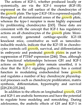
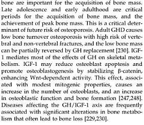
From the Technical Book “Pediatric Bone: Biology & Diseases” pg. 72 by Francis H. Glorieux, John M. Pettifor, Harald Jüppner I have taken 2 clips of the information. It shows that the receptors of the IGF-1 is found on the cell surface of the chondrocytes of the growth plate.

There is 2 types of IGF1R, Type 1 and Type 2. The type 1 is found mostly in the proliferative zone of the growth plate while the Type 2 type is found in all of the zones of maturation in the growth plate. It would seem that there is some ability in the IGF1R in suppresion PTHrP (Parathyroid hormone related protein) expression.