Monthly Archives: October 2012

Increase Height And Grow Taller Using cGMP

Me: I have written two related posts linking the ligand CNP with the receptor NPR2 resulting in increased cGMP production resulting in height increase. The study linked HERE which I have already analyzed the abstract. The link with Guanyl Cyclase B is that it acts as a catalyst for it. For most biological catalysts, they are not minerals or metals but proteins being enzymes. So…

CNP + NPR2 (+ GC-B as catalyst) –> increased levels of cGMP

It seems that elevated levels of cGMP in growth plates lead to the elongation of long bones. It is kind of sad that I have to quote HeightQuest again because of the research that has already been previously done on the link between cGMP and possible height increase HERE.

one critical formula to take away from the post again is 

GTP (+ GC-B as catalyst) –> cGMP

It seems that test subjects (mice) which are bred with the mutation in their genes which causes higher CNP and GC-B expression leads to all of the resting zone, the proliferation , and the hypertropy layer being increased in thickness. 

cGMP stands for Cyclic Guanosine Monophosphate. From the wikipedia article on cGMP HERE… (As always the most important points are highlighted)

is a cyclic nucleotide derived from guanosine triphosphate (GTP). cGMP acts as a second messenger much like cyclic AMP. Its most likely mechanism of action is activation of intracellular protein kinases in response to the binding of membrane-impermeable peptide hormones to the external cell surface.[1]

Synthesis

Guanylate cyclase (GC) catalyzes cGMP synthesis. This enzyme converts GTP to cGMP. In turn, peptide hormones such as the atrial natriuretic factor activate membrane-bound GC, while soluble GC is typically activated by nitric oxide to stimulate cGMP synthesis.

Effects

cGMP is a common regulator of ion channel conductance, glycogenolysis, and cellular apoptosis. It also relaxes smooth muscle tissues. In blood vessels, relaxation of vascular smooth muscles lead to vasodilation and increased blood flow.

cGMP is a secondary messenger in phototransduction in the eye. In the photoreceptors of the mammalian eye, the presence of light activates phosphodiesterase, which degrades cGMP. The sodium ion channels in photoreceptors are cGMP-gated, so degradation of cGMP causes sodium channels to close, which leads to the hyperpolarization of the photoreceptor’s plasma membrane and ultimately to visual information being sent to the brain.[2] GMP and a number of its derivatives also have an umami taste.[3]

cGMP is also seen to mediate the switching on of the attraction of apical dendrites of pyramidal cells in cortical layer V towards semaphorin-3A (Sema3a).[4]Whereas the axons of pyramidal cells are repelled by Sema3a, the apical dendrites are attracted to it. The attraction is mediated by the increased levels of soluble guanylate cyclase (SGC) that are present in the apical dendrites. SGC generates cGMP, leading to a sequence of chemical activations that result in the attraction towards Sema3a. The absence of SGC in the axon causes the repulsion from Sema3a. This strategy ensures the structural polarization of pyramidal neurons and takes place in embryonic development.

cGMP, like cAMP, gets synthesized when olfactory receptors receive odorous input. cGMP is produced slowly and has a more sustained life than cAMP, which has implicated it in long-term cellular responses to odor stimulation, such as long-term potentiation cGMP in the olfactory is synthesized by both membrane guanylyl cylcase (mGC) as well as soluble guanylyl cyclase (sGC). Studies have found that cGMP synthesis in the olfactory is due to sGC activation by nitric oxide, a neurotransmitter. cGMP also requires increased intracellular levels of cAMP and the link between the two second messengers appears to be due to rising intracellular calcium levels.[5]

Degradation

Numerous cyclic nucleotide phosphodiesterases (PDE) can degrade cGMP by hydrolyzing cGMP into 5′-GMP. PDE 5, -6 and -9 are cGMP-specific while PDE1, -2, -3, -10 and -11 can hydrolyse both cAMP and cGMP.

Phosphodiesterase inhibitors prevent the degradation of cGMP, thereby enhancing and/or prolonging its effects. For example, Sildenafil (Viagra) and similar drugs enhance the vasodilatory effects of cGMP within the corpus cavernosum by inhibiting PDE 5 (or PDE V). This is used as a treatment for erectile dysfunction. However, the drug can inhibit PDE6 in retina (albeit with less affinity than PDE5). This has been shown to result in loss of visual sensitivity but is unlikely to impair common visual tasks, except under conditions of reduced visibility when objects are already near visual threshold.[6] This effect is largely avoided by other PDE5 inhibitors, such as tadalafil.[7]

Protein kinase activation

cGMP is involved in the regulation of some protein-dependent kinases. For example, PKG (protein kinase G) is a dimer consisting of one catalytic and one regulatory unit, with the regulatory units blocking theactive sites of the catalytic units.

cGMP binds to sites on the regulatory units of PKG and activates the catalytic units, enabling them to phosphorylate their substrates. Unlike with the activation of some other protein kinases, notably PKA, the PKG is activated but the catalytic and regulatory units do not disassociate.

What Are Growth Hormone Secretagogues?

Me: It seems the hypothalamic-pituitary pathway system not only releases growth hormones (GH) but also growth hormone secretagogues (GHS). The human body releases growth hormones throughout life, which decreases to a lower rate later in life. At the same time the growth hormone is being released, the growth hormone inhibiting somatostatin is also released at the same time. It would appear that as time moves forward and we grow older, the rate of somatostatin release increase, until it overtakes the rate of GH release. remember that the release of GH by the pituitary is controlled by growth hormone releasing hormone and somatostatin in the hypothalamus.

The synthetic type of GH we could take after reaching physical maturity would have to be in injection form. However, this method is not in the pulsating fashion that GH is supposed to act in the body. Naturally, the hypothalamic-pituitary system releases GH in a pulsation way into the body. The secretagogues are peptides made of 6 amino acids linked together. These GHS can be taken orally without the stomach digesting the peptide and breaking it up effectively destroying it.

It seems that GHS like MK-677 in experiments have mixed results. The link says that the authors think GHS works because it mimics ghrelin towards the body’s GH receptors. If I was asked whether GHS can be used to increase height, I would say it could work for people who are of short stature due to low growth hormones in the system but probably little else. THe IGF-1 level was shown to increase from GHS intake from the first study below. 

From the website Iron Man Magazine, a well written article on GHS.

From the website on The Journal Of Endocrinology & Metabolism

Effects of an Oral Growth Hormone Secretagogue in Older Adults

  1. Heidi K. White, Charles D. Petrie, William Landschulz, David MacLean, Ann Taylor,Kenneth Lyles, Jeanne Y. Wei, Andrew R. Hoffman, Roberto Salvatori, Mark P. Ettinger,Miriam C. Morey, Marc R. Blackman, George R. Merriam and for the Capromorelin Study Group

Author Affiliations


  1. Duke University School of Medicine and Geriatric Research Education and Clinical Center (GRECC), Durham Veterans Affairs (VA) Medical Center (H.K.W., K.L., M.C.M.), Durham, North Carolina 27710; Pfizer Global Research and Development (C.D.P.), Groton, Connecticut 06340; Endocrine Clinical Research (W.L.), Eli Lilly and Co., Indianapolis, Indiana 46285; Brown University Medical School (D.M.), Providence, Rhode Island 02912; Novartis (A.T.), Cambridge, Massachusetts 02139; GRECC, Central Arkansas Veterans Affairs (VA) Healthcare System, and University of Arkansas for Medical Sciences (J.Y.W.), Little Rock, Arkansas 72205; VA Palo Alto Health Care System and Stanford University (A.R.H.), Palo Alto, California 94304; The Johns Hopkins University School of Medicine (R.S.), Baltimore, Maryland 21205; Radiant Research and the Regional Osteoporosis Center (M.P.E.), Stuart, Florida 34996; Washington DC VA Medical Center (M.R.B.), Washington, D.C. 20422; and VA Puget Sound Health Care System and University of Washington School of Medicine (G.R.M.), Seattle and Tacoma, Washington 98493
  1. Address all correspondence and requests for reprints to: Heidi K. White, M.D., M.H.S., Duke University School of Medicine, Box 3003, Durham, North Carolina 27710. E-mail:White031@mc.duke.edu.

Abstract

Context: GH secretion declines with age, possibly contributing to reduced muscle mass, strength, and function. GH secretagogues (GHS) may increase muscle mass and physical performance.

Objectives/Design: We conducted a randomized, double-masked, placebo-controlled, multicenter study to investigate the hormonal, body composition, and physical performance effects and the safety of the orally active GHS capromorelin in older adults with mild functional limitation.

Intervention/Participants: A total of 395 men and women aged 65–84 yr were randomized for an intended 2 yr of treatment to four dosing groups (10 mg three times/week, 3 mg twice a day, 10 mg each night, and 10 mg twice a day) or placebo. Although the study was terminated early according to predetermined treatment effect criteria, 315 subjects completed 6 months of treatment, and 284 completed 12 months.

Results: A sustained dose-related rise in IGF-I concentrations occurred in all active treatment groups. Each capromorelin dose prompted a rise in peak nocturnal GH, which was greatest with the least frequent dosing. At 6 months, body weight increased 1.4 kg in subjects receiving capromorelin and decreased 0.2 kg in those receiving placebo (P = 0.006). Lean body mass increased 1.4 vs. 0.3 kg (P = 0.001), and tandem walk improved by 0.9 sec (P = 0.02) in the pooled treatment vs. placebo groups. By 12 months, stair climb also improved (P = 0.04). Adverse events included fatigue, insomnia, and small increases in fasting glucose, glycosylated hemoglobin, and indices of insulin resistance.

Conclusions: In healthy older adults at risk for functional decline, administration of the oral GHS capromorelin may improve body composition and physical function.

From the Dr. Lam website article on Growth Hormones And Growth Hormone Secretagogues located HERE

B. Amino Acid Secretagogues

A secretagogue (pronounced se-cre’-ta-gog) is a natural polyamino acid chain that is postulated to initiate the pituitary gland to release growth hormone. It is the precursor to hGH. While hGH causes the body to act as if the pituitary has released growth hormone, a secretagogue actually causes the release of it. Hence a secretagogue causes the bodies own natural processes to produce growth hormones. Secretagogues do not act as growth hormones at all as they stimulate the pituitary gland to secrete growth hormone.

Interestingly, the inconvenience of hGH injections first led to the discovery of Secretagogues. For years, it was believed that the pituitary gland, which produces growth hormones, dries up as a natural effect of aging. Science has recently discovered that growth hormones reside in the pituitary gland, which stops the release due to aging. Scientists then discovered that certain combinations of amino acids could actually spur the pituitary gland to release the growth hormones. Experiments soon led to the right combinations.

Natural secretagogue is the most practical approach because there are no side effects. In comparison with hGH, their potency and efficacy are low. Since these are orally taken, they can be a first line approach for those who may not choose hGH injections.

This category of hGH products uses amino acids as “secretagogues,” which stimulate the pituitary gland to produce hGH. Other proprietary agents are usually part of the powder/tablet mix, which provide each product with a presumed marketing advantage. Studies show that certain amino acid combinations such as L-lysine, L-arginine, L-ornithine and L-glutamine can stimulate pituitary hGH. While this is theoretically plausible and positive clinical results have been widely reported, published double blind controlled studies that show evidence that these other proprietary factors provide additional pituitary hGH secretion is still incomplete at best. Most studies reveal at least two grams of amino acids are needed to have any effect on pituitary hGH stimulation.

Glutamine

Glutamine is the most abundant amino acid in the body and causes GH secretion. It is a conditional amino acid as the body may not be able to synthesize it under stressful conditions. Traditionally it has been used to strengthen the immune system. The standard anti-aging intake is 50 mg to 1 gm twice daily.

Glutamine is a neurotransmitter in the brain. It is essential for proper brain functions, immune functions, kidneys, pancreas, bladder, and liver functions.

Glutamine becomes one of our body’s most powerful antioxidants in high quantities. Many people, especially those in weight training, add this amino acid due to its benefit in muscle metabolism. Supplementation of two to three gm/day is quite common. For those who plan to take extra doses, it is best to divide the doses throughout the day with up to four servings daily.

Two grams of glutamine was shown to cause a four fold increase in Growth Hormone levels.

Lysine

Lysine is an essential amino acid, which affects bone formation, height, and genital function. It also boosts the effects of arginine. The recommended dosage is one gram on an empty stomach one hour before bedtime and before exercise.

Ornithine

Ornithine is a non-essential amino acid. It is used to potentate the effect of Arginine. The suggested dosage is one gram at bedtime. Doses of more than two to five grams have been known to cause diarrhea.

C. Oral Peptide Secretagogues

hGH is a hormone made up of a long chain of amino acids. Only a portion of the long chain of amino acids makes up the active ingredient. Researchers have been able to identify and extract these active peptides, which are usually five to ten amino acids linked together in a chain. These are then stabilized and formulated into a power or tablet effervescent form. The oral tablets are dissolved in water to be taken before bedtime on an empty stomach. This is to stimulate the release of hGH from the anterior pituitary, which peaks during the early phase of sleep. The effervescent form is best to draw the peptide away from the gastric juice closer to the mucous for better absorption. Gastric juices are highly acidic. Peptides are proteins that are easily denatured when exposed to an acidic environment. Extraction of the peptide is a tedious process. Peptides are not stable enough to maintain its activity in an aqueous environment. Thus, the peptide is formulated in the oral tablet format. Secretagogues using peptides are abundant in the marketplace. They are sold as a natural nutritional supplement and no FDA approval is required. However, some unscrupulous operators simply use ground bovine pituitary gland and pass them off as secretagogues. The consumer is often faced with the arduous task of identifying which is the real secretagogue.

Secretagogues can also work at multiple sites leading to growth hormone release. For example, a secretagogue targeted towards the hypothalamus would stimulate the hypothalamus to release Growth Hormone Releasing Factor (GHRF) that in turn stimulates the pituitary gland to release growth hormone. An oral peptide pituitary secretagogue, on the other hand, stimulates the pituitary gland directly to effect the release of growth hormone.

An effective secretagogue could easily raise IGF-1 levels, although the result is not as significant as growth hormone injections. Clinically many users have reported better sleep, increased alertness during the day, and less joint pain.

IGF-1 levels may not be the best indicator of how effective a secretagogue is for the GH receptor sites may be damaged. A low IGF-1 level does not mean that the body’s growth hormone level has not increased. It may simply mean that that the level is not accurately measured, or that there is a defective receptor site. If your IGF-1 does not increase, do not be despair. Talk to your health care practitioner. How you feel is just as important and sometimes even more important than laboratory studies alone.

D. Growth Factors

Growth factors (GF) are small protein chains, commonly known as polypeptides, which bind to cell surface receptor sites and exert actions directly on the target cells. This is generally done through cellular proliferation and or differentiation.

Some GFs exert generalized effect, while others are cell and action specific. There are many different classes of GFs. Some common ones include: Insulin-like Growth Factor (IGF-1) that is responsible for much of Growth Hormones (GH) action in the body; Interleukins (IL); Fibroblast Growth Factors (FGF); Transforming Growth Factor (TGF); Tumor Necrosis Factor (TNF); Epidermal Growth Factor (EGF); and Transforming Growth Factors-b (TGFs-b).

GFs come from a wide variety of sources. Epithelial Growth Factors (EGF) comes from sub maxillary gland, and FGF comes from a wide range of cells. A unique family of growth factors that is secreted primarily by leukocytes (white blood cells) is called cytokines. When such cytokines are secreted by lymphocytes, they are called lymphokines. Many of the lymphokines are also known as interleukins (ILs). Not only are interleukins secreted by leukocytes, they are also able to affect the cellular responses of leukocytes.

What Do Growth Factors Do? 

Different GFs have different jobs to do. Generally, all of them work at the cellular level to:

Repair damaged cells

Enhance cellular proliferation

Maintain optimum function of the target organ

Rejuvenate aging tissues

While hormones generally are more specific and sometimes work through other mediations elicited from its simulation of intermediate organs, GFs often act directly on the target tissue and have a wide range of effects. Its action is mostly stimulatory. It can also work synergistically with other GFs or hormones to elicit a biological effect. Growth hormone, for example, exerts its effect in the body via Insulin-like Growth Factor (IGF-1). In other words, it is the IGF-1 that actually carries out the function of growth hormone and not growth hormone itself.

The Connection Between mTOR, Rapamycin, Leucine And Height

Me: Throughout the other boards I have come across on the occasional thread which talks about the possibility of using mTor or Leucine to increase height.

Now as I have shown in previous posts, there is a link between tall stature and increased chance for cancer and faster aging rate. It seems that what makes you taller, makes it more likely that you develop cancer and age faster.

With mTor it has been shown in studies to possibly increase the rate of aging in people and also increase the chance for cancer. So using sort of a reverse causality type of logic, (which is not really logic but correlation) I would guess that Leucine and mTor might have some connection with height and might contribute to growth somehow.

Analysis: From the first article I found, we learn that mTor Stands for mammalian target of rapamycin (didn’t know that) which is a nutrient sensing protein kinase that regulates numerous cellular processes. In an experimental chondrogenic cell line, rapamycin seems to regulate and inhibit proteoglycan accumulation and collagen X exprsesion. This would suggest that the overall affect of Rapamycin is to inhibit the creation of chondrocytes. It decreases the amount of Ihh, which regulates chondrocyte differentiation If you added more ihh into the culture, it reverses the effect of mTor. The researchers concluded that if you can control and manipulate the mTOR signalling, you can control at least part of the mechanism causing chondrocyte differentiation, thus also longitudinal growth. 

In the 2nd article the researchers found that if you inactivate TOR or its substrate s6 kinase you cause cells to be smaller in size and also die. This shows that the TOR pathway controls cell growth. They did a homogolous recombination of the deletion of the C-terminal six amino acids of mTOR, which are essential for kinase activity, resulted in reduced cell size and proliferation arrest in embryonic stem cells. mTOR controls cell size and proliferation at least in mouse stem cells. mTOR can make cells produce chemicals such as cyclins that trigger cell growth.

In the 3rd article, the writer states from the beginning, ” Amino acids, in particular leucine, have been shown to regulate cell growth, proliferation, and differentiation through the mammalian target of rapamycin (mTOR), a nutrient-sensing protein kinase”…plus “we hypothesized that leucine restriction, acting through mTOR, would inhibit growth plate chondrocyte proliferation and differentiation. The effect of leucine restriction was compared with that of the specific mTOR inhibitor, rapamycin. Leucine restriction produced a dose-dependent inhibition of fetal rat metatarsal explant growth. This was accounted by reduced cell proliferation and hypertrophy but not apoptosis. mTOR activity, as reflected by ribosomal protein S6 phosphorylation, was only partially inhibited by leucine restriction, whereas rapamycin abolished S6 phosphorylation. In chondrogenic ATDC5 cells, leucine restriction inhibited cell number, proteoglycan accumulation, and collagen X expression despite minimal inhibition of mTOR

So we can say that leucine can influence mTOR which influences chondrogenesis which influeneces longitudinal growth. Rapamycin also influences mTOR but negatively. increases in leusin increase mTOR which increase chondrogenesis. increase in rapamycin leads to reduced mTOR which reduces chondrogenesis. If you see a decrease in chondrogenesis from leucin restriction you get clumping of proteoglycans and increased production of collagen X. The issues is that leucin and rapamycin both have a small effect on the overall genes.

I did a very quick search on Google for any evidence and this is what i found…

From link HERE

Dev Dyn. 2008 Mar;237(3):702-12.

mTOR signaling contributes to chondrocyte differentiation.

Phornphutkul C, Wu KY, Auyeung V, Chen Q, Gruppuso PA.

Source

Department of Pediatrics, Division of Pediatric Endocrinology and Metabolism, Rhode Island Hospital and Brown University, Providence, Rhode Island 02903, USA. chanika_phornphutkul@brown.edu

Abstract

The mammalian Target Of Rapamycin (mTOR) is a nutrient-sensing protein kinase that regulates numerous cellular processes. Fetal rat metatarsal explants were used as a physiological model to study the effect of mTOR inhibition on chondrogenesis. Insulin significantly enhanced their growth. Rapamycin significantly diminished this response to insulin through a selective effect on the hypertrophic zone. Cell proliferation (bromodeoxyuridine incorporation) was unaffected by rapamycin. Similar observations were made when rapamycin was injected to embryonic day (E) 19 fetal rats in situ. In the ATDC5 chondrogenic cell line, rapamycin inhibited proteoglycan accumulation and collagen X expression. Rapamycin decreased content of Indian Hedgehog (Ihh), a regulator of chondrocyte differentiation. Addition of Ihh to culture medium reversed the effect of rapamycin. We conclude that modulation of mTOR signaling contributes to chondrocyte differentiation, perhaps through its ability to regulate Ihh. Our findings support the hypothesis that nutrients, acting through mTOR, directly influence chondrocyte differentiation and long bone growth.

Me: It seems that mTOR may be one of the things that regulates Ihh.

From link HERE

mTOR Is Essential for Growth and Proliferation in Early Mouse Embryos and Embryonic Stem Cells

  1. Mirei Murakami1,2, Tomoko Ichisaka1,2, Mitsuyo Maeda3, Noriko Oshiro2,4,Kenta Hara2,5, Frank Edenhofer6, Hiroshi Kiyama3, Kazuyoshi Yonezawa2,4,* and Shinya Yamanaka1,2,*

+Author Affiliations

  • 1Research and Education Center for Genetic Information, Nara Institute of Science and Technology
  • 2CREST, Japan Science and Technology Agency, Nara 630-0192
  • 3Department of Anatomy and Neurobiology, Osaka City University Medical School, Osaka 545-8585
  • 4Biosignal Research Institute, Kobe University, Hyogo 657-8501
  • 5Fourth Department of Internal Medicine, Kobe University School of Medicine, Hyogo 650-0017, Japan
  • 6Institute of Reconstructive Neurobiology, University of Bonn Medical Center, D-53105 Bonn, Germany

ABSTRACT

TOR is a serine-threonine kinase that was originally identified as a target of rapamycin in Saccharomyces cerevisiae and then found to be highly conserved among eukaryotes. In Drosophila melanogaster, inactivation of TOR or its substrate, S6 kinase, results in reduced cell size and embryonic lethality, indicating a critical role for the TOR pathway in cell growth control. However, the in vivo functions of mammalian TOR (mTOR) remain unclear. In this study, we disrupted the kinase domain of mouse mTOR by homologous recombination. While heterozygous mutant mice were normal and fertile, homozygous mutant embryos died shortly after implantation due to impaired cell proliferation in both embryonic and extraembryonic compartments. Homozygous blastocysts looked normal, but their inner cell mass and trophoblast failed to proliferate in vitro. Deletion of the C-terminal six amino acids of mTOR, which are essential for kinase activity, resulted in reduced cell size and proliferation arrest in embryonic stem cells. These data show that mTOR controls both cell size and proliferation in early mouse embryos and embryonic stem cells.

Me: So mTOR in general controls cell size and proliferation in stem cells and embryos. That sounds pretty important in longitudinal growth to me.

From link HERE

mTOR inhibitors

mTOR is a kinase protein. It can make cells produce chemicals such as cyclins that trigger cell growth. It may also trigger cells to produce proteins which trigger the development of new blood vessels that cancers need in order to grow. In some types of cancer mTOR is switched on, which makes the cancer cells grow and produce new blood vessels. mTOR inhibitors are a new type of cancer growth blocker being used to try to stop the growth of some cancers. mTOR inhibitors include temsirolimus (Torisel), everolimus (Afinitor) and deforolimus.

As for Leucine, from link HERE

Leucine restriction inhibits chondrocyte proliferation and differentiation through mechanisms both dependent and independent of mTOR signaling

  1. Mimi S. Kim1,*, Ke Ying Wu1,*, Valerie Auyeung2, Qian Chen2,Philip A. Gruppuso2, and Chanika Phornphutkul1

+Author Affiliations

  1. Division of Pediatric Endocrinology and Metabolism, Departments of 1Pediatrics and 2Orthopaedic Surgery, Rhode Island Hospital/Warren Alpert School of Medicine of Brown University, Providence, Rhode Island
  1. Address for reprint requests and other correspondence: C. Phornphutkul, Division of Pediatric Endocrinology and Metabolism, Rhode Island Hospital, 593 Eddy St., Providence, RI 02903 (e-mail: Chanika_Phornphutkul@brown.edu)
  • Submitted 19 December 2008.
  • Accepted in final form15 April 2009.

Abstract

Linear growth in children is sensitive to nutritional status. Amino acids, in particular leucine, have been shown to regulate cell growth, proliferation, and differentiation through the mammalian target of rapamycin (mTOR), a nutrient-sensing protein kinase. Having recently demonstrated a role for mTOR in chondrogenesis, we hypothesized that leucine restriction, acting through mTOR, would inhibit growth plate chondrocyte proliferation and differentiation. The effect of leucine restriction was compared with that of the specific mTOR inhibitor, rapamycin. Leucine restriction produced a dose-dependent inhibition of fetal rat metatarsal explant growth. This was accounted by reduced cell proliferation and hypertrophy but not apoptosis. mTOR activity, as reflected by ribosomal protein S6 phosphorylation, was only partially inhibited by leucine restriction, whereas rapamycin abolished S6 phosphorylation. In chondrogenic ATDC5 cells, leucine restriction inhibited cell number, proteoglycan accumulation, and collagen X expression despite minimal inhibition of mTOR. Microarray analysis demonstrated that the effect of leucine restriction on ATDC5 cell gene expression differed from that of rapamycin. Out of 1,571 genes affected by leucine restriction and 535 genes affected by rapamycin, only 176 genes were affected by both. These findings indicate that the decreased chondrocyte growth and differentiation associated with leucine restriction is only partly attributable to inhibition of mTOR signaling. Thus nutrient restriction appears to directly modulate bone growth through unidentified mTOR-independent mechanisms in addition to the well-characterized mTOR nutrient-sensing pathway.

UNDERNUTRITION IS A WELL-DOCUMENTED cause of poor linear growth, whereas obesity produces accelerated linear growth in children. Primary causes of undernutrition are complex. They can range from inadequate availability of calories to inadequate specific nutrients, such as protein or amino acids. Despite the many causes and forms of undernutrition, one universal outcome is poor long bone growth, which can be presumed to be an effect on endochondral bone elongation (1, 6). Over the last several decades, the mechanisms by which nutritional status affects bone growth have focused on indirect effects via changes in insulin-like growth factor I (IGF-I) biological effect (3, 7). Impaired IGF-I production in undernutrition is in part associated with impaired hepatic growth hormone (GH) sensitivity, which is associated with decreased hepatic GH receptor expression, hepatic IGF-I mRNA, and circulating IGF-I levels (31). Similarly, overnutrition is associated with upregulation of the GH/IGF-I axis (23).

The effect of IGF-I on chondrocyte growth and differentiation within the growth plate has been well documented (14, 24). We and others have demonstrated that IGF-I has an important role in chondrocyte proliferation and differentiation (25, 28). In addition to the effects of IGF-I on chondrocytes, we have demonstrated that insulin at physiological concentration has a direct effect on chondrocyte differentiation (27), thus providing for another mechanism by which nutritional status can modulate bone growth.

In recent years, mechanisms by which nutrients exert a direct effect on cell growth and function have been elucidated (17). Although restriction of essential amino acids has been viewed as limiting because of their requirement as substrates for protein synthesis, essential amino acids also act as signaling factors in several regulatory pathways (15,16). Perhaps the most well-characterized signaling pathway regulated by amino acids has at its center the mammalian target of rapamycin (mTOR) (17). mTOR is a nutrient-sensing kinase that integrates input from amino acids, growth factors, and the energy status of the cell (15). It acts as a central controller of translation, controlling ribosomal biogenesis and global protein synthesis (4). The TOR protein is highly conserved from yeast to mammals (5). Rapamycin, a widely used immunosuppressive agent, directly inhibits mTOR activity and has been key to understanding the role of mTOR in cell regulation (22, 35).

We have recently demonstrated the effect of rapamycin on chondrocyte growth and differentiation in ATDC5 cells (26), fetal rat metatarsal explants (26), and the rabbit growth plate (unpublished observation). mTOR inhibition with rapamycin results in significantly decreased chondrocyte differentiation, a modest decrease in chondrocyte proliferation, and decreased total bone growth in physiological systems.

Leucine is the most potent nutrient regulator of mTOR signaling (29, 30). We therefore hypothesized that leucine availability would have a direct effect on chondrocyte growth and differentiation, resulting in decreased longitudinal bone growth when restricted. We further hypothesized that the effect of leucine restriction would be a direct result of mTOR inhibition, although mTOR-independent pathways have been described. One of these involves the mammalian general control nonderepressible 2 (GCN2) kinase (32). GCN2 is a stress kinase that is activated by amino acid starvation to modulate protein synthesis. GCN2 phosphorylates the α-subunit of the eukaryotic initiation factor (eIF2). The response that allows organisms to tolerate amino acid deprivation in states of malnutrition and starvation involves repression of protein synthesis and upregulation of amino acid biosynthesis and transport (10) with a net effect of decreased cell growth.

Based on our hypothesis and well-defined mechanisms that control chondrocyte growth and differentiation, we have performed studies using embryonic day 19 (E19) fetal rat metatarsal explants. The benefit of this model is the intact bone maintains its cell-cell and cell-matrix interaction. We have also used the ATDC5 chondrogenic cell line to extend our observations to an in vitro model. Last, using microarray analysis, we have explored other potential mechanisms accounting for the effect of leucine restriction on chondrocyte growth and differentiation.

DISCUSSION

The present studies were aimed at characterizing the effect of restricting a key nutrient, leucine, on two models of bone growth: metatarsal explants and growth and a chondrogenic cell line. Amino acids, particularly the branched-chain amino acids, regulate protein synthesis beyond the level of their own availability as the substrate for peptide-chain elongation (15, 17). They do so by functioning as signaling molecules. Leucine appears to be the most potent of the branched-chain amino acids in this regard, having a potent effect on signaling via two important signaling kinases, mTOR and GCN2 (8, 10,32, 33).

The mTOR signaling pathway has been shown to mediate the effects of leucine on mRNA translation initiation. We recently demonstrated the important role of mTOR in the regulation of chondrocyte growth and differentiation (26). Our previous findings provided for a mechanism whereby nutrients, acting through mTOR, can directly modify linear growth. In the present study, we performed analogous experiments comparing the effect of leucine restriction in the fetal metatarsal explant model as well as ATDC5 cells.

The present studies were undertaken to test the hypothesis that leucine restriction would exert its effects on bone growth through its ability to signal via mTOR. Using the more physiological bone explant model, we confirmed that leucine affected growth in a dose-dependent manner. The observed growth inhibition was associated with decreased chondrocyte proliferation as measured by decreased BrdU incorporation. Decreased chondrocyte proliferation presumably results in fewer chondrocytes that are available to differentiate and become hypertrophic cells, consistent with the decreased hypertrophic zone height that we observed.

Chondrocytes that differentiate into prehypertrophic and hypertrophic chondrocytes undergo a 4- to 10-fold increase in cytoplasmic volume, making distal hypertrophic cells a significant component of longitudinal bone growth. In rapidly growing bones, ∼10% of bone length is contributed by proliferating cells, a one-third by matrix synthesis throughout the growth plate, and nearly two-thirds by the contribution of hypertrophic cells (34).

Although leucine restriction appears to have an effect on chondrocyte proliferation and differentiation, we did not observe an increase in apoptosis as assessed by TUNEL staining. Premature cell death can result in reduced bone growth, as observed in humans with skeletal dysplasia (12) and in mice with disruption of genes important to chondrogenesis, including those encoding filamin B, matrilin-3, or components of the β-catenin signaling pathway (11, 21). Our results indicate that the effect of leucine restriction occurs during the early phase of the chondrocyte growth and differentiation process, not as a result of increased apoptosis.

Because mTOR is a signaling target for leucine, we examined the effect of leucine restriction on mTOR signaling in metatarsals by assessing the phosphorylation state of ribosomal protein S6. We observed only a modest decrease in phospho-S6 staining in the explants grown in 0.02 mM leucine. This was in sharp contrast to rapamycin, which abolished phospho-S6 staining. This raised the possibility that leucine response was mediated through a mechanism other than one involving modulation of mTOR activity.

Using the ATDC5 chondrogenic cell line, we extended our observations in an attempt to support or refute conclusions drawn from the metatarsal explant studies. Leucine restriction decreased chondrocyte differentiation as measured by accumulation of proteoglycan and expression of collagen X. Total cell mass measured by Neutral Red accumulation was also decreased. Again, markers of mTOR activity, S6 phosphorylation and the pattern of 4E-BP1 phosphorylation, were modestly inhibited under conditions of leucine restriction relative to the marked effect of rapamycin. The expression of Ihh, a key contributor to chondrocyte proliferation and differentiation (9, 18, 20), was also decreased under conditions of leucine restriction. We previously demonstrated that mTOR inhibition may directly regulate Ihh expression (26).

In an effort to identify an mTOR-independent pathway to account for the effects of leucine restriction, we examined the regulation of GCN2 in the ATDC5 cell line. The GCN2 pathway is activated by the accumulation of uncharged tRNAs during amino acid starvation as shown in myoblasts (10). This leads to the phosphorylation of eIF2α, resulting in inhibition of translation initiation of cellular proteins and a global reduction in protein synthesis (10, 13). We observed only a modest increase in the phosphorylation of eIF2α under conditions of leucine restriction.

Microarray analysis of the effects of leucine restriction vs. rapamycin on gene expression in ATDC5 cells revealed that only a small proportion of genes was affected by both leucine restriction and rapamycin. These genes, which numbered 176, accounted for 11.2% of the genes that were affected in leucine restriction. The large effect of leucine restriction relative to rapamycin (1,571 vs. 535 genes) may indicate that leucine restriction has a broader effect on chondrocyte growth and differentiation than does targeted inhibition of mTOR. Using very stringent criteria for significance, gene ontology and pathway analysis further supported marked differences in the effects of the two conditions.

In summary, our studies show that leucine restriction affects proliferation and differentiation in the ATDC5 chondrogenic cell line. Results using the fetal metatarsal explant model are consistent with the ATDC5 studies. Our findings support the conclusion that both mTOR and GCN2 signaling may contribute to the effect of leucine restriction on chondrocyte proliferation and differentiation and, therefore, on long bone growth. However, our studies are also consistent with the possibility that the effects of leucine restriction are mediated by pathways that are independent of effects on these two signaling kinases. We are left to conclude that the mechanism by which leucine restriction inhibits chondrogenesis and attenuates linear bone growth is complex, likely involving modulation of multiple pathways that may involve mTOR and GCN2, but that may also be independent of both of these well-characterized pathways.

Who Is The Real Superman? Comparing The Height And Size Of Dwight Howard And Shaquille O’neal

I had stated before once (or a few times before) that I used to really be obsessed with basketball as a teenager. I would practice for 4-5 hours everyday in the Summer heat. I wanted to possibly be a NBA player but I never did gain the size or the skills no matter how much I practiced. I always got my ass handed to me when going up against guys, even ones smaller than me.

I guess I realize now that my fate probably belongs more in the research lab than the court. So for today I wanted to write a rather fun post where I compare the height and size of Dwight Howard and Shaquille O’neal and see what could really claim to be the real “Superman” which they both have been labeled at some point in their professional basketball career. Of course, this will be similar in style and tone as a previous older post I did where I compared the height of Lebron James vs Kevin Durant, who I consider to be the two best basketball players on the planet today.

So let’s first see what the figures seem to say about the Ex-NBA star Shaquille O’neal.

If you have ever been involved in the NBA or know much about Shaq, it is that he has been almost always quoted and listed at a height of 7′ 1″ (2.16 meters) tall.

From a quick search on the internet from WikipediaBasketball ReferenceYahoo, NBA, they have all listed Shaq at 7′ 1″ (2.16 meters) in height.

From the NBA Predraft Measurements Profile on him on DraftExpress.Com (found from this link HERE) we see that Shaq was listed with these numbers….

He was drafted in the 1992 NBA as the #1 pick to the Orlando Magic.

  • Height Without Shoes: 7′ 1″
  • Height With Shoes: N/A
  • Weight: 303 lb
  • Wingspan: 7′ 7″
  • Standing Reach: 9′ 5″
  • Body Fat: 12.2
  • Maximum Vertical: 36 “
  • Maximum Vertical Reach: 12′ 5″
  • Birthday: 03/06/1972 (40 years old by this time in October 2012)
  • NBA time: 18 years with Orlando Magic, Los Angeles Lakers, Phoenix Suns, Miami Heat, Cleveland Cavaliers, Boston Celtics

Shaq’s weight has been far more volatile and has been listed from 325 lb (147 kg) on Wikipedia to claims that he was 360 lbs at one point.

From this source link HERE we find out that Shaq’s biological father named Joseph Toney who worked at a Newark Home Shelter is 6′ 1″ who used to be a local basketball start. Shaq’s mother Lucille has been claimed to have been 6′ 3″ which is extremely tall for any female. I remember years ago reading from a article that both of Shaq’s grandfathers from his mother’s and father’s side were 6′ 9″ in height, which is really insane.

We can see from the picture on the right which is of the 1996 Olympic Basketball Team that Shaq with his abnormally large shoes is standing on a podium with the other basketball All-Stars. It is sort of hard to make out which players are standing where but if you look close enough, you can make out the fact that Shaquille and David Robinson are standing on the same level height, which is three podiums off the ground. I would guess the reason why they took a picture of the team in this way was that they wanted to get a picture of all of the players in the Olympics team and get a good picture of all of the faces. That is not possible if certain people are taller than others which would block the face of some people. If you look closer, you would see that John Stockton and Hakeem Olajuwon are the other two players standing on the same ledge height. Stocken is listed at 6′ 1″ which would put his head at around the shoulder height area of Shaq, which he is. Olajuwon has been listed at 7 feet when he was playing for the Houston Rockets in the 90s but he admitted that his height was lower. I would guess his height was probably 6′ 11″ or maybe even 6′ 10″ (which is a very loweestimate for him). Olajuwon’s height reaches the eyebrow of Shaq and Robinson so his height seems to be right. Robinson was well known for being tall at 7′ 1″ as well. He was a very respected and kind, honest man so his listed height would be honest an accurate. If we look at the height of Robins and Shaq then, their height are almost the same, although Shaq may have slightly thicker shoes than Robinson.

There is a common story told by Shaq that at the age of 13, he was already 6’6″ or 6′ 7″ when he was stationed in Germany because he was what some people called a “military brat”. It was there where he met Coach Brown, who would eventually become his coach at LSU many years later.

His shoe size has also varied throughout his career being either listed at 22 or 23, which seems to be the largest feet of all the NBA players in history. Shaq is a very big guy so the heel and thickness in his sneakers must be large as well. The normal athlete has shoes or sneakers that are about 1-1.5″ thick. I would guess that since Shaq’s feet are so big, he has to get his shoes specifically designed so his shoe’s heel might be ever thicker, up to 2 inches maybe.

What is really strange is that on the CelebHeights.Com website for his profile HERE, there are actually people who claim that Shaq might “only” be 6′ 11″ which I have found really hard to believe. Most people who have ever met Shaq have all stated that he is a very big man, a true giant and almost no one doubts that Shaq is the 7′ 1″ he has been claiming for the last 20 years in basketball. I remember reading from a article post that Chris Kaman who is 7′ 0″ himself had stated that Shaq is probably the biggest man he has ever met, in terms of size, because there are a few guys in the NBA who might have been taller like Yao Ming.

Here is a picture of Shaq with his current girlfriend Nicole “Hoopz” Alexander who was a winner in a reality show (Flavor of Love). Alexander has been claimed to be around 5′ 2″ in height. This pictures gives a person a very good perspective at just how big Shaq is. There is only supposed to be a 2 feet or 24 inch difference in their heights but the picture makes it look like the height difference is far larger. If I was to make a guess, it would be maybe 3 feet difference, not 2. But, one should note that Alexander is not wearing any high heels or thick heeled shoes and shaq seems to be in his shoes, which give 1-2 inches of extra height. Still, the difference in size of the couple is something that I would do a double take on if I ever walked past the couple in the street.

One poster on the celebheights website posts this…

RisingForce says on 18/Jan/12 

RP says on 13/Jul/11 
OK….found out a couple of facts on Shaq. He was measured & weighed twice leading up to the ’92 draft. At one he was 7’0 5/8″ & 301 lbs….at the other he was 7’0 7/8″ & 303 lbs. In 2000-2001 season with the Lakers he weighed in at 327 lbs at training camp. Also, at one point after a injury kept him from practicing & performing any cardio for about 6 weeks in one season with the Lakers, he got as heavy as 368 pounds!!!

Can anyone verify this? Oddly specific figures to make up so that could be accurate, but when I’ve searched, nothing comes up to back this stuff up.

Me: I can only say that the facts might be true. When Shaq was photographed with certain other guys who were profiled at 7′ 1″ like Wilt Chamberlain or Kareem Abdul Jabbar, Chamberlain did look like he was taller than Shaq.


Now let’s see what Dwight Howard profile shows.

From sources like

Dwight himself states clearly in this Youtube video HERE (uploaded in 2008) that he is 6′ 11″. He also states that he weighs 270 lbs in the video.

From the NBA Predraft Measurements Profile on him on DraftExpress.Com (found from this link HERE) we see that Dwight was listed with these numbers….

High School:Southwest Atlanta Christian Academy
Hometown:Atlanta
Drafted: Rnd 1, Pick 1 in 2004 by Magic

  • Height Without Shoes: 6′ 9″
  • Height With Shoes: 6′ 10.25″
  • Weight: 240 lb
  • Wingspan: 7′ 4.5″
  • Standing Reach: 9′ 3.5″
  • Body Fat: N/A
  • No Step Vertical: 30.5″
  • Maximum Vertical: 35.5 “
  • Maximum Vertical Reach: N/A
  • Birthday: 12/08/1985 (26 years old by this time on October 2012)
To see whether Dwight is either the 6′ 9″ listed in the predraft measurements page or the 6′ 11″ he claims, let’s look at a few photos I have found around the internet. These two pictures are the photos of the 2008 Olympic Men’s Basketball Team. In the first picture, you can see that Coach K decided to line up all of the players by height, from tallest on the lef to the shortest on the right.
We can now see just how immense the  variability and change in size is of the basketball team. Keep in mind that Chris Paul has been listed at 6 ‘ 0″ which is slightly taller than the average US male citizen. From the picture and maybe the eye perspective it looks like Dwight could be maybe even a 1 foot and a half taller than Paul but he is supposed to be either 9 inches or 11 inches taller than Paul. We can estimate that the height of Dwight Howard is probably the same as Chris Bosh. From the sources and links found, Chris Bosh has been listed at 6′ 11″ on the NBA and basketball sites. I remember seeing a Youtube video once where Jay Leno made the obvious proclamation while talking to Chris Bosh that he is very tall. Leno asked Bosh how tall he is and Bosh said he was 6′ 11″. I seem to remember that earlier in Bosh’s career with the Raptors (I’m not sure about this fact) he was listed at 6′ 10″. The next tallest in the lineup would be Tayshuan Prince who has been listed at 6’ 9″. From the picture, I would easily guess that Prince is at least 2 inches shorter than Howard. It is important to note that Bosh is well known to have a rather long neck, which has been compared to a giraffe by other people. Howard has been talked about on basketball forums for having a rather small head. This shows that in terms of where the shoulder line is and wher ethe neck starts, Howards has a clear higher shoulder level than anyone else. This probably creates the illusion that Howard is taller than what he might actually be.

Here is another picture of Howard with the 2008 Olympic team, in their white uniforms.

I think it is rather clear now that Dwight Howard is probably not the 6′ 9″ that was measured on the predraft. Dwight was a few select players who came to the NBA directly from high school. I think Diwght was only 18 and as I have shown in previous posts, a male does not really stop growing until he is around 19-21. Even the way how NBA players have had growth spurts, it would not be that wild to state that after Howard was in the NBA, he continued to grow another 2 inches.

Most people have stated that the athleticism of Dwight is extremely high, and people like 4 time NBA champion Tim Duncan at 6′ 11″ has admitted that Dwight is far more talented than he was during his development.

Dwight is know around the NBA for his chiseled muscular build, his shoulders which are very big which has had many people wonder whether he has been using steroids to gain that big, and and his amazing jumping ability. In the  2008 Sprite Slam Dunk Competition which happens usually during the Halftime break for the annual All-Star game, Dwight jumped up to a reach of 12′ 6″ which is 1 inch higher than Shaq’s old record of 12′ 5″. That event can be validated from this Youtube Video of the event HERE.

As for the real question “Who is the real superman?” I would have to look into more than just the physical attributes that make up a person. Superman was not only big and strong, he also had strong character and cared about other people. When the character of Superman was first created in the 30s around the Great Depression, his listed height was 6′ 2″ and I think around 220 lbs. Back then 6′ 2″ was considered very big, but these days, there are many people who are taller than that. Some of the actors who played Superman in the movies like Christopher Reeves at 6′ 4″ and Brandon Routh at 6′ 3″ are taller than the original superman. When it comes to size, Shaq would be the clear choice. In terms of size, Shaq may have been the heaviest, and strongest NBA player in history, although Wilt Chamberlain has been shown to be super strong as well. For Wilt and Shaq, the very rules of how the game of basketball had to be changed because they were so dominant at one point.

For Dwight, most NBA greats have talked about ihs insane athleticism and jumping ability. It is very rare to find a man who is almost 7 feet be able to jump that high in the air. At his best, he is around 270 lbs of almost pure muscle, chiseled features, and can jump higher than shaq.

When you give the title or nickname of “Superman” to a person in any profession, they have to be great at almost everything when you combine and analysis all of the elements of the game. Offensively Dwight has been rather lacking in his career. Because of his jumping ability, he is a great shot blocker.

Personality wise, Shaq and Dwight both have a very friendly, outgoing, cheerful persona. Towards the media, Shaq has ventured into more venues than Dwight at this time. Shaq has been a musician producing a few records, had his own reality television show (even if Dwight was on it once), Has been in a few movies that he starred in, had been shown to be able to break dance on TV, has a Ph.D, has had his own statue unveiled at his alma mater, has being buying and investing in a diversified portfolio of companies, been a big social media star with twitter, and many other projects. Dwight has been on many commercials, TV appearances, has a youtube profile.

In terms of professional accomplishments, Shaq has won 4 NBA championships with a few MVPs while Dwight has been Defensive player of the year a few times ans is still chasing after his first championships.

When it comes to be the question of  title of superman to either Shaq or Dwight, I would have to give it to Shaq at this point because of his professional accomplishments and also his many projects and ventures out of his basketball life. He has been accomplished in academics, athletics, music, and television.

Determining Skeletal Maturity And Bone Age Using The Greulich Pyle Method Or Tanner Whitehouse Method

From this source link HERE

How does your endocrinologist determine whether you have stopped growing? Let’s find out.

There seems to be two main ways to determine skeletal maturity and bone age, using either the Greulich Pyle Method or the Tanner and Whitehouse Method. You get an X-ray or a radiograph done on your left hand. The reason it is your left hand has raised some questions and so far I have not found a reasonable answer for it. It seems the Greulich Pyle method is only used in the Netherlands from the source and it is far faster but not as reproducible and accurate as the Tanner and Whitehouse method.

For the Greulich Pyle Method…

There is a description for each of the development stages seen in the growth of the hand radiograph. The description is a guideline on how the physician should examine and diagnose the state of development of the bone.

Quoted from the source link…

The first step in an analysis is to compare the given radiograph with the image in the atlas that corresponds closest with the chronological age of the patient. Next one should compare it with adjacent images representing both younger and older children. When comparing the radiograph against an image in the atlas there are certain features a physician should use as maturity indicators. These features vary with the age of the child. In younger children the presence or absence of certain carpal or epiphyseal ossification centers are often pointers for the physician about the skeletal age of a child. In older children the shape of the epiphyses and the amount of fusion with the metaphysis is a good indicator of skeletal age. Once the atlas image that most resembles the radiograph is found the physician should conduct a more detailed examination of the individual bones and epiphyses. When the physician is sure that the matching radiograph has been found, she can find the skeletal age printed at the top of the page.

Me: When I read how this Method for determining bone age was done I couldn’t but felt slightly let down on how it is performed. I sort of expected the procedure to be more complex with some calculations. It seems to show that endocrinologists today might not even have a real way of calculating growth plate maturity, but sort of guess at it with an eyeball estimate which is not very accurate. 

For the Tanner and Whitehouse Method…

The tanner whitehouse method seems to be based on the bone standard maturity of certain areas in the hand instead of age. The method uses 20 regions of interest. Each region of interest is broken up into 3 parts, the epiphysis, diaphysis, and metaphysis, and the sections can be determined in the phalanx promixity.

Quoted from the source link…

The development of each ROI is divided into discrete stages and each stage is given a letter (A,B,C,D, . . ., I). A numerical score is associated with each stage of each bone. By adding the scores of all ROIs, an overall maturity score is obtained. This score is correlated with the bone age differently for males and females. The method has a modular structure which makes it suitable for automation. For the tanner whitehouse method, three score systems have been developed.

  • TW2 20 Bones: characterized by twenty bones including the bones of the first, third and fifth finger and the
  • carpal bones.
  • RUS: considers the same bones of the TW2 method except the carpal bones;
  • CARPAL: considers only the carpal bones.

For the finer details on the method, please refer to the source link above. I hope I gave you an idea on how pediatricians and endocrinologists try to evaluate whether you can still grow or not.

Graphical Outline Of The Endocrine System For Growth Development , For the Visual Learner

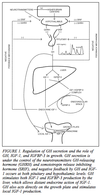
I was doing more research and I found this rather lovely graphical representation of the basic theoretical model on how the general growth hormones like GH and IGF are linked to accelerate and deccelerate the growth process. The source of the picture where it was taken from was from BCM on their website HERE

As you can see there appears to be two main negative feedback mechanisms causing and telling the hypothalamus to decrease the release of the growth hormone releasing hormone and increase somatotropin release inhibiting hormone. Ordinarily the hypothalamus would be releasing more GHRH and using less SRIF when the person is growing.

Overall there is actually 4 negative feedback mechanisms (but two mains ones). As you can see from the smaller graph, the GH release rate is far higher during sleep than waking periods. So kids, sleep more if you want to increase the growth rate.

What is surprising for me is that the GH seems to get to the IGFBP-3 as well as IGF-1 . Somehow I forgot that you need a binding protein to work on the actual protein.

The other odd thing from the diagram is that it suggests that there may be actually something else controlling the hypothalamus, which the graph termed “higher brain centers” which seems to be releasing neurotransmitters to the hypothalamus. What type of neurotransmitters is not stated in the document.

From the article…

Recently, a new class of orally active GH-secreting compounds (peptidyl and nonpeptidyl GH secretagogues) have been developed. GH secretagogue receptors are located in the hypothalamus
and pituitary. Their endogenous ligand, although not yet identified, may be part of a novel neuroendocrine system in the regulation of GH secretion.

Me: so there are more than just the old GHRH and SRIF controlling the release the release of GH, but now there is discovered the GH secretagogues

From the article…

IGFs influence fetal growth independently of GH. IGFBP-3 is highly GH dependent, and its circulating concentrations correlate closely with GH secretory status. Circulating concentrations of both IGF-1 and
IGFBP-3 are low during the first 5 years of life; they slowly increase to peak concentrations during ado-lescence. Both circulating concentrations and the biologic actions of IGF-1 are regulated closely by GH, but they also are influenced by malnutrition, chronic renal and liver disease, and hypothyroidism

Me: I know that IGF indeed influence fetal growth independent of GH. I did not know that IGFBP-3 is dependent on release rate of GH. It does seem that postnatally, the IGF influence on growth does become affected by GH. In addition, the GH can skip the liver production of IGF-1 stage and go directly to the growth plates and just start producing IGF-1 in the local zone.

I hope this visual diagram helped some people get a better overall picture of the basic endocrinology steps going on the growth development.