Monthly Archives: November 2012

Natural Height Growth Podcast, Episode 1: My Personal Story And Research

Finally, Finally, I have managed to get the 1st podcast up. It should only get easier from here since they always say that for almost everything, the first time is always the hardest, when it comes to learning any new skill or activity. For me this process of putting up a podcast series for this website has been a huge learning experience dealing with all types of challenges including learning about new hardware considerations, software issues, buying new services, and many other things.

For the last 2 weeks I have been putting a few hours in every other day to figure out how to get the first podcast episode up. Now it has been done! Woo hoo!!

Episode #1: My Personal Story And Research

For the first one, it is where I introduce myself and explain my story, where I come from, why I am doing this project. I then explain the few areas of research which seems the most promising.

Forgive me for my lags in the beginning a little. I got a little nervous putting this episode up and I felt my voice tonality change a little and I often stopped to think about what to say next sometimes but I finished it and got it done. And execution is what matters most. It will only get better from this point on.

Note: I am still having difficulty putting this episode on itunes because of a .jpg issue. That will be done within a few days though once I get that figured out.

Click Here to Subscribe via iTunes and/or leave a review for the podcast!

Length of time: 16 mins

Listen Here – (to download, right click and click ‘save’)

Mind Hack XVII: Decode Patterns Of Success With Cal Newport At Study Hacks

Something that I have been getting more interested in recent years is in the possible ways to increase one’s cognitive abilities. For this mind hacks edition, I present to the readers the writings and thinking of Cal Newport.

I was originally informed of Cal’s blog and books by Ramit Sethi on a webinar interview he did with Tim Ferris. Cal’s blog is Study Hacks – Decoding Patterns of Success. His website is at calnewport.com .

As he states on his “about” section….

What is Study Hacks?

Study Hacks was launched in the summer 2007 by me (aka., Cal Newport). At the time I was a computer science PhD candidate at MIT. Now I’m an assistant professor at Georgetown University.

I’m interested in why some people end up leading successful, enjoyable, meaningful lives, while so many others do not. Being a geek, I’m not satisfied with simplistic slogans (e.g., “follow your passion!”) or conventional wisdom (e.g., student success requires stress). Instead, I dive deeper, looking to decode underlying patterns of success.

When I started this project, I was a student. Therefore, much of my early writing concerns the patterns of success followed by remarkable students. I reject the idea that doing well in school requires stressful overwork, and instead promote a philosophy of simplicity: do less, but do what you do much better.

During this period I also wrote three books on my student philosophy: How to Be a High School Superstar (Random House, 2010), How to Become a Straight-A Student (Random House, 2006) and How to Win at College (Random House, 2005).

Over 125,000 copies of these books are currently in print, so people must be finding them useful.

Recently, as I’ve moved beyond my student years, I’ve turned more of my attention toward decoding patterns of success in the working world.  In September 2012, my new book on this topic, So Good They Can’t Ignore You (Hachette/Grand Central), was published. It lays out my case for why “follow your passion” is a dangerous suggestion and then chronicles my quest to figure out what works instead.


Me: What makes him a little more different in thinking than most self help, mind enhancing, blogs out there is his training. He went to MIT to study Computer Science, he is an assistant professor at Georgetown University. He tries to breakdown the process of “success” to figure out why some people become successful and how other people can learn from these already successful people and emulate their results. As he states very clearly in his profile section “…Instead, I dive deeper, looking to decode underlying patterns of success, in all their nuanced glory.

So far he has written three books, one of which I have bought.

The book I bought main thesis is that the commonly quoted phrase “Follow your passion” is horrible advice to give to people and for people to take. They should instead focus on what they are good at and try to hone their skills for competence. So far, the book is on my iphone4s as an audio book for about $24. Since I live a mobile lifestyle I try to have minimal things and be “light” so I carry few actual real books to read. The audio book saves in terms of energy lugging that book around, paper thus saving the trees, and effort since the transmission and consumption of the information is convenient. I highly recommend to most people that they try to get audio books since it saves on so many forms of resources. As always, those links above are Amazon Associate Links so the site will be getting a commision if you guy anything from the Amazon using the link.
I may no longer be in a formal school listening to lecturers teach but I know that I will definitely buy his other two books when I go back to graduate school. It will help make me more productive and effectives in my work quality and quantity.
For a sample of his writings, one the most read posts entitled “Beyond Passion: The Science of Loving What You Do“. His main point is this “I argued that mastering a rare and valuable skill is the key to generating a remarkable life — much more important than following your “passions” or matching your career (or academic major) to your personality.

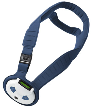
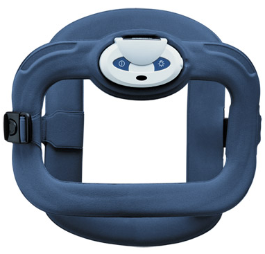
Cervical And Spinal Stim – Noninvasive Pulsed Electromagnetic Bone Growth Stimulator By Orthofix

 [Note: On the Orthofix website there is a very similar product to the Cervical Stim called the Spinal Stim. However for this post, I will be primarily talking about the Cervical Stim Product]

Analysis & Interpretation:

The two products being sold, the Cervical Stim and the Spinal Stim operate on the same pulse electromagnetic field theory (PEMF) as the products we have seen before with other products which are supposed to be easily worn and cause accelerated bone non-union healing. There are a few PubMed studies cited which show that the pulsed electromagnetic fields generated can increase the mTOR level and increase osteogenic activity. The science is weak in proof and there are not enough studies done to show that E&M fields really cause faster bone healing, except from studies done in the 30s and 40s of the last century. The devices are easy to wear and comfortable but slightly on the expensive side.

Implications For Height Increase

While the cervical stim is supposed to be worn around the neck touching the cervical vertebrate, the spinal stim is supposed to be worn around the torso touching the thoracic vertebrates. Each can possible lead to some increased vertebrate lengthening if there is really a fracture in the vertebrate in the right direction. However the increase in any possible height would be negligible since the healing if it does cause accelerated bone growth and spinal fusion would cause the vertebrate to be slightly curved from one side of the vertebrate being having more bone thus stunting whatever few millimeters of height increase the device can give to that one side.


For the Cervical Stim product main page HERE

cervicalProduct Description

Cervical-Stim® offers the security of proven, safe and effective bone growth stimulation to significantly increase the rate of cervical fusion success in high-risk patients.

It offers the comfort of an external device that patients tell us is the most effortless treatment they have ever experienced. Not to mention the comfort enjoyed with improved clinical outcomes: In a PMA study of 323 high-risk ACDF patients, 84% achieved fusion within six months using Cervical-Stim vs. 69% for those who did not receive treatment. Additionally, the data from this study prove for the first time that fusion has a positive effect on VAS and NDI clinical outcomes after ACDF.

Since 1990, more than 200,000 spine fusion patients have healed with bone growth stimulation from Orthofix. We brought you the first and most effective external bone growth stimulator for lumbar fusion, and now offer you the only FDA approved device to increase the rate of cervical fusion success.

  • Comfortably worn during daily activities, with or without a cervical brace, four hours per day
  • Proven to overcome risk factors for nonfusion
  • Next generation design with smart technology and patient friendly functionality

Indication

The Cervical-Stim is a noninvasive, pulsed electromagnet bone growth stimulator indicated as an adjunct to cervical fusion surgery in patients at high risk for nonfusion.

Physician Benefits

  • Safe and effective bone growth stimulation to significantly increase the rate of cervical fusion success in high-risk patients
  • Smart technology can provide outcomes reports and hardcopy patient compliance monitoring
  • Written guarantee for bone growth stimulation products
  • The only FDA approved bone growth stimulator for cervical fusion
  • Patient friendly, comfortably worn during daily activities with or with a cervical brace, four hours per day

Clinical References:

  • Exposure of Murine Cells to Pulsed Electromagnetic Fields Rapidly Activates the mTOR Signaling Pathway. Patterson T et al. Bioelectromagnetics 27: 535-544, 2006. Science Sheet (PDF 174KB)
  • Pulsed Electromagnetic Fields Rapidly Modulate Intracellular Signaling Events in Osteoblastic Cells: Comparison to Parathyroid Hormone and Insulin. Schnoke Matthew and Ronald J Midura. Journal of Orthopaedic Research 933-940, July 2007. Science Sheet (PDF 211KB)
  • Effects of BMP-2 and Pulsed Electromagnetic Field (PEMF) on Rat Primary Osteoblastic Cell Proliferation and Gene Expression. Selvamurugan N et al.Journal of Orthopaedic Research 1213-1220, Sep 2007. Science Sheet (PDF 129KB)
  • Randomized, Prospective, and Controlled Clinical Trial of Pulsed Electromagnetic Field Stimulation for Cervical Fusion. Foley Kevin T et al. The Spine Journal 8: 436-442, 2008.

Patient Benefits

  • Lightweight, all-in-one unit allows unrestricted patient mobility – no separate cables, control units or battery packs
  • Comfortably worn around the neck, with or without a cervical brace, four hours per day
  • Patient friendly functionality: Instant information panel with LCDs for ongoing operational status
  • Easy buckle for on/off
  • Designed for more flexibility and comfort
  • Internal countdown shuts off automatically when daily treatment is complete
  • Normally poses no issues when traveling through airport security (user should bring product manual with their PEMF device and turn the device off if it must be put through an airport x-ray scan.)

Contraindications

There are no known contraindications for the Cervical-Stim as an adjunct to cervical spine fusion surgery.

Warnings

  • The Cervical-Stim may interfere with the operation of a cardiac pacemaker or defibrillator. Consultation with the attending cardiologist is recommended
  • The Cervical-Stim should be removed prior to any imaging procedures (e.g., CT scan, MRI, etc.)

Precautions

  • The Cervical-Stim should not be used if there are mental or physical conditions that may preclude compliance with physician or device instructions
  • The Cervical-Stim has not been evaluated in treating patients with the following conditions: osseous or ligamentous spinal trauma, spondylitis, Paget’s disease, moderate to severe osteoporosis, metastatic cancer, renal disease, rheumatoid arthritis, uncontrolled diabetes mellitus, patients prone to vascular migraine headache, seizure, epilepsy, thyroid conditions or neurological diseases
  • Animal teratological studies performed with this device did not show any adverse effects in animals. However, the safety of this device for use on patients who are pregnant or nursing has not been established

Footnotes

  1. Results from Orthofix Inc. PMA Randomized, Prospective Controlled Clinical Trial of Pulsed Electromagnetic Field Stimulation for Cervical Fusion, 2004.
  2. Anterior Cervical Discectomy and Fusion: Correlation of Fusion Status with Clinical Outcome, Kevin Foley, M.D., Neurosurgery. Presented at the NASS 20th Annual Meeting, Philadelphia, PA

For the Spinal Stim product main page HERE

spinalstimProduct Description

Spinal-Stim delivers the highest success rates in bone growth stimulation for spine fusion and nonoperative salvage. In fact, 92% of patients receiving Spinal-Stim adjunctively have achieved fusion success1. When treating failed fusion with Spinal-Stim, 67% of patients achieve successful fusion with no additional surgery. Spinal-Stim offers:

  • Proven Field Coverage
  • Risk Factor Coverage
  • Guarantee Program
  • Insurance Acceptance
  • FDA Approved Models for Both Cervical and Lumbar Spine

Indication

The Spinal-Stim is a noninvasive electromagnetic bone growth stimulator indicated as a spinal fusion adjunct to increase the probability of fusion success and as a nonoperative treatment of salvage of failed spinal fusion, where a minimum of nine months has elapsed since the last surgery.

Physician Benefits

  • Proven Field Coverage
  • Risk Factor Coverage
  • Guarantee Program
  • Insurance Acceptance
  • Smart technology can provide outcomes analysis and hardcopy patient compliance monitoring

Footnotes

  1. Spinal-Stim Summary of Safety and Effectiveness. P850007/S6. February 7, 1990. One-Year Postoperative Pre-Marketing Data From Randomized Double-Blind Placebo-Controlled Clinical Trial.
  2. Spinal-Stim Summary of Safety and Effectiveness. P850007/S6. February 7, 1990. One Year Follow-up, Pre-Marketing Data, Open Trial, Pseudoarthrosis Stratum Clinical Study.

Patient Benefits

  • Lightweight, all-in-one unit allows unrestricted patient mobility – no separate cables, control units or battery packs
  • Comfortably worn with or without a brace, two hours per day
  • Patient friendly functionality: Instant information panel with LCDs for ongoing operational status
  • Easy buckle for on/off
  • Designed for more flexibility and comfort
  • Internal countdown shuts off automatically when daily treatment is complete
  • Normally poses no issues when traveling through airport security (user should bring product manual with their PEMF device and turn the device off if it must be put through an airport x-ray scan.)


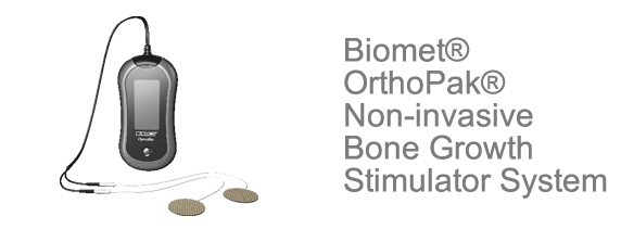
Biomet OrthoPak Non-invasive Bone Growth And Spine Fusion Stimulator System

What I will be posting in this and the next post are devices I have found being sold from different companies which have devices which can accelerate the healing rate of bone fractures. The devices are something that can be attached to the skin of the area of the body the person is trying to stimulate bone growth in.

Analysis & Interpretation

This device seems to be able to use the PEMF (pulsed electromagnetic field) concept to heal bones. This is similar to the idea of using the LIPUS theory for bone fracture healing.

With the Bone Growth Stimulator System (OrthoPak), the device is used to help accelerate the rate of bone non-unions heal. You have two round electrodes you attach to the skin about 2-3 inches apart from each other and then turn on the device to cause a current to go under the skin connecting the electrodes into a closed circuit (I am guessing). The electrodes are supposed to be disposable and easy to adhere to the skin (with some type of weak glue I am guessing). The electrical signal is supposed to be of high frequency.

With the Spine Fusion Stimulator System (SpinePak) the device is exactly the same but the application of it is different. The main difference seems to be wear you are supposed to attach the electrodes around the lower back region, and maybe also the magnitude of the current the wires are supposed to induce.

Implications For Height Increase

This device which requires a doctor prescription to get is one of the easy to use, lightweight devices which can possible cause a little bit of height increase if the theory makes any sense. Somehow there have been a few sporadi studies and articles published even from 80 years ago which suggest that inducing a low magnitude electrical signal. An old post about the possibility of using the PEMF theory as a way to increase height was written “Increase Height And Grow Taller Using Pulsed Electromagnetic Field Therapy, PEMF“. To iterate the message I had written in that post and add some more content to that old message, maybe the application of a slightly stronger electrical field at a more dynamic but consisten usage to the interverterbal disk area can cause the annulus to increase in width and lead to some temporary increase height of a few millimeters.

From the company Biomet website HERE

For the Biomet Orthopak Non-Invasive Bone Growth Stimulator System FAQ Broshure

The Biomet® OrthoPak®System promotes normal healing by inducing weak pulsing electrical signals at the nonunion fracture site. These signals are generated by a low energy electromagnetic field created by passing specific electrical current pulses through a flexible therapeutic treatment coil. A specific electrical current is delivered to the coil by the control unit. The coil then delivers the therapeutic electromagnetic signal to the nonunion fracture site.

Easy To Use and Operate

The Biomet® OrthoPak® System is portable, easy to use and operate; thus allowing you to treat your fracture nonunion while going about your daily routine. You can use the system with a cast or with a brace. The Biomet® OrthoPak® System capacitive coupling technology is the only non-invasive technology that promotes normal healing at the nonunion site 24 hours per day as recommended. The Biomet® OrthoPak® System can only be obtained with a doctor’s prescription. Upon receiving your Biomet® OrthoPak® System you will receive a Patient’s Guide containing complete instructions. Your compliance with these instructions will contribute to the successful outcome of your treatment.

System Advantages

  • The Biomet® OrthoPak® System is especially appropriate for treating proximal femur (center of the thigh bone), metatarsal/tarsal (arch of the foot or ankle) and clavicle (collarbone) fractures
  • The Biomet® OrthoPak® System is the smallest and lightest, low-profile non-invasive bone growth stimulator on the market today
  • The Biomet® OrthoPak® System electrode placement is easy and universal to all application areas – one versatile system can treat various fracture nonunions
  • The Biomet® OrthoPak® System is supplied with a unique ergonomic, rotating holster designed for active lifestyles
  • Patient usage and therapeutic treatment time may be downloaded to a PC for viewing, storage or printout via use of the Biomet compliance Data Download Software and USB Cable
  • Two different types of disposable electrodes

The Bone Healing Process

Bone is one of the tissues within the human body capable of healing itself when damaged, in much the same way as skin and other tissues. When you fracture a bone, the body naturally produces low level electric impulses that help to heal the fracture site. Occasionally, the fracture may not heal on its own. This will result in what is called a nonunion. The Biomet® OrthoPak® Non-invasive Bone Growth Stimulator System is a proven, safe, and effective method for promoting the healing of fractures that have progressed to a nonunion.

How does the Biomet® OrthoPak® Non-invasive Bone Growth Stimulator System Work?

The Biomet® OrthoPak® Non-invasive Bone Growth Stimulator System delivers an electrical treatment signal that is intended to help your fracture nonunion heal. Two lightweight electrodes, which look like round bandages are dermally applied and placed 2-3 inches apart and over from the fracture nonunion site. The electrodes are easy to apply and are extremely lightweight. The Biomet® OrthoPak® Non-invasive Bone Growth Stimulator System is battery operated with a rechargeable battery. Upon insertion of a charged battery, the Biomet® OrthoPak® Non-invasive Bone Growth Stimulator System is automatically activated and ready to deliver therapeutic treatment. The Biomet® OrthoPak® Non-invasive Bone Growth Stimulator System is portable and easy to operate. You will be able to treat your fracture nonunion while going about your daily routine, relaxing or while sleeping.


Biomet® SpinalPak® Non-invasive Spine Fusion Stimulator System

Click for the Biomet SpinalPak Non-invasive Spine Fusion Stimulator System FAQ Broshure 

By prescribing the Biomet® SpinalPak® Non-invasive Spine Fusion Stimulator System, your doctor has taken advantage of an adjunctive non invasive treatment to aid in the healing of your primary lumbar spine fusion surgery. The Biomet® SpinalPak®System delivers a low intensity electrical signal that promotes normal healing at the fusion site. The therapeutic signal operates at a high frequency; therefore, you should not feel anything during your treatment. Two small, lightweight disposable electrodes (conductors of the electrical signal) are placed on your lower back, inches apart from one another, at the level of your surgery. These electrodes are easy to apply and adhere to the skin.

Easy To Use and Operate

The Biomet® SpinalPak® System is portable, easy to use and operate; thus, allowing to help you heal while going about your daily routine. The system’s lightweight, low-profile, disposable electrodes allow treatment to occur in conjunction with wearing a back brace or just stand alone. The Biomet® SpinalPak® System’s capacitive coupling technology is the only non-invasive technology that promotes normal healing at the fusion site 24 hours per day as recommended. The Biomet® SpinalPak® System can only be obtained with a doctor’s prescription. Upon receiving your Biomet® SpinalPak® System you will receive a Patient’s Guide containing complete instructions. Your compliance with these instructions will contribute to the successful outcome of your treatment.

System Advantages

  • The Biomet® SpinalPak® System is the smallest and lightest, low-profile non-invasive spine fusion stimulator on the market today
  • The Biomet® SpinalPak® System is supplied with a unique ergonomic, rotating holster designed for active lifestyles
  • Patient usage and therapeutic treatment time may be downloaded to a PC for viewing, storage or printout via use of the Biomet Compliance Data Download Software and USB Cable
  • Two different types of disposable electrodes

How does the Biomet® SpinalPak® Non-invasive Spine Fusion Stimulator  System work?

Spinal fusion joins one or more lumbar vertebrae to  eliminate motion, increase stability and to try to reduce  pain. Following spinal fusion surgery, in the lower (lumbar) spine, the Biomet® SpinalPak® Non-invasive Spine Fusion Stimulator System may be prescribed to  assist in healing the fusion by sending electrical impulses directly to the spine that mimics your body’s natural  healing process. Two lightweight dermally applied electrodes similar in size to a quarter are placed on your back adjacent to the surgical site. The electrodes are easy to apply and are extremely lightweight. The Biomet® SpinalPak® Non-invasive Spine Fusion Stimulator System is battery operated with a rechargeable battery pack. Upon connection of the charged battery pack, the Biomet® SpinalPak® Non-invasive Spine Fusion Stimulator System is automatically activated and ready to deliver therapeutic treatment.

How long will I have to wear the device? 

The Biomet® SpinalPak® Non-invasive Spine Fusion Stimulator System is designed to deliver 270 days of  continuous therapeutic treatment. The indicated wear time  is 24 hours per day. Certain factors such as smoking, instrumentation and multiple level fusions may factor into the duration of your treatment. Biomet recommends that your doctor closely monitor your healing progress to determine how long you should treat with the device. It is important that you use the device as prescribed.

Comparing The Potential Of Lordoplasty, Kyphoplasty, Or Vertebroplasty To Restore Lost Height

A while back I had looked at the types of surgery that are done on the spinal vertebrate which have been shown to lead to some height increase. The three types of surgical procedures I had looked at are…

  1. Lordoplasty
  2. Kyphoplasty
  3. Vertebroplasty

The posts are…

From a quick look at the abstract of the articles below I will try to summarize the different surgical processes on their effectiveness in producing some type of heihgt increase.

Summary & Analysis:

Overall there was just more information on vertebroplasty and kyphoplasty than lordoplasty. It seems that lordoplasty is derived from a special type of vertebroplasty known as percutaneous vertebroplasty. Overall it seems that all the 3 different vertebral surgical types are supposed to reduce vertebral and spinal compression which would crush vertebrate and might pinch down on nerves causing acute and chronic pain. The percutaneous vertebroplasty can be simple, relatively inexpensive, safe, and effective. However the vertebroplasty doesn’t seem to resolve the issue of spinal deformity from kyphosis. It seems that PVP can’t solve the height loss and spinal deformity issue. This requires kyphoplasty. Kyphoplasty involves getting a wedge placed into one of the vertebrate to stop vertical height loss and vertebral compression.
From article #5, the researchers conclude that “Lordoplasty is more useful than kyphoplasty in terms of the improved anatomic restoration and postoperative maintenance.” While the wedges would eventually still lead to vertebral compression, the anterior vertebral height of the lordoplasty showed less decreased height/height loss.
While from article #1, it suggest that the kyphoplasty and vertebroplasty results in the same amount of overall height restoration in the vertebrate after surgery but notes that in kyphoplasty, there is less leakage of the cement to other softer tissues than the vertebroplasty. Article #2, we find that the vertebral height restoration is about 5. 1 mm for the kyphoplasty while it is only 2.3 mm for the vertebroplasty. The effectiveness to achieve some height restoration was also higher using kyphoplasty. Article #3 only shows that percutaneous vertebroplasty does add the wedge angle to cause some height restoration. Article #4 states the same thing, that vertebroplasty results in vertebral height increase. The results are ,”The average increase in vertebral body height was 2.5 mm anteriorly, 2.7 mm centrally, and 1.4 mm posteriorly”. article #5 does not do much real comparison on the vertebral height restoration of kyphoplasty and lordoplasty. However we find that article #7 does show that kyphoplasty is slightly better than vertebroplasty. The thing to conclude at this point is that from the few articles I’ve seen so far kyphoplasty may be more effective in restoring height that vertebroplasty. As for lordoplasty, it seems to be also better than lordoplasty. The injection of cement into the vertebrate does lead to a height increase, but that increase will eventually be reduced due to normal compression of the vertebrate over time.

The Library

Update #1: I’ve decided to turn this section into a library of scientific articles that are EXTREMELY relevant to our purpose. Almost all the articles have a link to the complete PDF file for the entire published paper. This is going to house some of the biggest collection of papers, books, articles, and magazine that we need.

Update #2: This post was originally entitled “10 Scientific Papers On The Human Growth Process And Mechanics The Reader Should Definitely Read“. As you can see, I’ve had to expand on this list by A LOT to house for any serious height increase researcher the tools they need or are looking for.


A Complete Collection Of All The “Height Increase” And “Grow Taller” Books

This will be another one of those posts which will eventually turn into a section/webpage of the website which will be occasionally edited and added upon. I was not aware of just how big and many books of this type there are. When I was first doing my research, I had no idea that there was this many books all devoted to this topic. The Primary source where I found this list of books was from the “Grow Taller Step” website store section (HERE).

If you want to try out your luck and buy one of the books from the links, then that is your choice. I am not promoting these books on Amazon, only to say that they do exist. The good news for you the website visitor is that a nice number of the books below are already uploaded to the “Downloads” section of this website for you to take for free.

[Note: These books are NOT in alphabetical order, yet]


  1. How to Get Taller – Grow Taller By 4 Inches In 8 Weeks, Even After Puberty! – David Taylor – (Amazon Link)
  2. How to Get Taller – The Complete Exercise Guide (Grow Taller) – David Taylor – (Amazon Link)
  3. How To Grow Taller: Guaranteed Increase Your Height Within 8 Weeks – Peter Douglas (Amazon Link)
  4. Growing Taller Secrets: Journey Into The World Of Human Growth And Development, or How To Grow Taller Naturally And Safely. Second Edition – Robert Grand (No Amazon Link)
  5. How to Naturally Increase your Height (Grow Taller Guide) – Russian Sports Authority (Amazon Link)
  6. How to Grow Taller — The Amazing Secrets to Quickly and Easily Grow Taller! — Get the Respect of Being Stronger, Confident, Taller and More Attractive Today! – Mike Summers (Amazon Link)
  7. Grow Taller book – Hayden Carter (Amazon Link)
  8. How Can I Grow Taller: Learn How To Increase Height And How To Grow Taller Naturally And Artificially. Height Increase Tips, How To Get Taller With Yoko And More. So Is There A Way To Get Taller? Yes! – Jared D. Carlson (Amazon Link)
  9. Increasing Height Through Exercise – Steven C. Cummings (Amazon Link)
  10. Increase Your Height – Krishna Gopal Vikal (No Amazon Link)
  11. Grow Taller – Statton, Burk, Cunningham Serrano
  12. How to Grow Taller “Master Secrets to Growing at Least 4 Inches in 2 Months! – Monica Heart (Amazon Link)
  13. This Tall Grow Taller and Taller – The Answer to the Fear of Height [In Japanese Language] – Aiyoshi Kawahata (Amazon Link)
  14. How To Grow Taller: A Comprehensive Guide And Revolutionary Exercise Program To Make You Grow – Kanwaljit Singh Kalsi (Amazon Link)
  15. Grow Taller Proven Insider Secret Tips and Techniques That Will Help You Grow (Body Image Solutions) – Dr. Jesus Serrano (Amazon Link)
  16. The Most Effective Way to Grow Taller (Chinese Edition) – Yang Shu Wen (No Amazon Link)
  17. Small Children, Children Grow Taller Quiz – SHI DING PING BIAN ZHU (Amazon Link)
  18. Children Naturally Grow Taller (Chinese Edition) – PU XIU SHENG CAO FANG (Amazon Link)
  19. How Quickly Children Grow Taller 10 cm (Chinese Edition) – GONG SUI (Amazon Link)
  20. You Will Grow Taller at 23 (Chinese Edition) – Jin Yang Xiu (Amazon Link)
  21. Natural Human Growing Taller Method (Modul) – Jorge Garcia (Amazon Link)
  22. Gaining Height Through Exercise : 100 Straightening and Stretching Exercises to Make You Grow – Pierre Berthelet (Amazon Link)
  23. How to Grow Taller Naturally with the Power of Your Mind – Joan M Rivera (No Amazon Link)
  24. Grow Taller Now – Unknown (No Amazon link, source link)
  25. How To Grow Taller, A Comprehensive Guide And Revolutionary Exercise Program To Make You Grow – Kanwaljit Singh Kalsi (source link)
  26. Maximize Your Height: Four Keys to Growing as Tall as Your Genetics Will Allow – Ben Kim (source link)
  27. Height Enhancement Bible

Medical, Clinical, Experimental, Professional, Textbook, Technical Books

  1. A History of the Study of Human Growth” [Paperback] – Author: James Mourilyan Tanner (Amazon Link)
  2. Absolute Ultimate Guide for Lehninger Principles of Biochemistry (Study Guide & Solutions Manual), 5th Ed.” (Softcover) – Author: David L. Nelson, Michael M. Cox (Amazon Link)
  3. Atlas of Human Anatomy (Netter Basic Science)” – Author: Frank H. Netter (Amazon Link)
  4. Cancer Stem Cells Theories And Practice” – Author: Edited by Stanley Shostak, InTech, 429 pp; 19 MByte , ISBN 978-953-307-225-8 (source link(Note: Free Full Text is the source link)
  5. Embryonic Stem Cells – Recent Advances in Pluripotent Stem Cell-Based Regenerative Medicine” – Author: Edited by Craig Atwood, ISBN 978-953-307-198-5, Hard cover, 410 pages, Publisher: InTech, Published: April 26, 2011 under CC BY-NC-SA 3.0 license (source link(Note: Free Full Text is available for download at 23 MB through source link)
  6. Endocrinology, 2-Volume Set: Adult and Pediatric, Expert Consult Premium Edition – Enhanced Online Features and Print, 6e (Endocrinology (deGroot))” [Hardcover] – Author: J. Larry Jameson MD PhD, Leslie J. De Groot MD (Amazon Link)
  7. Endocrinology, An Integrated Approach” – Author: Stephen Nussey, Saffron Whitehead , 2001 – :Taylor & Francis, 358 pp, ISBN 9781859962527 (source link)
  8. General, Organic, & Biological Chemistry, 2nd edition” [Print Replica] [Kindle Edition] – Author: Janice Smith (Amazon Link)
  9. Genetics: Analysis and Principles, 3rd Ed.” [Hardcover] – Author: Robert Brooker (Amazon Link)
  10. Genetics: From Genes to Genomes (Hartwell, Genetics), 4th Ed. 2010″ [Hardcover] – Author: Leland Hartwell, Leroy Hood, Michael Goldberg, Ann Reynolds, Lee Silver (Amazon Link)
  11. “Gray’s Anatomy for Students” [Kindle Edition] – Author: Richard Drake, A. Wayne Vogl, Adam W. M. Mitchell (Amazon Link)
  12. Greenspan’s Basic and Clinical Endocrinology, Ninth Edition (LANGE Clinical Medicine)” [Paperback] – Author: David Gardner, Dolores Shoback (Amazon Link)
  13. Growth, Maturation and Body Composition: The Fels Longitudinal Study 1929-1991” [Hardcover] – Author: Alex F. Roche (Amazon Link)
  14. Hand Bone Age, A Digital Atlas For Skeletal Maturity” – Author: Vicente Gilsanz, Osman Ritab (source link)
  15. Human Body Size and the Laws of Scaling” – Author: editor is Thomas T. Samaras (source link)
  16. Lehninger Principles Of Biochemistry & eBook, 5th Ed.” (Hardcover), Author – Albert Lehninger, David L. Nelson, Michael M. Cox  (Amazon Link)
  17. Limb Lengthening And Reconstruction Surgery” – Author: S. Robert Rozbruch, M.D., Svetlana Ilizarov, M.D (Amazon Link)
  18. Molecular Biology of the Cell , 5th Ed. [Hardcover]” – Author: Bruce Alberts, Alexander Johnson, Julian Lewis, Martin Raff, Keith Roberts, Peter Walter (Amazon Link)
  19. Multiple Epiphyseal Dysplasia” [Paperback] – Author: Edited by Iustinus Tim Avery (Amazon Link)
  20. Netter’s Concise Orthopaedic Anatomy, 2e (Netter Basic Science)” [Paperback] – Author: Jon C. Thompson MD (Amazon Link)
  21. Organic Chemistry” [Print Replica] [Kindle Edition] – Author: Francis A Carey (Amazon Link)
  22. Orthopedics of the Upper and Lower Limb” [Kindle Edition] – Author: (Amazon Link)
  23. Pediatric Bone: Biology & Diseases” – Author: Francis H. Glorieux, John M. Pettifor, Harald Jüppner (source link)
  24. Perspectives in Human Growth, Development and Maturation” [Paperback] – Author: Parasmani Dasgupta (Editor), Roland Hauspie (Editor) (Amazon Link)
  25. Saltation and Stasis in Human Growth and Development: Evidence, Methods and Theory (Auxology)” [Paperback] – Author: Michelle Lampl (Editor) (Amazon Link)
  26. School Of Height” – Author: A. S. Palko , Translated by Dr. Michael Yessis – $25 – Flexcart.com
  27. The Cambridge Encyclopedia of Human Growth and Development” [Hardcover] – Author: Stanley J. Ulijaszek (Editor), Francis E. Johnston (Editor), Michael A. Preece (Editor), James Tanner (Foreword) (Amazon Link)
  28. The Growth Plate (Biomedical and Health Research, 54)” – Editors: Irving M. Shapiro, Barbara Boyan, H. Clarke Anderson – $120 – Amazon Link
  29. The Truth About Your Height – Exploring the Myths and Realities of Human Size and its effects on Health, Performance, Pollution and Survival” – Author: Thomas T. Samaras (source link)
  30. Williams Textbook of Endocrinology: Expert Consult-Online and Print, 12e” [Hardcover] – Author: Shlomo Melmed, Kenneth S. Polonsky MD, P. Reed MD Larsen, Henry M. Kronenberg MD (Amazon Link)

National Census Height Database Papers

Mean Body Weight, Height, and Body Mass Index, United States 1960–2002 (Advanced Data, For Vital And Health Statistics) Number 347, October 27, 2004

Height And Weight Of Adult Ages 18-74 Years By Socioeconomic And Geographic Variables, United States (Data from the National Health Survey, Series 11 Number. 224, Vital And Health Statistics)

Weight, Height, And Selected Body Dimensions Of Adults United States 1960-1962, Data from the National Health Survey, Series 11 Number 8, National Center For Health Statistics


E-Book Downloads

All of the downloads available are just stuff I have found across the internet. Some are PDFs and some are books. They are all FREE to take and download and use. If you see something that should not be posted here in violation of copyright laws, inform me and I will promptly take it down. All I ask is that you give a thanks for the postings. If you really like the webpage with the E-Products then you could make a donation to the cause.


Video, Mpeg, Audio, & MP3 Downloads

  • Grow Taller With Me Part I – Author: Zixia (Nicole) – Update Jan 10th, 2014 – At Zixia’s request I have decided to no longer allow for this file to be available. We respect most people’s right for privacy on the internet to protect their real, personal lives. 
  • Grow Taller WIth Me Part 2 – Author: Zixia (Nicole) – Update Jan 10th, 2014 – At Zixia’s request I have decided to no longer allow for this file to be available. We respect most people’s right for privacy on the internet to protect their real, personal lives.
  • Dr. Laura De Giorgio – Grow Taller Program (This program is over 340 MB so it is too large to upload to the website. There is 11 CD Mp3 files in it, around 10-30 MB in size for each one. If you want the entire program, you will have to email me )
  • Grow Taller 4 You ProgramAuthor: Lance Ward (Inside the “Program” you will find 2 PDF Manuals and 5 Video files. Since the 5 Video files are too large to upload, I only uploaded the two manuals: Manual 1, Manual 2 – If you want the other 5 video files, you will have to email me)

Device, Techniques & Patents

Apparatus and method for invasive electrical stimulation of bone fractures – Eddie Adams (4602638)

Overview: An invasive electrical stimulation device includes a cathode an anode, each comprising a thin electrically conductive sheet having one exposed surface and the opposite surface covered with an insulation layer. A pair of insulated leads connect the cathode and anode to an implantable power pac which includes a source of electric power and an insulating encasement surrounding the same. The cathode may be equipped with a plurality of spaced-apart FM channel monitors which are electrically connected to an FM telemetry component within the power pac for transmitting signals indicative of the healing response of a fracture to electrical stimulation. A frequency modulated current regulator may be added to the power pac for adjusting the amperage in response to FM control signals from a remote transmitter or automatically by an implantable computer assisted device.

Automatic lengthening bone fixation device – Behrooz Akbarnia, et. al. (EP2244644 A1)

Overview: A bone fixation device adapted to be coupled to bone anchors that allows movement of rods to permit a screw-rod construct to lengthen in response to bone growth without necessitating post surgical installation adjustment of the device. The bone fixation device includes a locking mechanism that is operably associated with a housing to allow relative movement of a rod and a housing in a first direction and inhibiting relative movement of the rod and the housing in a second direction.

Bone and tissue lengthening device – R. Alan Spievack (EP0684793 B1)

Overview: A device for lengthening bone in a human or animal by incrementally extending the distance between discrete separated portions of the bone to permit continued bone growth between the separated portions comprising an intramedullary nail (4) having distal (6) and proximal (8) portions both of which are secured with the medullar canal of the bone. A hydraulic cylinder (2) comprises the proximal portion of the nail and a piston (9) comprises the distal portion of the nail. An implantable supply of operating fluid (30) communicates with the cylinder. A ratcheting mechanism (40), between the piston and cylinder, limits their relative movement. A shock absorber mechanism (52) permits limited lost motion between the piston and cylinder and ratcheting release mechanisms are employed to permit the piston and cylinder to reverse directions.

Combined tissue/bone growth stimulator and external fixation device – John C. Tepper, Richard M. Bryant (6678562)

Overview: A combined tissue/bone growth stimulator and external fixation device is provided to aid in the treatment of fractures, osteotomies, soft tissue injuries, and reconstructive surgery and to reduce the likelihood of complications. The tissue/bone growth stimulator apparatus includes an external fixation device for stabilization of a selected portion of a patient. The tissue/bone growth stimulator apparatus also includes an electrical circuit attached to and forming an integral component of the external fixation device and operable to generate an electrical drive signal. The tissue/bone growth stimulator apparatus also includes a cable adapted to connect the electrical drive signal to a stimulator portion operable to provide an electromagnetic field to stimulate the growth of bone and tissue at the selected portion of the patient and/or at other portions of the patient. More particularly, the electromagnetic field may be produced by directing a current through the external fixation…

Compact biomedical pulsed signal generator for bone tissue stimulation – James W. Kronberg (5217009)

Overview: An apparatus for stimulating bone tissue for stimulating bone growth or treating osteoporosis by applying directly to the skin of the patient an alternating current electrical signal comprising wave forms known to simulate the piezoelectric constituents in bone. The apparatus may, by moving a switch, stimulate bone growth or treat osteoporosis, as desired. Based on low-power CMOS technology and enclosed in a moisture-resistant case shaped to fit comfortably, two astable multivibrators produce the desired waveforms. The amplitude, pulse width and pulse frequency, and the subpulse width and subpulse frequency of the waveforms are adjustable. The apparatus, preferably powered by a standard 9-volt battery, includes signal amplitude sensors and warning signals indicate an output is being produced and the battery needs to be replaced.

Composition for increasing body height – Nakao, Kazuwa (EP1743653 A1)

Overview: This invention provides a composition for increasing a body height of a patient with short stature or an individual other than patients with short stature. More specifically, the invention provides: a composition for increasing the body height of an individual comprising a guanyl cyclase B (GC-B) activator as an active ingredient, the composition being to be administered to an individual free from FGFR3 abnormality; a method for increasing the body height of an individual free from FGFR3 abnormality which comprises activating GC-B; a method for screening an agent for increasing the body height of an individual which comprises selecting an agent for increasing the body height using GC-B activity as an indication; and a method for extending a cartilage bone free from FGFR3 abnormality which comprises activating GC-B in an individual.

Device for treating osteoporosis, hip and spine fractures and fusions with electric fields –  Carl T. Brighton et al. (7158835)

Overview: A technique and device for preventing and/or treating osteoporosis, hip and spine fractures, and/or spine fusions by incorporating at least one conductive coil (110) into a garment (90) adapted to be worn adjacent to the patient’s skin over a treatment area and applying an electrical signal to the coil effective to produce a magnetic flux the penetrates the treatment area so as to produce an electric field in the bones and the treatment area.

Electrical stimulation of articular chondrocytes – Carl T. Brighton (4487834)

Overview: Cell development in articular chondrocytes is enhanced by subjecting them to an alternating current field having a frequency of about 60/khz and a current density in the order of 30-40 .mu.amps/cm.sup.2

External bone fixation device – Bruce Henry Ide, Anthony Philip Pohl (EP0566561 A1)

Overview: This invention relates to external bone fixation devices used in the treatment of bone fractures and more specifically to a unilateral bone fixation device suitable for use in carrying out bone transport procedures and also bone lengthening procedures.

Human growth gene and short stature gene region. Therapeutic uses – Dr. Ercole Rao (EP1260228 A3)

Overview: Subject of the present invention is an isolated human nucleic acid molecule encoding polypeptides containing a homeobox domain of sixty amino acids having the amino acid sequence of SEQ ID NO: 1 and having regulating activity on human growth.

Three novel genes residing within the about 500kb short stature critical region on the X and Y chromosome were identified. At least one of these genes is responsible for the short stature phenotype. The cDNA corresponding to this gene may be used in diagnostic tools, and to further characterize the molecular basis for the short stature-phenotype. In addition, the identification of the gene product of the gene provides new means and methods for the development of superior therapies for short stature

Human growth hormone for treating children with abnormal short stature and kits and methods for diagnosing gs protein dysfunctions – Francis De Zegher (EP1370281 A2)

Overview: The present invention presents the use of human Growth Hormone for the manufacture of a medicament for the treatment of abnormal short stature, said short stature being characterized by a Gs protein pathway dysfunction. The invention further presents diagnostic methods and diagnostic kits for the determination of Gs pathway dysfunction.

Hyaluronan used in improvement of anti-oxidation and proliferation in chondrocytes – Huan-Ching Hsu et al (US 2008/0051367 A1)

Overview: The present invention relates to the use of hyaluronan in protecting chondrocytes against oxidative damages and further promoting their proliferation. Particularly, the present invention relates to the use of hyaluronan with a molecular weight of 600,000-1,200,000 Dalton in protecting chondrocytes against reactive oxygen damages and further promoting their proliferation.

Implantable bone growth stimulator –  Carl T. Brighton (4549547)

Overview: Disclosed is an implantable bone growth stimulator which needs no internal battery or large buffer capacitor. A receiving coil in a preferred embodiment, a ferrite core coil, receives RF energy from a transmitter external to the patient. The received RF energy is voltage doubled and rectified and provided to a constant current generator which in turn supplies a constant amount of current between implanted cathodes and an implanted anode. In preferred embodiments, the receiving coil, power supply, and cathodes are one unit which is located in place on an internal fixation device attached to the bones near the fracture site. In another embodiment, only the cathodes are located in the vicinity of the fracture site and the receiver coil voltage multiplier circuit and constant current source are located remote from the fracture site. Additionally, the internal fixation device in a preferred embodiment may have a plastic insert therein having one or more holes therethrough for the…

Implantable bone growth stimulator and method of operation – John H. Erickson, et. al (5441527)

Overview: A method for the therapeutic stimulation of bone growth of a bone site is disclosed comprising the steps of implanting first and second electrodes into the tissue near the base site. The electrodes are coupled to a bone growth stimulator which generates an alternating current.

Implantable bone-lengthening device –  James McCarthy (Publication number:US 2005/0234448 A1)

Overview: The invention relates to implantable bone-lengthening devices that are not placed intra-medullarily, but are placed extramedullarily, i.e., outside of the bones, but under the skin. The devices do not require exposed hardware (that leads to infection) or skin and muscle penetration from the pins (that cause pain), and produce minimal scarring from pin sites because the devices are placed under the skin of a patient using minimally invasive techniques. The devices may be designed with smooth contours to enable implantation using minimally invasive techniques. The devices may be actuated using an actuator that is externally or internally powered. In the case of external power, the devices may be powered remotely through high frequency transmission of power through the skin. Also included are bone-lengthening devices having fluid reservoirs and conduits for storing and delivering therapeutic fluids to treatment sites.

Increasing bone fracture resistance by repeated application of low magnitude forces resembling trauma forces – Gary S. Beaupre, Dennis R. Carter, Wilson C. Hayes (5752925)

Overview: The invention presents a method and device for increasing the fracture resistance of a bone tissue to a traumatic force. The method includes the step of selecting a nonphysiological impulse force having a location and direction resembling that of the traumatic force, but having a magnitude significantly smaller than the magnitude of the traumatic force. The impulse force is then repeatedly applied to the bone tissue, thereby stimulating the bone tissue to grow bone mass in critical areas where stresses from the traumatic force are largest. A device for applying the method includes an impulse force applicator for repeatedly applying the impulse force and a positioner for positioning the impulse force relative to the bone tissue.

Method and apparatus for cartilage growth stimulation –  Roger J. Talish et al (Patent number: 7108663)

Overview: The invention relates to apparatus and method for ultrasonically stimulating cartilage growth. The apparatus includes at least one ergonomically constructed ultrasonic transducer configured to cooperate with a placement module or strip for placement in proximity to an area where cartilage growth is desired. The apparatus also utilizes a portable, ergonomically constructed main operating unit constructed to fit within a pouch worn by the patient. In operation, at least one ultrasonic transducer positioned in proximity to an osteochondral site is excited for a predetermined period of time. To ensure that at least one ultrasonic transducer is properly positioned, and to insure compliance with a treatment protocol, a safety interlock is provided to prevent inadvertent excitation of the at least one ultrasonic transducer.

Method and apparatus for controlling the growth of cartilage – Abraham R. Liboff, Bruce R. McLeod, Stephen D. Smith (Patent number: 5067940)

Overview: An apparatus and method for regulating the growth of cartilage in vivo are provided. The apparatus includes a magnetic field generator and a magnetic field detector for producing a controlled, fluctuating, directionally oriented magnetic field parallel to a predetermined axis projecting through the target tissue. The field detector samples the magnetic flux density along the predetermined axis and provides a signal to a microprocessor which determines the average value of the flux density. The applied magnetic field is oscillated at predetermined frequencies to maintain a preselected ratio of frequency to average flux density. This ratio is maintained by adjusting the frequency of the fluctuating magnetic field and/or by adjusting the intensity of the applied magnetic field as the composite magnetic flux density changes in response to changes in the local magnetic field to which the target tissue is subjected. By maintaining these precise predetermined ratios of frequency to average…

Method for non-invasive electrical stimulation of epiphyseal plate growth – Carl T. Brighton

Overview: Epiphyseal growth plate stimulation in the bone of a living body is achieved by applying electrodes non-invasively to a body and supplying to said electrodes an AC signal in the range of about 2.5 to 15 volts peak-to-peak at a frequency of about 20-100 KHz.

Method for the promotion of growth, ingrowth and healing of bone tissue and the prevention of osteopenia by mechanical loading of the bone tissue – Kenneth J. McLeod, Clinton T. Rubin (5103806)

Overview: A method for preventing osteopenia, promoting bone tissue growth, ingrowth, and healing of bone tissue includes the step of applying a mechanical load to the bone tissue at a relatively low level on the order of between about 50 and about 500 microstrain, peak-to-peak, and at a relatively high frequency in the range of about 10 and about 50 hertz. Mechanical loading at such strain levels and such frequencies has been found to prevent bone loss and enhance new bone formation.

Method for treating cartilage related diseases –  Kai Yu et al (Patent number: 6713513)

Overview: The present invention relates to a method for preventing or treating of cartilage related diseases, such as osteoarthritis, rheumatoid arthritis, articular cartilage damage, multiple chondritis, osteochondrosis, etc., which comprises administering an effective amount of calcium L-threonate to a subject in need of such prevention or treatment. It had been demonstrated by experiments that calcium L-threonate could improve significantly the positive expression percentage of collagen I mRNA in the chondrocyte and osteoblast, improved significantly the positive expression percentage of articular cartilage and epiphyseal cartilage, facilitated the growth of chondrocyte, increased the quantity of bone collagen, and promoted the formation of bone and cartilage matrix and the synthesis of proteoglycan. It could also promote the formation of bone nourishing blood vessels and improve the microcirculation of bone.

Method for treatment of non-union bone fractures by non-invasive electrical stimulation – Carl T. Brighton, Solomon R. Pollack (Patent number: 4535775)
Overview: Bone fractures previously considered incurable are healed non-invasively by applying to electrodes coupled to the skin of a living body in the vicinity of a bone fracture an alternating voltage having a wave form that is symmetrical with respect to the axis, a frequency in the range 20-100 KHz and a value in the range from about 2 to 10 volts peak to peak.

Overview: The invention relates to methods and articles of manufacture for treating short stature disorders related to the Short Stature Homeobox-containing (SHOX) gene which, in the human genome, is located in the pseudoautosomal region (PAR1) on the short arm of the X chromosome (Xp22.3) and Y chromosome (Yp11.3). More particularly, the invention relates to methods for treating a SHOX gene disorder, other than Turner syndrome, due to deficiency of one copy (haploinsufficiency) of the SHOX gene, by administering a growth hormone, particularly by administering human growth hormone to a human subject with a SHOX gene disorder.
Methods for modulating chondrocyte proliferation using pulsing electric fields –  James W. Kronberg et al (US 2008/0039901 A1)
Overview: Compositions and methods are provided for modulating the growth, development and repair of cartilage, bone or other connective tissue. Devices and stimulus waveforms are provided to differentially modulate the behavior of chondrocytes, osteoblasts and other connective tissue cells to promote proliferation, differentiation, matrix formation or mineralization for in vitro or in vivo applications. Continuous-mode and pulse-burst-mode stimulation of cells with charge-balanced signals may be used. Cartilage, bone and other connective tissue growth is stimulated in part by nitric oxide release through electrical stimulation and may be modulated through co-administration of NO donors and NO synthase inhibitors. The methods and devices described are useful in promoting repair of bone fractures, cartilage and connective tissue repair as well as for engineering tissue for transplantation.
MODULATING BONE GROWTH IN TREATING SCOLIOSIS – Kieran Murphy (Patent application number: 20100221229)

Overview: The present specification provides, amongst other things, a method of treating scoliosis comprising delivering a therapeutically acceptable amount of a growth modulator to an epiphyseal growth plate to correct or compensate for disproportionate growth.

Novel cartilage cell proliferation and differentiation inducer – Yuriko Furuya, Hisataka Yasuda (EP2343087 A1)

Overview: This invention provides a method for administration of an effective amount of RANKL-binding molecules that act on prechondrocytes and/or mesenchymal stem cells, accelerate cartilage differentiation, proliferation, and maturation of such cells, enhance chondrocyte differentiation, and induce chondrocyte proliferation to induce chondrocyte proliferation and differentiation or increase cartilage matrix production and a pharmaceutical composition used for inducing chondrocyte proliferation and differentiation or increasing cartilage matrix production. The pharmaceutical composition used for treatment or prevention of a chondropathies comprises, as an active ingredient, a compound that acts on prechondrocytes and/or mesenchymal stem cells and induces at least one of the following: (a) acceleration of prechondrocyte and/or mesenchymal stem cell differentiation; (b) acceleration of prechondrocyte and/or mesenchymal stem cell proliferation; (c) acceleration of prechondrocyte and/or mesenchymal stem cell maturation; (d) enhancement of chondrocyte differentiation; (e) chondrocyte proliferation; and (f) increased production of the cartilage matrix.

Non-invasive method for in-vivo bone-growth stimulation –  Kenneth J. McLeod et al. (Patent number : 5376065)

Overview: A method and apparatus for preventing osteopenia, promoting bone tissue growth, ingrowth, and healing of bone tissue includes the step of and means for applying a mechanical load to the bone tissue at a relatively low level on the order of between about 50 and about 500 microstrain, peak-to-peak, and at a relatively high frequency in the range from about 10 and about 100 Hz. Mechanical loading at such strain levels and such frequencies has been found to prevent bone loss and enhance new bone formation.

Regulation of matrix metalloproteinase gene expression using specific and selective electrical and electromagnetic signals – T. Carl Brighton (EP1643953 A2)

Overview: Methods and devices are provided for the targeted treatment of injured or diseased cartilage tissue that include generating specific and selective electric and electromagnetic signals that generate fields optimized for reduction of matrix metalloproteinase gene expression and exposing cartilage tissue to the fields generated by specific and selective signals so as to regulate matrix metalloproteinase gene expression in such cartilage tissue. The resulting methods and devices are useful for the targeted treatment of osteoarthritis, rheumatoid arthritis, cartilage injury, cartilage defects, and tumor metastasis.

REGULATION OF STEM CELL GENE PRODUCTION WITH SPECIFIC AND SELECTIVE ELECTRIC AND ELECTROMAGNETIC FIELDS – Carl T. Brighton (US 2012/0149968 A1)

Overview: Methods and devices are described for the regulation of BMP 2 and 4, TGF-beta 1, 2, and 3, FGF-2, osteocalcin, and alkaline phosphatase mRNA in stem cells via capacitive coupling or inductive coupling of specific and selective electric and/or electromagnetic fields to the bone cells or other tissues containing the stem cells, where the specific and selective fields are generated by application of specific and selective signals to field generating devices disposed with respect to the stem cells so as to facilitate the treatment of diseased or injured bone and other tissues. The resulting methods and devices are useful for the targeted treatment of osteoporosis, osteopenia, osteonecrosis, fresh bone fractures, fractures at risk, nonunion, bone defects, spine fusion, and/or other conditions in which BMP 2 and 4, TGF-beta 1, 2, and 3, FGF-2, osteocalcin, and alkaline phosphatase mRNA and/or protein deficiencies in stem cells has been implicated.

REGULATION OF TRANSFORMING GROWTH FACTOR-BETA (TGF- ß) GENE EXPRESSION IN LIVING CELLS VIA THE APPLICATION OF SPECIFIC AND SELECTIVE ELECTRIC AND ELECTROMAGNETIC FIELDST. Carl Brighton (EP2024003 A1)

Overview: Methods and devices are described for the regulation of Transforming Growth Factor (TGF)- βl, β2, and/or β3 protein gene expression in bone cells and other tissues via the capacitive coupling or inductive coupling of specific and selective electric fields to the bone cells or other tissues, where the specific and selective electric fields are generated by application of specific and selective electric and electromagnetic signals to electrodes or one or more coils or other field generating device disposed with respect to the bone cells or other tissues so as to facilitate the treatment of diseased or injured bone and other tissues.

Regulation of type II collagen gene expression using specific and selective electrical and electromagnetic signals – Carl T. Brighton (RE41391)

Overview: Methods and devices for the regulation of type II collagen gene expression in cartilage cells via the application of specific and selective fields generated by specific and selective electric and electromagnetic signals in the treatment of diseased or injured articular cartilage. By gene expression is meant the up regulation or down regulation of the process whereby specific portions (genes) of the human genome (DNA) are transcribed into mRNA and subsequently translated into protein. Methods and devices are provided for the targeted treatment of injured or diseased cartilage tissue that include generating specific and selective electric and electromagnetic signals that generate specific and selective fields optimized for type II collagen gene expression and exposing cartilage tissue to the specific and selective fields generated by specific and selective signals so as to regulate type II collagen gene expression in such cartilage tissue. The resulting methods and devices are useful…

Resolution of aggrecan gene expression using specific and selective electrical and electromagnetic signals – T. Carl Brighton (EP1560553 A2)

Overview: Methods and devices for the regulation of aggrecan gene expression in cartilage cells via the application of fields generated by specific and selective electric and electromagnetic signals in the treatment of diseased or injured articular cartilage. By gene expression is meant the up regulation or down regulation of the process whereby specific portions (genes) of the human genome (DNA) are transcribed into mRNA and subsequently translated into protein. Methods and devices are provided for the targeted treatment of injured or diseased cartilage tissue that include generating specific and selective electric and electromagnetic signals that generate fields optimized for aggrecan gene expression and exposing cartilage tissue to the fields generated by specific and selective signals so as to regulate aggrecan gene expression in such cartilage tissue. The resulting methods and devices are useful for the targeted treatment of osteoarthritis, rheumatoid arthritis, cartilage injury, and cartilage defects.

System and method of up-regulating bone morphogenetic proteins (BMP) gene expression in bone cells via the application of fields generated by specific and selective electric and electromagnetic signals – Carl T Brighton (8017369)

Overview: Methods and devices are described for the regulation of bone morphogenetic protein gene expression in bone cells via the application of fields generated by specific and selective electric and electromagnetic signals in the treatment of diseased or injured bone. By gene expression is meant the up-regulation or down-regulation of the process whereby specific portions (genes) of the human genome (DNA) are transcribed into mRNA and subsequently translated into protein. Methods and devices are provided for the targeted treatment of injured or diseased bone tissue that include generating specific and selective electric and electromagnetic signals that generate fields optimized for increase of bone morphogenetic protein gene expression and exposing bone to the fields generated by specific and selective signals so as to regulate bone morphogenetic protein gene expression in such bone tissue. The resulting methods and devices are useful for the targeted treatment of bone fractures, fractures…

Therapeutic agent for cartilaginous diseases – Masahiro Iwamoto et al. (6756358)

Overview: The present invention relates to a therapeutic agent for cartilaginous diseases, an accelerator for chondrocyte proliferation and an accelerator for proteoglycan production comprising HGF (hepatocyte growth factor) as an active component, and a treatment method for cartilaginous diseases of human or mammals comprising administering an effective amount of HGF. The active component HGF has an effect to promote the proliferation of chondrocytes and to promote the production of proteoglycan. Therefore, the therapeutic agent and accelerator of the present invention are useful for the prevention and treatment of various disorders caused by cartilaginous diseases.

Therapeutic method – Jillian Cornish, Reginald Ian Reid (EP1027027 A2)

Overview: This invention is directed to new therapeutic uses which involve the stimulation of chondrocyte proliferation. More particularly, it is directed to the use of amylin and adrenomedullin as agents which stimulate chondrocyte proliferation and which therefore have utility in the treatment of cartilage disorders and/or cartilage mediated bone growth.

Use of natriuretic peptides for the treatment of stature disorders related to the shox gene – Beate Haecker, Gudrun Rappold-Hoerbrand (EP1583554 A2)

Overview: The invention relates to the use of natriuretic peptides (ANP or BNP) for the preparation of pharmaceutical compositions for the treatment of short stature in a subject being suspected of having a genetic defect in the human SHOX gene. Further, the invention relates to use of natriuretic peptides in combination with a growth protein and/or a SHOX protein for the preparation of pharmaceutical compositions for the treatment of patients having a SHOX gene disorder. The invention also relates to the use of natriuretic peptides (ANP and BNP) in combination with a SHOX protein for the preparation of pharmaceutical compositions for the treatment of patients with cardiovascular diseases. The invention also comprises an article of manufacture comprising packaging material and a pharmaceutical composition comprising natriuretic peptides.

Scientific Articles, Journal Publishings, Papers, Etc.

These are the PDF documents I have found which detail the human growth process and mechanics which I would highly advice the reader read to get a better understanding on what our research will eventually focus on later on in this website. As always just getting through the required texts and papers to reach full understanding is not enough. Just like in school where there was required textbooks and readings, there will also be included papers who are recommended as suggested reading for the reader to get a better understanding.

Note: As always the papers are listed in alphabetical order.

  1. 1,25-(OH)2D3 modulates growth plate chondrocytes via membrane receptor-mediated protein kinase C by a mechanism that involves changes in phospholipid metabolism and the action of arachidonic acid and PGE2.” – Author: Boyan BD, Sylvia VL, Dean DD, et. al. (source link)
  2. 24,25-(OH)(2)D(3) regulates cartilage and bone via autocrine and endocrine mechanisms.” – Author: Boyan BD, Sylvia VL, Dean DD, Schwartz Z. (source link)
  3. A comparison of three cell types as potential candidates for intervertebral disc therapy: annulus fibrosus cells, chondrocytes, and bone marrow derived cells.” – Author: Kuh SU, Zhu Y, Li J, Tsai KJ, Fei Q, Hutton WC, Yoon TS. (source link)
  4. A method of assessing skeletal maturity from radiographs” – Author: Roy M. Acheson (source link)
  5. A novel growth-promoting factor derived from fetal bovine cartilage, chondromodulin II. Purification and amino acid sequence.” – Author: Hiraki Y, Inoue H, Kondo J, Kamizono A, Yoshitake Y, Shukunami C, Suzuki F. (source link)
  6. A novel in vivo model to study endochondral bone formation; HIF-1alpha activation and BMP expression.” – Author: Emans PJ, Spaapen F, Surtel DA, et. al. (source link)
  7. Accelerated Growth Plate Mineralization and Foreshortened Proximal Limb Bones in Fetuin-A Knockout Mice” – Author: Jong Seto, et. al. (source link)
  8. Acceleration of regenerate ossification during distraction osteogenesis with recombinant human bone morphogenetic protein-7.” – Author: Mizumoto Y, Moseley T, Drews M, Cooper VN 3rd, Reddi AH. (source link)
  9. Activation of collagen type II expression in osteoarthritic and rheumatoid cartilage.” – Author: Aigner T, Stöss H, Weseloh G, Zeiler G, von der Mark K. (source link)
  10. Adult mesenchymal stem cells and cell-based tissue engineering” – Author: Rocky S Tuan, Genevieve Boland and Richard Tuli (source link)
  11. [Advance on repair of growth plate injury].” – Author: Wang J, Yang ZM. (source link)
  12. ALK2 functions as a BMP type I receptor and induces Indian hedgehog in chondrocytes during skeletal development.” – Author: Zhang D, Schwarz EM, Rosier RN, Zuscik MJ, Puzas JE, O’Keefe RJ. (source link)
  13. Alterations of the growth plate in chronic renal failure.” – Author: Santos F, Carbajo-Pérez E, Rodríguez J, Fernández-Fuente M, Molinos I, Amil B, García E. (source link)
  14. An electromagnetic compressive force by cell exciter stimulates chondrogenic differentiation of bone marrow-derived mesenchymal stem cells.” – Author: Park SH, Sim WY, Park SW, Yang SS, Choi BH, Park SR, Park K, Min BH. (source link)
  15. [Application of extracorporeal shock-wave therapy (ESWT) in delayed unions and non-unions].” – Author: Liu MQ, Guo X, Kuang SC, Wang SH, Rong GW. (source link)
  16. APPLICATION OF NON-LINEAR ELASTIC WAVE SPECTROSCOPY (NEWS) TO IN VITRO DAMAGEASSESSMENT IN CORTICAL BONE” – Author: Marie Muller (source link)
  17. Arachidonic acid directly mediates the rapid effects of 24,25-dihydroxyvitamin D3 via protein kinase C and indirectly through prostaglandin production in resting zone chondrocytes.” – Author: Schwartz Z, Sylvia VL, Curry D, Luna MH, Dean DD, Boyan BD. (source link)
  18. Arms and the Man: The Problem of Symmetric Growth” – Author: (source link)
  19. Articular cartilage collagen: an irreplaceable framework?” – Author: Eyre DR, Weis MA, Wu JJ. (source link)
  20. Articular cartilage regeneration using periosteum.” – Author: O’Driscoll SW. (source link)
  21. Assessment of epiphyseal plate allograft viability and function after ex vivo storage in University of Wisconsin Solution.” – Author: Ravindran S, Boyer MI, Martens E, Ntouvali H, McAlinden A. (source link)
  22. Basic Science of Articular Cartilage and Osteoarthritis” – Author: Andrew D. Pearle, et. al. (source link)
  23. Bioengineering and characterization of physeal transplant with physeal reconstruction potential.” Author: Lee KM, Cheng AS, Cheung WH, Lui PP, Ooi V, Fung KP, Leung PC, Leung KS (source link)
  24. BIOPHYSICAL STIMULATION OF BONE FRACTURE REPAIR, REGENERATION AND REMODELLING” – Authors:  Edmund Y.S. Chao* and Nozomu Inoue (source link)
  25. Biological repair of the degenerated intervertebral disc by the injection of growth factors.” – Author: Masuda K. (source link)
  26. Biology Of Bone” (Powerpoint File .pdf) – Author: Dr. Mark A.Birch (source link)
  27. BMP and Ihh/PTHrP signaling interact to coordinate chondrocyte proliferation and differentiation.” – Author: Minina E, Wenzel HM, Kreschel C, Karp S, Gaffield W, McMahon AP, Vortkamp A. (source link)
  28. BMP-2 enhances TGF-beta3-mediated chondrogenic differentiation of human bone marrow multipotent mesenchymal stromal cells in alginate bead culture.” – Author: Shen B, Wei A, Tao H, Diwan AD, Ma DD. (source link)
  29. BMP2, but not BMP4, is crucial for chondrocyte proliferation and maturation during endochondral bone development.” – Author: Shu B, et. al. (source link)
  30. BMP-6 and BMPR-1a are up-regulated in the growth plate of the fractured tibia.” – Author: Fischerauer EE, et. al. (source link)
  31. BMP-6 is an autocrine stimulator of chondrocyte differentiation.” – Author: Grimsrud CD, Romano PR, D’Souza M, Puzas JE, Reynolds PR, Rosier RN, O’Keefe RJ. (source link)
  32. BMP signaling stimulates chondrocyte maturation and the expression of Indian hedgehog.” – Author: Grimsrud CD, Romano PR, D’Souza M, et. al. (source link)
  33. Bone formation via cartilage models: the “borderline” chondrocyte.” – Author: Bianco P, Cancedda FD, Riminucci M, Cancedda R. (source link)
  34. Bone Growth in Length and Width: The Yin and Yang of Bone Stability” – Author:  F. Rauch  (source link)
  35. Bone morphogenetic protein-2 facilitates expression of chondrogenic, not osteogenic, phenotype of human intervertebral disc cells.” – Author: Kim DJ, Moon SH, Kim H, Kwon UH, Park MS, Han KJ, Hahn SB, Lee HM. (source link)
  36. Bone morphogenetic protein 2 stimulates articular cartilage proteoglycan synthesis in vivo but does not counteract interleukin-1alpha effects on proteoglycan synthesis and content.” – Author: Glansbeek HL, van Beuningen HM, Vitters EL, Morris EA, van der Kraan PM, van den Berg WB. (source link)
  37. Bone quality and growth characteristics of growth plates following limb transplantation between animals of different ages – Results of an experimental study in male syngeneic rats” – Author: Hitesh N Modi,, et. al. (source link)
  38. Bone regeneration and fracture healing. Experience with distraction osteogenesis model.” – Author: Richards M, Goulet JA, Weiss JA, Waanders NA, Schaffler MB, Goldstein SA. (source link)
  39. Breaking bony bridges by physeal distraction. A new approach.” – Author: Canadell J, de Pablos J. (source link)
  40. Brief exposure to high-dose transforming growth factor-beta1 enhances periosteal chondrogenesis in vitro: a preliminary report.” – Author: Miura Y, Parvizi J, Fitzsimmons JS, O’Driscoll SW. (source link)
  41. Calcium citrate: a new biomaterial that can enhance bone formation in situ.” – Author: Li-Ming Wang, Wei Wang, Xiu-Cui Li, et. al. (source link)
  42. Can TGF-beta1 and rhBMP-2 act in synergy to transform bone marrow stem cells to discogenic-type cells?” – Author: Kuh SU, Zhu Y, Li J, Tsai KJ, Fei Q, Hutton WC, Yoon ST. (source link)
  43. Carboxypeptidase Z (CPZ) links thyroid hormone and Wnt signaling pathways in growth plate chondrocytes.” – Author: Wang L, Shao YY, Ballock RT. (source link)
  44. Catch-up growth after hypothyroidism is caused by delayed growth plate senescence.” – Author: Marino R, Hegde A, Barnes KM, Schrier L, Emons JA, Nilsson O, Baron J. (source link)
  45. Cellular mechanisms for methotrexate chemotherapy-induced bone growth defects.” – Author: Xian CJ, Cool JC, Scherer MA, Macsai CE, Fan C, Covino M, Foster BK. (source link)
  46. Cervical vertebral bone age in girls.” – Author: Mito T, Sato K, Mitani H. (source link)
  47. Changes in the tibial growth plates of chickens with thiram-induced dyschondroplasia.” – Author: Rath NC, Richards MP, Huff WE, Huff GR, Balog JM. (source link)
  48. Chondrocyte allograft transplantation for damaged growth plate reconstruction.” – Author: Park JS, Ahn JI, Oh DI. (source link)
  49. Chondrocyte injection in distraction epiphysiolysis (rabbit model).” – Author: Ahn JI, Erdin RA, Smith R, Canale ST, Hasty KA. (source link)
  50. Chondrocytes and longitudinal bone growth: the development of tibial dyschondroplasia.” – Author: Farquharson C, Jefferies D. (source link)
  51. Chondrogenic differentiation of bovine bone marrow mesenchymal stem cells (MSCs) in different hydrogels: influence of collagen type II extracellular matrix on MSC chondrogenesis.” – Author: Bosnakovski D, Mizuno M, Kim G, Takagi S, Okumura M, Fujinaga T. (source link)
  52. Chondrogenic differentiation of bovine synovium: bone morphogenetic proteins 2 and 7 and transforming growth factor beta1 induce the formation of different types of cartilaginous tissue.” – Author: Shintani N, Hunziker EB. (source link)
  53. Chondrogenic differentiation of growth factor-stimulated precursor cells in cartilage repair tissue is associated with increased HIF-1alpha activity.” – Author: Gelse K, Mühle C, Knaup K, Swoboda B, Wiesener M, Hennig F, Olk A, Schneider H. (source link)
  54. Chondrogenic differentiation of mesenchymal stem cells and its clinical applications.” Author: Lee JW, Kim YH, Kim SH, Han SH, Hahn SB. (source link)
  55. Chondromodulin-I as a novel cartilage-specific growth-modulating factor.” – Author: Hiraki Y, Shukunami C. (source link)
  56. Chondromodulin-I expression in the growth plate of young uremic rats.” – Author: Amil B, Fernandez-Fuente M, Molinos I, et. al. (source link)
  57. Chondromodulin 1 stabilizes the chondrocyte phenotype and inhibits endochondral ossification of porcine cartilage repair tissue.” – Author: Klinger P, Surmann-Schmitt C, Brem M, et. al. (source link)
  58. Chronologic age and skeletal maturation of the cervical vertebrae and hand-wrist: is there a relationship?” – Author: Uysal T, Ramoglu SI, Basciftci FA, Sari Z. (source link)
  59. Coculture of synovium-derived stem cells and nucleus pulposus cells in serum-free defined medium with supplementation of transforming growth factor-beta1: a potential application of tissue-specific stem cells in disc regeneration.” – Author: Chen S, Emery SE, Pei M. (source link)
  60. Collagen of articular cartilage.” – Author: Eyre D. (source link)
  61. Combination of transforming growth factor-beta2 and bone morphogenetic protein 7 enhances chondrogenesis from adipose tissue-derived mesenchymal stem cells.” – Author: Kim HJ, Im GI. (source link)
  62. Combined effects of insulin-like growth factor-1 and transforming growth factor-beta1 on periosteal mesenchymal cells during chondrogenesis in vitro.” – Author: Fukumoto T, Sperling JW, Sanyal A, Fitzsimmons JS, Reinholz GG, Conover CA, O’Driscoll SW. (source link)
  63. Comparative review of growth factors for induction of three-dimensional in vitro chondrogenesis in human mesenchymal stem cells isolated from bone marrow and adipose tissue.” – Author: Puetzer JL, Petitte JN, Loboa EG. (source link)
  64. Comparative study of the ability of mesenchymal stem cells derived from bone marrow, periosteum, and adipose tissue in treatment of partial growth arrest in rabbit.” – Author: Hui JH, Li L, Teo YH, Ouyang HW, Lee EH. (source link)
  65. Co-ordination of TGF-beta and FGF signaling pathways in bone organ cultures.” – Author: Mukherjee A, Dong SS, Clemens T, Alvarez J, Serra R. (source link)
  66. CTGF/Hcs24, a hypertrophic chondrocyte-specific gene product, stimulates proliferation and differentiation, but not hypertrophy of cultured articular chondrocytes.” – Author: Nishida T, Kubota S, Nakanishi T, Kuboki T, Yosimichi G, Kondo S, Takigawa M. (source link)
  67. Cultured mesenchymal stem cell transfers in the treatment of partial growth arrest.” – Author: Chen F, Hui JH, Chan WK, Lee EH. (source link)
  68. Cyclic hydrostatic pressure enhances the chondrogenic phenotype of human mesenchymal progenitor cells differentiated in vitro” – Author: Angele P, Yoo JU, Smith C, Mansour J, Jepsen KJ, Nerlich M, Johnstone B. (source link)
  69. Cyclic, mechanical compression enhances chondrogenesis of mesenchymal progenitor cells in tissue engineering scaffolds.” – Author: Angele P, Schumann D, Angele M, et.al.(source link)
  70. Daily or intermittent calcitriol administration during growth hormone therapy in rats with renal failure and advanced secondary hyperparathyroidism.” – Author: Sanchez CP, He YZ. (source link)
  71. Delayed stimulatory effect of low-intensity shockwaves on human periosteal cells.” – Author: Tam KF, Cheung WH, Lee KM, Qin L, Leung KS. (source link)
  72. Depletion of resting zone chondrocytes during growth plate senescence.” – Author: Schrier L, Ferns SP, Barnes KM, Emons JA, Newman EI, Nilsson O, Baron J. (source link)
  73. Development and validation of new model for microvascular transplantation of epiphyseal plate allografts with minimal adjoining epiphyseal and metaphyseal bone.” – Author: Bray PW, Neligan PC, Bowen CV, Danska JS, Boyer MI. (source link)
  74. Developmental regulation of the growth plate” – Author: Kronenberg HM. (Full Text not available, source link HERE)
  75. Dexamethasone increases growth hormone receptor messenger ribonucleic acid levels in liver and growth plate.” – Author: Heinrichs C, Yanovski JA, Roth AH, Yu YM, Domené HM, Yano K, Cutler GB Jr, Baron J. (source link)
  76. [Dexamethasone up-regulates the expression of glucocorticoid receptor in growth plate and inhibits the longitudinal growth of bone: experiment with rats].” – Author: Zhang HF, Wang WP, Yang Y, Zhang YL, Pi YL, Tian XY, Jiang XF. (source link)
  77. Diaphyseal bone formation in murine tibiae in response to knee loading.” – Author: Zhang P, Tanaka SM, Jiang H, Su M, Yokota H. (source link)
  78. Differential effects of insulin-like growth factor I and growth hormone on developmental stages of rat growth plate chondrocytes in vivo.” – Author: Hunziker EB, Wagner J, Zapf J. (source link)
  79. Differential effects of local application of BMP-2 or TGF-beta 1 on both articular cartilage composition and osteophyte formation.” – Author: van Beuningen HM, Glansbeek HL, van der Kraan PM, van den Berg WB. (source link)
  80. Differential regulation of growth plate chondrocytes by 1alpha,25-(OH)2D3 and 24R,25-(OH)2D3 involves cell-maturation-specific membrane-receptor-activated phospholipid metabolism.” – Author: Boyan BD, Sylvia VL, Dean DD, Del Toro F, Schwartz Z. (source link)
  81. Differentiation of mesenchymal stem cells towards a nucleus pulposus-like phenotype in vitro: implications for cell-based transplantation therapy.” – Author: Risbud MV, Albert TJ, Guttapalli A, Vresilovic EJ, Hillibrand AS, Vaccaro AR, Shapiro IM. (source link)
  82. Differentiation of mesenchymal stem cells transplanted to a rabbit degenerative disc model: potential and limitations for stem cell therapy in disc regeneration.” – Author: Sakai D, Mochida J, Iwashina T, Watanabe T, Nakai T, Ando K, Hotta T. (source link)
  83. Direct inhibition of Indian hedgehog expression by parathyroid hormone (PTH)/PTH-related peptide and up-regulation by retinoic acid in growth plate chondrocyte cultures.” – Author: Yoshida E, Noshiro M, Kawamoto T, Tsutsumi S, Kuruta Y, Kato Y. (source link)
  84. Distinct roles of type I bone morphogenetic protein receptors in the formation and differentiation of cartilage.” – Author: Zou H, Wieser R, Massagué J, Niswander L. (source link)
  85. Distraction Osteogenesis and Its Challenges in Bone Regeneration” – Authors:  Reggie C. Hamdy, Juan S. Rendon and Maryam Tabrizian
  86. Dual roles of Wnt signaling during chondrogenesis in the chicken limb.” – Author: Hartmann C, Tabin CJ. (source link)
  87. Effect of extracorporeal shock waves on callus formation during bone lengthening.” – Author: Narasaki K, Shimizu H, Beppu M, Aoki H, Takagi M, Takashi M. (source link)
  88. Effect of high-energy extracorporeal shock waves on the immature epiphysis in a rabbit model.” – Author: Ozturk H, Bulut O, Oztemur Z, Kaloglu C, Kol IO. (source link)
  89. Effect of hypoxia and reoxygenation on gene expression and response to interleukin-1 in cultured articular chondrocytes.” – Author: Martin G, Andriamanalijaona R, Grässel S, Dreier R, Mathy-Hartert M, Bogdanowicz P, Boumédiene K, Henrotin Y, Bruckner P, Pujol JP. (source link)
  90. Effect of IGF-I in the chondrogenesis of bone marrow mesenchymal stem cells in the presence or absence of TGF-beta signaling.” – Author: Longobardi L, O’Rear L, Aakula S, Johnstone B, Shimer K, Chytil A, Horton WA, Moses HL, Spagnoli A. (source link)
  91. Effects of cyclic compressive loading on chondrogenesis of rabbit bone-marrow derived mesenchymal stem cells.” – Author: Huang CY, Hagar KL, Frost LE, Sun Y, Cheung HS. (source link)
  92. Effects of distraction and compression on proliferation of growth plate chondrocytes. A study in rabbits.” – Author: Alberty A, Peltonen J, Ritsilä V. (source link)
  93. Effects of estrogen on growth plate senescence and epiphyseal fusion.” – Author: Weise M, De-Levi S, Barnes KM, Gafni RI, Abad V, Baron J (source link)
  94. Effects of mechanical factors on the fracture healing process.” – Author: Claes LE, et. al. (source link)
  95. Effect of plant extract YGF on inducement of IGF-1” – Author: Cheol Seok Choi, et. al. (source link)
  96. Effects of Jaoga-Yukmiwon(R), a Korean herbal medicine, on chondrocyte proliferation and longitudinal bone growth in adolescent male rats.” – Author: Leem K, Park SY, Lee DH, Boo YM, Cho KH, Lim J, Jeon H, Park HJ, Chung JH, Kim H.(source link)
  97. Effects of timing of low-intensity pulsed ultrasound on distraction osteogenesis.” – Author: Sakurakichi K, Tsuchiya H, Uehara K, Yamashiro T, Tomita K, Azuma Y. (source link)
  98. Efficient chondrogenic differentiation of mesenchymal cells in micromass culture by retroviral gene transfer of BMP-2.” – Author: Carlberg AL, Pucci B, Rallapalli R, Tuan RS, Hall DJ. (source link)
  99. Elbow loading promotes longitudinal bone growth of the ulna and the humerus” – Author: Ping Zhang, Hiroki Yokota (source link)
  100. Endocrine Regulation of Longitudinal Bone Growth” – Author: Jan M. Wit,  Cecilia Camacho- Hübner (source link)
  101. Endocrine regulation of the growth plate” – Author: Nilsson O, et. al. (source link)
  102. Enhancement of bone formation with a synthetic matrix containing bone morphogenetic protein-2 by the addition of calcium citrate.” – Author: Wei Wang, Qingyu Chen, Xiucui Li, et. al. (source link)
  103. Epiphyseal plate transplantation: an historical review.” – Author: Boyer MI, Bray PW, Bowen CV. (source link)
  104. Epiphyseal plate transplantation between sites of different growth potential.” – Author: Glickman AM, Yang JP, Stevens DG, Bowen CV. (source link)
  105. Estrogen deficiency leads to decrease in chondrocyte numbers in the rabbit growth plate.” – Author: Takano H, Aizawa T, Irie T, Kokubun S, Itoi E. (source link)
  106. Estrogen Signaling in Growth Plate Cartilage” – Authors:  Elham Karimian & Lars Sävendahl
  107. Evidence supporting dual, IGF-I-independent and IGF-I-dependent, roles for GH in promoting longitudinal bone growth.” – Author: Wang J, Zhou J, Cheng CM, Kopchick JJ, Bondy CA. (source link)
  108. [Experimental and clinical research on repair of growth plate injury].” – Author: Wang J, Yang Z. (source link)
  109. Exposure to pulsed low intensity ultrasound stimulates extracellular matrix metabolism of bovine intervertebral disc cells cultured in alginate beads.” – Author: Miyamoto K, An HS, Sah RL, Akeda K, Okuma M, Otten L, Thonar EJ, Masuda K. (source link)
  110. Expression of bone morphogenic proteins and receptors at the injured growth plate cartilage in young rats.” – Author: Ngo TQ, Scherer MA, Zhou FH, Foster BK, Xian CJ. (source link)
  111. Expression of bone-specific genes by hypertrophic chondrocytes: implication of the complex functions of the hypertrophic chondrocyte during endochondral bone development.” – Author: Gerstenfeld LC, Shapiro FD. (source link)
  112. Expression of cartilage-specific functional matrix chondromodulin-I mRNA in rabbit growth plate chondrocytes and its responsiveness to growth stimuli in vitro.” – Author: Shukunami C, Hiraki Y. (source link)
  113. Expression of Indian hedgehog, parathyroid hormone-related protein, and their receptors in the postnatal growth plate of the rat: evidence for a locally acting growth restraining feedback loop after birth.” – Author: van der Eerden BC, Karperien M, Gevers EF, Löwik CW, Wit JM. (source link)
  114. Expression of proinflammatory cytokines and growth factors at the injured growth plate cartilage in young rats.” – Author: Zhou FH, Foster BK, Sander G, Xian CJ. (source link)
  115. Expression of the cartilage derived anti-angiogenic factor chondromodulin-I decreases in the early stage of experimental osteoarthritis.” – Author: Hayami T, Funaki H, Yaoeda K, (source link)
  116. Expression of type X collagen, Indian hedgehog and parathyroid hormone related-protein in normal and tibial dyschondroplastic chick growth plates.” – Author: Webster SV, Farquharson C, Jefferies D, Kwan AP. (source link)
  117. Extracorporeal shock wave therapy for nonunion of the tibia.” – Author: Elster EA, Stojadinovic A, Forsberg J, Shawen S, Andersen RC, Schaden W. (source link)
  118. Extracorporeal shockwave therapy shows time-dependent chondroprotective effects in osteoarthritis of the knee in rats.” – Author: Wang CJ, Sun YC, Wong T, Hsu SL, Chou WY, Chang HW. (source link)
  119. Factors regulating condylar cartilage growth under repeated load application.” – Author: Ng AF, Yang YO, Wong RW, Hägg EU, Rabie AB. (source link)
  120. Feasibility of a stem cell therapy for intervertebral disc degeneration.” – Author: Sobajima S, Vadala G, Shimer A, Kim JS, Gilbertson LG, Kang JD. (source link)
  121. FGF-2 enhances TGF-beta1-induced periosteal chondrogenesis.” – Author: Stevens MM, Marini RP, Martin I, Langer R, Prasad Shastri V. (source link)
  122. FGFR3 down-regulates PTH/PTHrP receptor gene expression by mediating JAK/STAT signaling in chondrocytic cell line.” – Author: Li M, Seki Y, Freitas PH, et. al. (source link)
  123. Fibroblast growth factor-2 maintains the differentiation potential of nucleus pulposus cells in vitro: implications for cell-based transplantation therapy.” – Author: Tsai TT, Guttapalli A, Oguz E, Chen LH, Vaccaro AR, Albert TJ, Shapiro IM, Risbud MV. (source link)
  124. Fibroblast Growth Factor Expression In The Postnatal Growth Plate” – Author: Jacob E. Lazarus, Anita Hegde, Anenisia C. Andrade, Ola Nilsson, Jeffrey Baron (source link)
  125. Fibroblast growth factor receptor 3 effects on proliferation and telomerase activity in sheep growth platechondrocytes.” – Author: Smith LB, Belanger JM, Oberbauer AM. (source link)
  126. Four distinct chondrocyte populations in the fetal bovine growth plate: highest expression levels of PTH/PTHrP receptor, Indian hedgehog, and MMP-13 in hypertrophic chondrocytes and their suppression by PTH (1-34) and PTHrP (1-40).” – Author: Weisser J, Riemer S, Schmidl M, Suva LJ, Pöschl E, Bräuer R, von der Mark K. (source link)
  127. Free physeal transplantation in the rabbit. An experimental approach to focal lesions.” – Author: Olin A, Creasman C, Shapiro F. (source link)
  128. Frequency-dependent enhancement of bone formation in murine tibiae and femora with knee loading.” – Author: Zhang P, Tanaka SM, Sun Q, Turner CH, Yokota H. (source link)
  129. Fundamental limits on longitudinal bone growth: growth plate senescence and epiphyseal fusion.” – Author: Nilsson O, Baron J. (source link)
  130. Gene expression and tibial dyschondroplasia.” – Author: Praul CA, Ford BC, Gay CV, Pines M, Leach RM. (source link)
  131. Graphic Representation of Skeletal Maturity Determinations” – Author: M. Ines Boechat1, David Choen Lee2 (source link)
  132. Growth factor stimulation of bone healing. Effects on osteoblasts, osteomies, and implants fixation.” – Author: Lind M. (source link)
  133. Growth Hormone, Insulin-Like Growth Factors, and the Skeleton” – Author: Andrea Giustina, Gherardo Mazziotti, and Ernesto Canalis (source link)
  134. Growth hormone in vivo potentiates the stimulatory effect of insulin-like growth factor-1 in vitro on colony formation of epiphyseal chondrocytes isolated from hypophysectomized rats.” – Author: Lindahl A, Isgaard J, Isaksson OG. (source link)
  135. Growth-inhibiting conditions slow growth plate senescence.” – Author: Forcinito P, Andrade AC, Finkielstain GP, Baron J, Nilsson O, Lui JC. (source link)
  136. Growth of long bones in renal failure: roles of hyperparathyroidism, growth hormone and calcitriol.” – Author: Sanchez CP, Salusky IB, Kuizon BD, Abdella P, Jüppner H, Goodman WG. (source link)
  137. Growth-plate cartilage in chronic renal failure.” – Author: Sanchez CP. (source link)
  138. Growth Plate Mechanics and Mechanobiology. A Survey of Present Understanding” – Author: Isabelle VILLEMURE, Ian A.F. STOKES – (source link)
  139. Growth plate reconstruction using chondrocyte allograft transplants.” – Author: Kawabe N, Ehrlich MG, Mankin HJ. (source link)
  140. Growth plate senescence and catch-up growth.” – Author: Julian C. Lui,1 Ola Nilsson,2 and Jeffrey Baron(source link)
  141. Growth plate senescence is associated with loss of DNA methylation.” – Author: Nilsson O, Mitchum RD Jr, Schrier L, Ferns SP, Barnes KM, Troendle JF, Baron J. (source link)
  142. Growth promotion by insulin-like growth factor I in hypophysectomized and diabetic rats.” – Author: Zapf J. (source link)
  143. Histologic and Dynamic Changes Induced by Chronic Metabolic Acidosis in the Rat Growth Plate” – Author: EDUARDO CARBAJO*, JOSÉ MANUEL LÓPEZ, FERNANDO SANTOS, FLOR ANGEL ORDÓÑEZ, PILAR NIÑO* and JULIÁN RODRÍGUEZ‡ (source link)
  144. Human articular chondrocytes secrete parathyroid hormone-related protein and inhibit hypertrophy of mesenchymal stem cells in coculture during chondrogenesis.” – Author: Fischer J, Dickhut A, Rickert M, Richter W. (source link)
  145. Hyaluronic acid and autologous synovial fluid induce chondrogenic differentiation of equine mesenchymal stem cells: a preliminary study” – Author A.A. Hegewald, J. Ringe, J. Bartel, I. Kruger, et. al. (source link)
  146. Hypertrophy in mesenchymal stem cell chondrogenesis: effect of TGF-beta isoforms and chondrogenic conditioning.” – Author: Mueller MB, Fischer M, Zellner J, et. al. (source link)
  147. Identification of an autocrine chondrocyte colony-stimulating factor: chondromodulin-I stimulates the colony formation of growth plate chondrocytes in agarose culture.” – Author: Inoue H, Kondo J, Koike T, Shukunami C, Hiraki Y. (source link)
  148. IGF-binding proteins are multifunctional and act via IGF-dependent and -independent mechanisms.” – Author: Mohan S, Baylink DJ. (source link)
  149. Impact of growth factors and PTHrP on early and late chondrogenic differentiation of human mesenchymal stem cells.” – Author: Weiss S, Hennig T, Bock R, Steck E, Richter W. (source link)
  150. Impact of growth plate senescence on catch-up growth and epiphyseal fusion.” – Author: Nilsson O, Baron J. (source link)
  151. Impact of mutations of cartilage matrix genes on matrix structure, gene activity and chondrogenesis.” – Author: So CL, Kaluarachchi K, Tam PP, Cheah KS. (source link)
  152. Impaired growth, delayed ossification, and reduced osteoclastic activity in the growth plate of calcium-supplemented rats with renal failure.” – Author: Sanchez CP, Kuizon BD, Abdella PA, Jüppner H, Salusky IB, Goodman WG. (source link)
  153. Implantation of perichondrium-derived chondrocytes in physeal defects of rabbit tibiae.” – Author: Yoo WJ, Choi IH, Chung CY, Cho TJ, Kim IO, Kim CJ. (source link)
  154. In vitro cartilage tissue engineering with 3D porous aqueous-derived silk scaffolds and mesenchymal stem cells.” – Author: Wang Y, Kim UJ, Blasioli DJ, Kim HJ, Kaplan DL. (source link)
  155. In vitro stage-specific chondrogenesis of mesenchymal stem cells committed to chondrocytes.” – Author: Chen WH, Lai MT, Wu AT, Wu CC, Gelovani JG, Lin CT, Hung SC, Chiu WT, Deng WP. (source link)
  156. Indian Hedgehog coordinates endochondral bone growth and morphogenesis via Parathyroid Hormone related-Protein-dependent and -independent pathways” – Author: Seth J. Karp1, Ernestina Schipani, Benoit St-Jacques, Joy Hunzelman2, Henry Kronenberg, and Andrew P. McMahon1 (source link)
  157. Inhibition of terminal chondrocyte differentiation by bone morphogenetic protein 7 (OP-1) in vitro depends on the periarticular region but is independent of parathyroid hormone-related peptide.” – Author: Haaijman A,, et. al. (source link) (source link 2)
  158. Inhibition of the proteasomal function in chondrocytes down-regulates growth plate chondrogenesis and longitudinal bone growth.” – Author: Wu S, De Luca F. (source link)
  159. Initial evidence for the involvement of bone morphogenetic protein-2 early during periosteal chondrogenesis.” – Author: Sanyal A, Sarkar G, Saris DB, Fitzsimmons JS, Bolander ME, O’Driscoll SW. (source link)
  160. Insulin-like growth factor I accelerates proliferation and differentiation of cartilage progenitor cells in cultures of neonatal mandibular condyles.” – Author: Maor G, Hochberg Z, Silbermann M. (source link)
  161. Insulin-like growth factors (IGFs): implications for aging.” – Author: Cohen P, Ocrant I, Fielder PJ, et. al. (source link)
  162. Interaction of growth factors regulating chondrocyte differentiation in the developing embryo.” – Author: Vortkamp A. (source link)
  163. Interactions between Sox9 and beta-catenin control chondrocyte differentiation.” – Author: Akiyama H, et. al. (source link)
  164. Interleukin-1beta and TNF-alpha Act in Synergy to Inhibit Longitudinal Growth in Fetal Rat Metatarsal Bones” – Author: Katarina Mårtensson, Dionisios Chrysis, and Lars Sa¨vendahl (source link)
  165. Intradiscal administration of osteogenic protein-1 increases intervertebral disc height and proteoglycan content in the nucleus pulposus in normal adolescent rabbits.” – Author: An HS, Takegami K, Kamada H, Nguyen CM, Thonar EJ, Singh K, Andersson GB, Masuda K. (source link)
  166. Joint Loading Modality: Its Application to Bone Formation and Fracture Healing” – Author: Ping Zhang1, George M. Malacinski2, and Hiroki Yokota1 (source link)
  167. Knee loading stimulates cortical bone formation in murine femurs.” – Author: Zhang P, Su M, Tanaka SM, Yokota H. (source link)
  168. Lengthening of mouse hindlimbs with joint loading.” – Author: Zhang P, Hamamura K, Turner CH, Yokota H. (source link)
  169. Leptin synergizes with thyroid hormone signaling in promoting growth plate chondrocyte proliferation and terminal differentiation in vitro.” – Author: Wang L, Shao YY, Ballock RT. (source link)
  170. Load application induces changes in the expression levels of Sox-9, FGFR-3 and VEGF in condylar chondrocytes.” – Author: Papadopoulou AK, et. al. (source link)
  171. LOAD-DRIVEN BONE LENGTHENING” – Project Number: 5R03AR055322-02 – Author: Ping Zhang (source link)
  172. LOCAL REGULATION OF GROWTH PLATE CHONDROCYTES: Molecular PLATE CHONDROCYTES: Molecular Mechanisms and Implications for Longitudinal Bone Growth” – Authors:  Anenisia Coelho de Andrade – Thesis for doctoral degree (Ph.D) 2010 at Karolinska Institutet
  173. Locally produced estrogen promotes fetal rat metatarsal bone growth; an effect mediated through increased chondrocyte proliferation and decreased apoptosis.” – Author: Chagin AS, Chrysis D, Takigawa M, Ritzen EM, Sävendahl L. (source link)
  174. Longitudinal growth rate following slow physeal distraction. The proximal tibial growth plate studied in rabbits.” – Author: Pereira BP, Cavanagh SP, Pho RW. (source link)
  175. Low intensity pulsed ultrasound accelerated bone remodeling during consolidation stage of distraction osteogenesis.” – Author: Chan CW, Qin L, Lee KM, Zhang M, Cheng JC, Leung KS. (source link)
  176. Low-intensity pulsed ultrasound accelerated callus formation, angiogenesis and callus remodeling in osteoporotic fracture healing.” – Author: Cheung WH, Chow SK, Sun MH, Qin L, Leung KS. (source link)
  177. Low-intensity pulsed ultrasound accelerates bone maturation in distraction osteogenesis in rabbits.” – Author: Shimazaki A, Inui K, Azuma Y, Nishimura N, Yamano Y. (source link)
  178. Low-intensity pulsed ultrasound does not enhance distraction callus in a rabbit model.” – Author: Taylor KF, Rafiee B, Tis JE, Inoue N. (source link)
  179. Low-intensity ultrasound inhibits apoptosis and enhances viability of human mesenchymal stem cells in three-dimensional alginate culture during chondrogenic differentiation.” – Author: Lee HJ, Choi BH, Min BH, Park SR. (source link)
  180. Low-intensity ultrasound stimulation enhances chondrogenic differentiation in alginate culture of mesenchymal stem cells.” – Auhor: Lee HJ, Choi BH, Min BH, Son YS, Park SR. (source link)
  181. Matrix Metalloproteinases Are Not Essential for Aggrecan Turnover during Normal Skeletal Growth and Development” – Author: Christopher B. Little,† Clare T. Meeker, Rosalind M. Hembry,‡ Natalie A. Sims,§ Kate E. Lawlor, Sue B. Golub, Karena Last, and Amanda J. Fosang* (source link)
  182. Mechanical means to improve bone strength: ultrasound and vibration.” – Author: Kasturi G, Adler RA (source link)
  183. Mechanical strain leads to condylar growth in adult rats.” – Author: Xiong H, Rabie AB, Hagg U. (source link)
  184. Mechanical tension-stress induces expression of bone morphogenetic protein (BMP)-2 and BMP-4, but not BMP-6, BMP-7, and GDF-5 mRNA, during distraction osteogenesis.” – Author: Sato M, Ochi T, Nakase T, Hirota S, Kitamura Y, Nomura S, Yasui N. (source link)
  185. Mechanobiology of skeletal regeneration.” – Author: Carter DR, Beaupré GS, Giori NJ, Helms JA. (source link)
  186. Mesenchymal stem cells as a potential pool for cartilage tissue engineering.” – Author: Csaki C, Schneider PR, Shakibaei M. (source link)
  187. Mesenchymal stem cells for cartilage engineering.” – Author: Huselstein C, Li Y, He X. (source link)
  188. Mesenchymal stem cells in connective tissue engineering and regenerative medicine: applications in cartilage repair and osteoarthritis therapy.” – Author: Mobasheri A, Csaki C, Clutterbuck AL, Rahmanzadeh M, Shakibaei M. (source link)
  189. Mesenchymal stem cells in regenerative medicine: opportunities and challenges for articular cartilage and intervertebral disc tissue engineering.” – Author: Richardson SM, Hoyland JA, Mobasheri R, Csaki C, Shakibaei M, Mobasheri A. (source link)
  190. Microarray expression analysis of genes and pathways involved in growth plate cartilage injury responses and bony repair.” – Author: Macsai CE, Georgiou KR, Foster BK, Zannettino AC, Xian CJ. (source link)
  191. Microelectrode study of stress-generated potentials obtained from uniform and nonuniform compression of human bone.” – Author: Iannacone W, Korostoff E, Pollack SR. (source link)
  192. Microvascular transplantation of epiphyseal plates: studies utilizing allograft donor material.” – Author: Boyer MI, Bowen CV. (source link)
  193. Mode of action of pituitary growth hormone on target cells.” – Author: Isaksson OG, Edén S, Jansson JO. (source link)
  194. Modified Greulich-Pyle, Tanner-Whitehouse, and Roche-Wainer-Thissen (knee) methods for skeletal age assessment in a group of Italian children and adolescents” – Author: M. Vignolo, S. Milani, E. DiBattista, A. Naselli, M. Mostert, G. Aicardi (source link)
  195. Modulating chondrocyte hypertrophy in growth plate and osteoarthritic cartilage.” – Author: Gauci SJ, Golub SB, Tutolo L, Little CB, Sims NA, Lee ER, Mackie EJ, Fosang AJ. (source link)
  196. Modulation of endochondral bone formation: roles of growth hormone, 1,25-dihydroxyvitamin D and hyperparathyroidism.” – Author: Sanchez CP. (source link)
  197. Molecular and biophysical mechanisms regulating hypertrophic differentiation in chondrocytes and mesenchymal stem cells.” – Author: Studer D, Millan C, Öztürk E, Maniura-Weber K, Zenobi-Wong M. (source link)
  198. [Molecular mechanism in the differentiation of chondrocytes].” – Author: Kitoh H, Ishiguro N. (source link)
  199. [Molecular mechanisms of cartilage formation and chondrocyte maturation].” – Author: Tamamura Y, Iwamoto M. (source link)
  200. Morphology and physiology of the epiphyseal growth plate.” – Author: Burdan F, et. al. (source link)
  201. Multiple pathways to cellular senescence: role of telomerase repressors.” – Author: Oshimura M, Barrett JC. (source link)
  202. Nanotechnology and mesenchymal stem cells with chondrocytes in prevention of partial growth plate arrest in pigs.” – Author: Planka L, Srnec R, Rauser P, Stary D, Filova E, Jancar J, Juhasova J, Kren L, Necas A, Gal P. (source link)
  203. New aspects of normal bone biology” (Ch. 2)- Author: Susan M. Ott (source link)
  204. New technologies for the enhancement of skeletal repair” – Author: T. William Axelrad, Sanjeev Kakar, Thomas A. Einhorn (Full Text no available, source link HERE)
  205. New use of a three-dimensional pellet culture system for human intervertebral disc cells: initial characterization and potential use for tissue engineering.” – Author: Lee JY, Hall R, Pelinkovic D, Cassinelli E, Usas A, Gilbertson L, Huard J, Kang J. (source link)
  206. Nicotine-induced chondrogenic differentiation of human bone marrow stromal cells in vitro.” – Author: Xiaozhou Ying, Wei Zhang, Shaowen Cheng, etc. (source link)
  207. Nicotine promotes proliferation and collagen synthesis of chondrocytes isolated from normal human and osteoarthritis patients.” – Author: Xiaozhou Ying, Shaowen Cheng, Yue Shen, et. al. (source link)
  208. Noninvasive loading of the rat ulna in vivo induces a strain-related modeling response uncomplicated by trauma or periostal pressure.” – Author: Torrance AG, Mosley JR, Suswillo RF, Lanyon LE. (source link)
  209. Normal bone growth requires optimal estrogen levels: negative effects of both high and low dose estrogen on the number of growth plate chondrocytes.” – Author: Takano H, Aizawa T, Irie T, Itoi E, Kokubun S, Roach HI. (source link)
  210. Normal Maturation of the Distal Femoral Epiphyseal Cartilage: Age-related Changes at MR Imaging” – Author: Laura J. Varich, et. al. (source link)
  211. Novel chondrogenic and chondroprotective effects of the natural compound harmine.” – Author Hara ES, Ono M, Kubota S, Sonoyama W, Oida Y, Hattori T, Nishida T, Furumatsu T, Ozaki T, Takigawa M, Kuboki T. (source link)
  212. Nuclear factor-kappaB p65 facilitates longitudinal bone growth by inducing growth plate chondrocyte proliferation and differentiation and by preventing apoptosis.” – Author: Wu S, Flint JK, Rezvani G, De Luca F. (source link)
  213. Nuclear factor-kappaB (NF-kappaB) p65 interacts with Stat5b in growth plate chondrocytes and mediates the effects of growth hormone on chondrogenesis and on the expression of insulin-like growth factor-1 and bone morphogenetic protein-2.” – Author: Wu S, Morrison A, Sun H, De Luca F. (source link)
  214. Nuclear localization of type I parathyroid hormone/parathyroid hormone-related protein receptors in deer antler osteoclasts: evidence for parathyroid hormone-related protein and receptor activator of NF-kappaB-dependent effects on osteoclast formation in regenerating mammalian bone.” – Author: Faucheux C, Horton MA, Price JS. (source link)
  215. Ontogeny of pituitary regulation of growth in the developing rat: comparison of effects of hypophysectomy and hormone replacement on somatic and organ growth, serum insulin-like growth factor-I (IGF-I) and IGF-II levels, and IGF-binding protein levels in the neonatal and juvenile rat.” – Author: Glasscock GF, Gin KK, Kim JD, Hintz RL, Rosenfeld RG. (source link)
  216. Osteogenesis imperfecta, rehabilitation medicine, fundamental research and mesenchymal stem cells.” – Author: Caplan AI. (source link)
  217. Osteogenic effects of low-intensity pulsed ultrasound, extracorporeal shockwaves and their combination – an in vitro comparative study on human periosteal cells.” – Author: Tam KF, Cheung WH, Lee KM, Qin L, Leung KS (source link)
  218. Osteogenic Growth Peptide Accelerates Bone Healing During Distraction Osteogenesis In Rabbit Tibia” – Author: Z. Y. Zhao, et. al. (source link)
  219. Osteogenic protein-1 enhances matrix replenishment by intervertebral disc cells previously exposed to interleukin-1.” – Author: Takegami K, Thonar EJ, An HS, Kamada H, Masuda K. (source link)
  220. Osteogenic protein-1 injection into a degenerated disc induces the restoration of disc height and structural changes in the rabbit anular puncture model.” – Author: Masuda K, Imai Y, Okuma M, Muehleman C, Nakagawa K, Akeda K, Thonar E, Andersson G, An HS. (source link)
  221. Parathyroid hormone [PTH(1-34)] and parathyroid hormone-related protein [PTHrP(1-34)] promote reversion of hypertrophic chondrocytes to a prehypertrophic proliferating phenotype and prevent terminal differentiation of osteoblast-like cells.” – Author: Zerega B, Cermelli S, Bianco P, Cancedda R, Cancedda FD. (source link)
  222. Parathyroid hormone-related peptide (PTHrP)-dependent and -independent effects of transforming growth factor beta (TGF-beta) on endochondral bone formation.” – Author: Serra R, Karaplis A, Sohn P. (source link)
  223. Parathyroid hormone-related peptide expression in the epiphyseal growth plate of the juvenile chicken: evidence for the origin of the parathyroid hormone-related peptide found in the epiphyseal growth plate.” – Author: Medill NJ, Praul CA, Ford BC, Leach RM. (source link)
  224. Parathyroid hormone stimulates proliferation of chondroprogenitor cells in vitro.” -Author: Lewinson D, Silbermann M. (source link)
  225. Pediatric assessment of skeletal growth” – Author: Vicente Gilsanz (source link)
  226. Periosteum responds to dynamic fluid pressure by proliferating in vitro.” – Author: Saris DB, Sanyal A, An KN, Fitzsimmons JS, O’Driscoll SW. (source link)
  227. Phosphorylation of SOX9 by cyclic AMP-dependent protein kinase A enhances SOX9’s ability to transactivate a Col2a1 chondrocyte-specific enhancer.” – Author: Huang W, Zhou X, Lefebvre V, de Crombrugghe B. (source link)
  228. Physeal bridge resection.” – Author: Khoshhal KI, Kiefer GN. (source link)
  229. Physeal distraction and cell proliferation in the growth plate.” – Author: Apte SS, Kenwright J. (source link)
  230. Physiologic and clinical relevance of the insulin-like growth factor binding proteins.” – Author: Cohen P, Rosenfeld RG. (source link)
  231. PHYSIOLOGICAL MECHANISMS ADOPTED BY CHONDROCYTES IN REGULATING LONGITUDINAL BONE GROWTH IN RATS” – Authors:  E.B.HUNZIKER and R.K.SCHENK  – Journal Of Physiology 1989 (source link)
  232. Platelet derived growth factor stimulates chondrocyte proliferation but prevents endochondral maturation.” – Author: Kieswetter K, Schwartz Z, Alderete M, Dean DD, Boyan BD. (source link)
  233. Premature induction of hypertrophy during in vitro chondrogenesis of human mesenchymal stem cells correlates with calcification and vascular invasion after ectopic transplantation in SCID mice.” – Author: Pelttari K, Winter A, Steck E, Goetzke K, Hennig T, Ochs BG, Aigner T, Richter W. (source link)
  234. Prevention of bone growth defects, increased bone resorption and marrow adiposity with folinic Acid in rats receiving long-term methotrexate.” – Author: Fan CM, Foster BK, Hui SK, Xian CJ. (source link)
  235. Prevention of disc degeneration with growth factors.” – Author: Masuda K, An HS. (source link)
  236. Proepithelin stimulates growth plate chondrogenesis via nuclear factor-kappaB-p65-dependent mechanisms.” – Author: Wu S, Zang W, Li X, Sun H (source link)
  237. [Progress of research in osteoarthritis. Pharmacological effects of hyaluronan].” – Author: Yasuda T. (source link)
  238. Progression and recapitulation of the chondrocyte differentiation program: cartilage matrix protein is a marker for cartilage maturation.” – Author: Chen Q, Johnson DM, Haudenschild DR, Goetinck PF. (source link)
  239. Proliferation of the hypertrophic chondrocytes of the growth plate after physeal distraction. An experimental study in rabbits.” – Author: Alberty A, Peltonen J. (source link)
  240. PTH/PTHrP receptor mRNA is down-regulated in epiphyseal cartilage growth plate of uraemic rats.” – Author: Ureña P, Ferreira A, Morieux C, Drüeke T, de Vernejoul MC.(source link)
  241. PTHrP and skeletal development.” – Author: Kronenberg HM. (source link)
  242. PTHrP prevents chondrocyte premature hypertrophy by inducing cyclin-D1-dependent Runx2 and Runx3 phosphorylation, ubiquitylation and proteasomal degradation.” – Author: Zhang M, et. al. (source link)
  243. Quantification Of Chondrocyte Performance In Growth Plate Cartilage During Longitudinal Bone Growth” – Author: Ernst B. Hunziker, et. al. (source link)
  244. Rapid chondrocyte maturation by serum-free culture with BMP-2 and ascorbic acid.” – Author: Leboy PS, Sullivan TA, Nooreyazdan M, Venezian RA. (source link)
  245. Recapitulation of the parathyroid hormone-related peptide-Indian hedgehog pathway in the regenerating deer antler.” – Author: Faucheux C, Nicholls BM, Allen S, Danks JA, Horton MA, Price JS. (source link)
  246. Recent studies on the biological action of parathyroid hormone (PTH)-related peptide (PTHrP) and PTH/PTHrP receptor in cartilage and bone.” – Author: Amizuka N, Henderson JE, White JH, Karaplis AC, Goltzman D, Sasaki T, Ozawa H. (source link)
  247. Recombinant bone morphogenetic protein (BMP)-2 regulates costochondral growth plate chondrocytes and induces expression of BMP-2 and BMP-4 in a cell maturation-dependent manner.” – Author: Erickson DM, Harris SE, Dean DD, Harris MA, Wozney JM, Boyan BD, Schwartz Z. (source link)
  248. Recombinant human osteogenic protein-1 upregulates proteoglycan metabolism of human anulus fibrosus and nucleus pulposus cells.” – Author: Imai Y, Miyamoto K, An HS, Thonar EJ, Andersson GB, Masuda K. (source link)
  249. Recombinant human parathyroid hormone (PTH 1-34) and low-intensity pulsed ultrasound have contrasting additive effects during fracture healing.” – Author: Warden SJ, Komatsu DE, Rydberg J, Bond JL, Hassett SM. (source link)
  250. Reduced chondrogenic potential of adipose tissue derived stromal cells correlates with an altered TGFbeta receptor and BMP profile and is overcome by BMP-6.” – Author: Hennig T, Lorenz H, Thiel A, Goetzke K, Dickhut A, Geiger F, Richter W. (source link)
  251. Regulation of cartilage growth by growth hormone and insulin-like growth factor I.” – Author: Isaksson OG, Ohlsson C, Nilsson A, Isgaard J, Lindahl A (source link)
  252. Regulation of mesenchymal stem cell and chondrocyte differentiation by MIA.” – Author: Tscheudschilsuren G, Bosserhoff AK, Schlegel J, Vollmer D, Anton A, Alt V, Schnettler R, Brandt J, Proetzel G. (source link)
  253. Regulation of phospholipase D (PLD) in growth plate chondrocytes by 24R,25-(OH)2D3 is dependent on cell maturation state (resting zone cells) and is specific to the PLD2 isoform.” – Author: Sylvia VL, Schwartz Z, Del Toro F. (source link)
  254. Regulation of rate of cartilage differentiation by Indian hedgehog and PTH-related protein.” – Author: Vortkamp A, Lee K, Lanske B, Segre GV, Kronenberg HM, Tabin CJ. (source link)
  255. Regulation of skeletal muscle mass in mice by a new TGF-p superfamily member” – Author: Alexandra C. McPherron*, Ann M. Lawler & Se-Jin Lee* (source link)
  256. Rejuvenation of periosteal chondrogenesis using local growth factor injection.” – Author: Reinholz GG, Fitzsimmons JS, Casper ME, Ruesink TJ, Chung HW, Schagemann JC, O’Driscoll SW. (source link)
  257. Repair of full-thickness articular cartilage defects by cultured mesenchymal stem cells transfected with the transforming growth factor beta1 gene.” – Author: Guo X, Zheng Q, Yang S, Shao Z, Yuan Q, Pan Z, Tang S, Liu K, Quan D. (source link)
  258. Repair of full-thickness cartilage defects in rabbit knees with free periosteal graft preincubated with transforming growth factor.” – Author: Hsieh PC, Thanapipatsiri S, Anderson PC, Wang GJ, Balian G. (source link)
  259. [Repair of growth plate defects of rabbits with cultured cartilage transplantation].” – Author: Wang J, Yang ZM, Xie HQ. (source link)
  260. Repair of injured articular and growth plate cartilage using mesenchymal stem cells and chondrogenic gene therapy.” – Author: Xian CJ, Foster BK. (source link)
  261. [Repair of upper tibial epiphyseal defect with engineered epiphyseal cartilage in rabbits].” – Author: Zhou Q, Li QH, Dai G. (source link)
  262. Repeated mechanical loading enhances the expression of Indian hedgehog in condylar cartilage.” – Author: Ng TC, Chiu KW, Rabie AB, Hägg U. (source link)
  263. Response of the growth plate to distraction close to skeletal maturity. Is fracture necessary?” – Author: Kenwright J, Spriggins AJ, Cunningham JL (source link)
  264. Restoration of disc height loss by recombinant human osteogenic protein-1 injection into intervertebral discs undergoing degeneration induced by an intradiscal injection of chondroitinase ABC.” – Author: Imai Y, Okuma M, An HS, Nakagawa K, Yamada M, Muehleman C, Thonar E, Masuda K. (source link)
  265. Restoration of longitudinal growth by bioengineered cartilage pellet in physeal injury is not affected by low intensity pulsed ultrasound.” – Author: Chow SK, Lee KM, Qin L, Leung KS, Cheung WH. (source link)
  266. Retardative effects of a corticosteroid hormone upon chondrocyte growth in the mandibular condyle of neonatal mice.” – Author: Silbermann M, Weiss A, Raz E. (source link)
  267. Retinoic acid induces rapid mineralization and expression of mineralization-related genes in chondrocytes.” – Author: Iwamoto M, Shapiro IM, Yagami K, Boskey AL, Leboy PS, Adams SL, Pacifici M. (source link)
  268. Retinoic acid is a major regulator of chondrocyte maturation and matrix mineralization.” – Author: Iwamoto M, Yagami K, Shapiro IM, Leboy PS, Adams SL, Pacifici M. (source link)
  269. Retinoic Acid Is a Potent Regulator of Growth Plate Chondrogenesis” – Author: FRANCESCO DE LUCA, et. al. (source link)
  270. Retinoic acid treatment induces type X collagen gene expression in cultured chick chondrocytes.” – Author: Pacifici M, Golden EB, Iwamoto M, Adams SL. (source link)
  271. Role of parathyroid hormone-related peptide and Indian hedgehog in skeletal development.” – Author: Jüppner H. (source link)
  272. Role of telomerase in cellular proliferation and cancer.” – Author: Holt SE, Shay JW. (source link)
  273. Roles of Growth Hormone and Insulin-like Growth Factor 1 in Mouse Postnatal Growth” – Authors: Floria Lupu,* Joseph D. Terwilliger,† Kaechoong Lee,‡ Gino V. Segre,‡ and Argiris Efstratiadis
  274. Roles of Wnt/β-catenin signalling pathway in the bony repair of injured growth plate cartilage in young rats.” – Author: Chung R, Wong D, Macsai C, Piergentili A, Del Bello F, Quaglia W, Xian CJ. (source link)
  275. Runx2 stabilizes hypoxia-inducible factor-1 alpha through competition with pVHL and stimulates angiogenesis in growth plate hypertrophic chondrocytes” – Author: Sun Hee Lee, Xiangguo Che, Jae-Hwan Jeong, Je-Yong Choi, Young-Joo Lee, Yong-Hee Lee, Suk-Chul Bae, and You Mie Lee (source link)
  276. Sex Steroids and Bone” – Author: JULIET E. COMPSTON (source link)
  277. Short-term glucocorticoid treatment of prepubertal mice decreases growth and IGF-I expression in the growth plate.” – Author: Smink JJ, Gresnigt MG, Hamers N, Koedam JA, Berger R, Van Buul-Offers SC. (source link)
  278. Signal transduction in electrically stimulated bone cells.” – Author: Brighton CT, Wang W, Seldes R, Zhang G, Pollack SR. (source link)
  279. Single cell enzyme activity and proliferation in the growth plate: effects of growth hormone.” – Author: Gevers EF, Milne J, Robinson IC, Loveridge N. (source link)
  280. Skeletal adaptations to mechanical usage: results from tibial loading studies in rats.” – Author: Forwood MR, Turner CH. (source link)
  281. Skeletal Growth and IGF Levels in Rats after HT042 Treatment.” – Author: Kim MY, Kim JY, Lim D, Lee D, Kim Y, Chang GT, Choi HY, Kim H. (source link)
  282. Smad-Runx interactions during chondrocyte maturation.” – Author: Leboy P, Grasso-Knight G, D’Angelo M, et. al. (source link)
  283. Smad2 and 3 mediate transforming growth factor-beta1-induced inhibition of chondrocyte maturation.” – Author: Ferguson CM, Schwarz EM, Reynolds PR, Puzas JE, Rosier RN, O’Keefe RJ. (source link)
  284. Spatiotemporal pattern of the mouse chondromodulin-I gene expression and its regulatory role in vascular invasion into cartilage during endochondral bone formation.” – Author: Shukunami C, Iyama K, Inoue H, Hiraki Y. (source link)
  285. Stages of Intramembranous Ossification” – Author: Copyright © 2006 Pearson Education, Inc., publishing as Benjamin Cummings (source link)
  286. Stanniocalcin 1 acts as a paracrine regulator of growth plate chondrogenesis.” – Author: Wu S, Yoshiko Y, De Luca F. (source link)
  287. “Steroid hormone action in musculoskeletal cells involves membrane receptor and nuclear receptor mechanisms.” – Author: Boyan BD, Dean DD, Sylvia VL, Schwartz Z. (source link)
  288. Stimulation of articular cartilage repair in established arthritis by local administration of transforming growth factor-beta into murine knee joints.” – Author: Glansbeek HL, van Beuningen HM, Vitters EL, van der Kraan PM, van den Berg WB. (source link)
  289. Stimulatory effects of insulin-like growth factor-I on growth plate chondrogenesis are mediated by nuclear factor-kappaB p65.” – Author: Shufang Wu, Doris Fadoju, Geoffrey Rezvani, and Francesco De Luca (source link)
  290. Strain magnitude related changes in whole bone architecture in growing rats.” – Author: Mosley JR, March BM, Lynch J, Lanyon LE. (source link)
  291. Strain rate and timing of stimulation in mechanical modulation of fracture healing.” – Author: Goodship AE, Cunningham JL, Kenwright J. (source link)
  292. Studies On The Development Of Rise With Plant Extracts For Enhancing Growth Rate” – Author: Jeong Chan R, et. al. (source link)
  293. Studies on the treatment of premature arrest of the growth plate with a novel engineered epiphyseal tissue” – Author N/A (source link)
  294. Systemic and Local Regulation of the Growth Plate” – Authors: B. C. J. Van Der Errden, M. Karperien, and J. M. Wit
  295. Tamoxifen impairs both longitudinal and cortical bone growth in young male rats.” – Author: Karimian E, Chagin AS, Gjerde J, Heino T, Lien EA, Ohlsson C, Sävendahl L. (source link)
  296. Tamoxifen induces permanent growth arrest through selective induction of apoptosis in growth plate chondrocytes in cultured rat metatarsal bones.” – Author: Chagin AS, Karimian E, Zaman F, Takigawa M, Chrysis D, Sävendahl L. (source link)
  297. Tanner-Whitehouse bone age reference values for North American children.” – Author: Tanner J, Oshman D, Bahhage F, Healy M. (source link)
  298. Temporal Analysis of Rat Growth Plates: Cessation of Growth with Age Despite Presence of a Physis” – Author: Helmtrud I. Roach, et. al. (source link)
  299. Temporal and spatial expression of bone morphogenetic protein-2, -4, and -7 during distraction osteogenesis in rabbits.” – Author: Rauch F, Lauzier D, Croteau S, Travers R, Glorieux FH, Hamdy R. (source link)
  300. Temporal exposure to chondrogenic factors modulates human mesenchymal stem cell chondrogenesis in hydrogels.” – Author: Buxton AN, Bahney CS, Yoo JU, Johnstone B. (source link)
  301. TGFbeta2 mediates the effects of hedgehog on hypertrophic differentiation and PTHrP expression.” – Author: Alvarez J, Sohn P, Zeng X, Doetschman T, Robbins DJ, Serra R. (source link)
  302. The adaptive remodeling of condylar cartilage—a transition from chondrogenesis to osteogenesis.” – Author: Shen G, Darendeliler MA. (source link)
  303. The chondrogenic potential of periosteum decreases with age.” – Author: O’Driscoll SW, Saris DB, Ito Y, Fitzimmons JS. (source link)
  304. The chondrogenic transcription factor Sox9 is a target of signaling by the parathyroid hormone-related peptide in the growth plate of endochondral bones.” – Author: Huang W, Chung UI, Kronenberg HM, de Crombrugghe B. (source link)
  305. The chondroprotective effects of ferulic acid on hydrogen peroxide-stimulated chondrocytes: inhibition of hydrogen peroxide-induced pro-inflammatory cytokines and metalloproteinase gene expression at the mRNA level.” – Author: Chen MP, Yang SH, Chou CH, Yang KC, Wu CC, Cheng YH, Lin FH. (source link)
  306. The development of the cervical vertebrae as an indicator of skeletal maturity: comparison with the classic method of hand-wrist radiograph.” – Author: Stiehl J, Müller B, Dibbets J. (source link)
  307. The distal femoral and proximal tibial growth plates: MR imaging, three-dimensional modeling and estimation of area and volume.” – Author: Craig JG, Cody DD, Van Holsbeeck M. (source link)
  308. The effect of 1,25-dihydroxyvitamin D3 on cartilage growth in neonatal mice.” – Author: Silbermann M, Mirsky N, Levitan S, Weisman Y. (source link)
  309. The effect of bone morphogenic protein-2-coated tri-calcium phosphate/hydroxyapatite on new bone formation in a rat model of femoral distraction osteogenesis.” – Author: Yang JH, Kim HJ, Kim SE, Yun YP, Bae JH, Kim SJ, Choi KH, Song HR. (source link)
  310. The effects of 17 beta-estradiol on chondrocyte differentiation are modulated by vitamin D3 metabolites.” – Author: Schwartz Z, Finer Y, Nasatzky E, Soskolne WA, Dean DD, Boyan BD, Ornoy A. (source link)
  311. The effects of delayed puberty on the growth plate.” – Author: Butler TA, Yingling VR. (source link)
  312. THE EFFECT OF HYPERBARIC OXYGEN ON THE TRANSPLANTATION OF EPIPHYSIAL GROWTH CARTILAGE IN THE RABBIT” – Author: J. W. CALDERWOOD (source link)
  313. The effects of oestrogen on linear bone growth” – Author: Anders Juul (source link)
  314. The enhancement of bone regeneration by ultrasound.” – Author: Claes L, Willie B. (source link)
  315. The enhancement of periosteal chondrogenesis in organ culture by dynamic fluid pressure.” – Author: Mukherjee N, Saris DB, Schultz FM, Berglund LJ, An KN, O’ Driscoll SW. (source link)
  316. The guidance of human mesenchymal stem cell differentiation in vitro by controlled modifications to the cell substrate.” – Author: Curran JM, Chen R, Hunt JA. (source link)
  317. The healing potential of the periosteum molecular aspects.” – Author: Malizos KN, Papatheodorou LK. (source link)
  318. The herbal formula HT042 induces longitudinal bone growth in adolescent female rats.” – Author: Kim MY, Park Y, Pandit NR, Kim J, Song M, Park J, Choi HY, Kim H. (source link)
  319. The long-term effects of extracorporeal shock waves on the epiphysis of the adolescent rat.” – Author: Oztemur Z, Ozturk H, Ozyurek S, Kaloglu C, Golge UH, Bulut O. (source link)
  320. The parathyroid hormone-related protein and Indian hedgehog feedback loop in the growth plate.” – Author: Kronenberg HM, Chung U. (source link)
  321. The perichondrium plays an important role in mediating the effects of TGF-beta1 on endochondral bone formation.” – Author: Alvarez J, Horton J, Sohn P, Serra R. (source link)
  322. The periosteum: what is it, where is it, and what mimics it in its absence?” – Author: Jerry R. Dwek (source link)
  323. The PTHrP–Ihh feedback loop in the embryonic growth plate allows PTHrP to control hypertrophy and Ihh to regulate proliferation” – Author: C. C. van Donkelaar · R. Huiskes (source link)
  324. The potential and limitations of a cell-seeded collagen/hyaluronan scaffold to engineer an intervertebral disc-like matrix.” – Author: Alini M, Li W, Markovic P, Aebi M, Spiro RC, Roughley PJ. (source link)
  325. The role of BMP-7 in chondrogenic and osteogenic differentiation of human bone marrow multipotent mesenchymal stromal cells in vitro.” – Author: Shen B, Wei A, Whittaker S, Williams LA, Tao H, Ma DD, Diwan AD. (source link)
  326. The role of collagen II and cartilage fibril-associated molecules in skeletal development.” – Author: Aszódi A, Hunziker EB, Olsen BR, Fässler R. (source link)
  327. The role of estrogen receptor α in growth plate cartilage for longitudinal bone growth.” – Author: Börjesson AE, et. al. (source link)
  328. The Role Of Growth Factors In Tendon And Ligament Healing” – Author: Timothy Molloy, Yao Wang and George A.C. Murrell (source link)
  329. The role of intra-articular hyaluronan (Sinovial) in the treatment of osteoarthritis.” – Author: Gigante A, Callegari L. (source link)
  330. The role of periosteum in cartilage repair.” – Author: O’Driscoll SW, Fitzsimmons JS. (source link)
  331. The role of sex hormones in the kinetics of chondrocytes in the growth plate. A study in the rabbit.” – Author: Irie T, Aizawa T, Kokubun S. (source link)
  332. The role of the resting zone in growth plate chondrogenesis.” – Author: Abad V, Meyers JL, Weise M, Gafni RI, Barnes KM, Nilsson O, Bacher JD, Baron J. (source link)
  333. ” [The treatment of premature arrest of growth plate with a novel engineered growth plate: experimental studies].” – Author: Zhou Y, Lu S, Wang J, Zhang Y, Huang J. (source link)
  334. Thyroid Hormone Acts Directly on Growth Plate Chondrocytes to Promote Hypertrophic Differentiation and Inhibit Clonal Expansion and Cell Proliferation” – Author: HELEN ROBSON, THOMAS SIEBLER†, DAVID A. STEVENS, STEPHEN M. SHALET, AND GRAHAM R. WILLIAMS (source link)
  335. Thyroid hormone interacts with the Wnt/beta-catenin signaling pathway in the terminal differentiation of growth plate chondrocytes.” – Author: Wang L, Shao YY, Ballock RT. (source link)
  336. Thyroid hormone-mediated growth and differentiation of growth plate chondrocytes involves IGF-1 modulation of beta-catenin signaling.” – Author: Wang L, Shao YY, Ballock RT. (source link)
  337. Thrombin peptide (TP508) treatment of rat growth plate cartilage cells promotes proliferation and retention of the chondrocytic phenotype while blocking terminal endochondral differentiation.” – Author: Schwartz Z, Carney DH, Crowther RS, Ryaby JT, Boyan BD. (source link)
  338. Thyroxine Is The Serum Factor That Regulates Morphogenesis Of Columnar Cartilage From Isolated Chondrocytes In Chemically Defined Medium” – Author: R. Tracy Ballock, & A. H. Reddit (source link)
  339. Tissue engineering of the synovial joint: the role of cell density.” – Author: Troken A, Marion N, Hollister S, Mao J. (source link)
  340. Transcriptional mechanisms of chondrocyte differentiation.” – Author: de Crombrugghe B, Lefebvre V, Behringer RR, Bi W, Murakami S, Huang W. (source link)
  341. Transient Activation of Wnt/β-Catenin Signaling Induces Abnormal Growth Plate Closure and Articular Cartilage Thickening in Postnatal Mice” – Author: Takahito Yuasa, et. al. (source link)
  342. Transplantation of epiphyseal plate allografts between animals of different ages.” – Author: Stevens DG, Boyer MI, Bowen CV. (source link)
  343. [Treatment of a bone bridge by transplantation of mesenchymal stem cells and chondrocytes in a composite scaffold in pigs: experimental study].” – Author: Plánka L, Nečas A, Crha M, Proks P, Vojtová L, Gál P. (source link)
  344. Treatment of growth arrest by transfer of cultured chondrocytes into physeal defects.” – Author: Lee EH, Chen F, Chan J, Bose K. (source link)
  345. Treatment of growth plate injury with autogenous chondrocytes: a study in rabbits.” – Author: Tobita M, Ochi M, Uchio Y, Mori R, Iwasa J, Katsube K, Motomura T. (source link)
  346. Treatment of human mesenchymal stem cells with pulsed low intensity ultrasound enhances the chondrogenic phenotype in vitro.” – Author: Schumann D, Kujat R, Zellner J, Angele MK, Nerlich M, Mayr E, Angele P. (source link)
  347. Treatment of rabbit growth plate injuries with an autologous tissue-engineered composite. An experimental study.” – Author: Jin XB, Luo ZJ, Wang J. (source link)
  348. Treatment of resting zone chondrocytes with 24,25-dihydroxyvitamin D3 [24,25-(OH)2D3] induces differentiation into a 1,25-(OH)2D3-responsive phenotype characteristic of growth zone chondrocytes.” – Author: Schwartz Z, Dean DD, Walton JK, Brooks BP, Boyan BD. (source link)
  349. Treatment of resting zone chondrocytes with bone morphogenetic protein-2 induces maturation into a phenotype characteristic of growth zone chondrocytes by downregulating responsiveness to 24,25(OH)2D3 and upregulating responsiveness to 1,25-(OH)2D3.” – Author: Schwartz Z, Sylvia VL, Liu Y, Dean DD, Boyan BD. (source link)
  350. Treatment of resting zone chondrocytes with transforming growth factor-beta 1 induces differentiation into a phenotype characteristic of growth zone chondrocytes by downregulating responsiveness to 24,25-(OH)2D3 and upregulating responsiveness to 1,25-(OH)2D3.” – Author: Schwartz Z, Sylvia VL, Liu Y, Dean DD, Boyan BD. (source link)
  351. Type X collagen expression in osteoarthritic and rheumatoid articular cartilage.” – Author: Aigner T, Reichenberger E, Bertling W, Kirsch T, Stöss H, von der Mark K. (source link)
  352. Ultrasound for fracture healing: current evidence.” – Author: Watanabe Y, Matsushita T, Bhandari M, Zdero R, Schemitsch EH. (source link)
  353. [Ultrastructure of cultured cartilage, articular cartilage, growth plate and meniscus].” – Author: Wang J, Yang ZM, Qin TW, Xie HQ. (source link)
  354. Unique and redundant roles of Smad3 in TGF-beta-mediated regulation of long bone development in organ culture.” – Author: Alvarez J, Serra R. (source link)
  355. Vertebrate skeletogenesis” – Author: Véronique Lefebvre and Pallavi Bhattaram (source link)
  356. Viscoelastic response of the rat loading model: implications for studies of strain-adaptive bone formation.” – Author: Hsieh YF, Wang T, Turner CH. (source link)
  357. Viscosupplementation with intra-articular hyaluronic acid for treatment of osteoarthritis in the elderly.” – Author: Abate M, Pulcini D, Di Iorio A, Schiavone C. (source link)
  358. What Makes the Permanent Articular Cartilage Permanent?” – Author: Murphy CL. (source link)
  359. Wnt/beta-catenin signaling in mesenchymal progenitors controls osteoblast and chondrocyte differentiation during vertebrate skeletogenesis.” – Author: Day TF, Guo X, Garrett-Beal L, Yang Y (source link)
  360. Wnt/β-catenin signaling interacts differentially with Ihh signaling in controlling endochondral bone and synovial joint formation” – Author: Kingston Kinglun Mak1, Miao-Hsueh Chen2, Timothy F. Day1, Pao-Tien Chuang2,*and Yingzi Yang1 – (source link)
  361. Wnt induction of chondrocyte hypertrophy through the Runx2 transcription factor.” – Author: Dong YF, Soung do Y, Schwarz EM (source link)
  362. Wnt signal transduction pathways” – Author: Yuko Komiya and Raymond Habas (source link)
  363. Zonal responsiveness of the human intervertebral disc to bone morphogenetic protein-2.” – Author: Kim H, Lee JU, Moon SH, et. al. (source link)