One of the most recent posts that I looked through entitled “Combining Growth Factors TGF-Beta1 And IGF-1 With Dynamic Deformational Loading On Chondrocyte Implanted Hydrogels” looked at the idea of combining the effects of a LSJL type device with the most well known growth factors we’ve looked at.
The real reason why I am still reserved about the LSJL method, at least in terms of the theory is because of how the force of the chondrocytes will have to be dispersed and diffused out in all 3 dimensions. The arguement made by Tyler about why the process of continuous MSCs differentiating into chondrocytes in the cavity of the epiphysis is that the chondrocytes in the epiphyseal plates are already strong enough to push apart the upper body of the body from the lower part of the body, assuming we are looking at growth plate cells in the legs. This is a rather good argument and does show that overall, the combined strength of the chondrocytes are strong enough to push against the weight of gravitational force pushing down. However, the cartilage is not completely surrounded by bone which is as strong as steel. It is only covered on the top and bottom by the bones but not on the sides. The sides are sovered by muscles, ligaments, and skin, all of which are far more elastic, stretchable, than the bones.
This is what lead to us looking at whether there are instances where growth plates were able to push past bone bridges which might develop from childhood injusries leading to fractures which lead to bone bridges which cover the gap/thickness of the cartilage/growth plate. So far there is evidence and cases where people’s growth plates did manage to still increase and overcome the resistance.
However, the LSJL is inducing chondrocytes inside a system that completely surrounds it. Every single side is covered in calcium fortified matrix strength material. Even if it was able to push at the bone, most loading forms, although mostly in the axial direction, lead to bone width increases, not lengthening. We saw in past posts like the one with the prepubertal, and post pubertal soccer female players that their cortical bones in terms of thickness increased. This is my real issue with the idea.
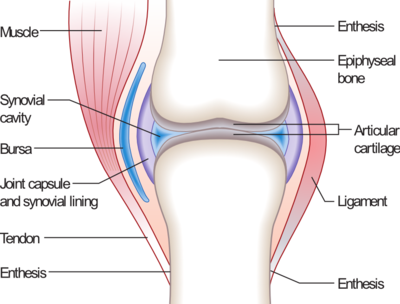
 However the most recent post gave me another idea on how it might be possible to increase height. We remember from our elementary anatomy courses that places like the knee are held in structure by cartilage touching cartilage. The articular cartilage of the femoral head is rubbing against the articular cartilage of the tibial head. Just see the picture on the right.
If I was to take a guess from a engineering degrees of freedom/constraints point of view, the epiphysis being subjected to just LSJL will not really be able to increase in the axial longitudinal direction as much as that we wish for. This is from a series of layers covering the bone. The bone itself does want to grow if you give it a chance. The key is to remove the contraints. Let’s assume that the LSJL technique does work in leading to chondrogenesis through progenitor cells differentiating.
We can do the dynamic loading but it seems to make the bones thicker in width and cause it to loss it’s mechanosensitivity and becomes even harder, stronger, and thicker as according to the the consequences of Wolff’s Law.
My new proposed height incease idea is really just the addition of one new step in using all three steps of LSJL< chondrocyte implants, and growth factor injection. Those will all be done, but the step to add is to make a surgical incision into the cartilage through also across the initial bone layer into the inside of the bone.
There is two possibilites I propose.
- We either cut into the femoral head articular cartilage around it until we make a complete closed pathway for the incision going all the way through to the bone’s inside. The direction will be from a top down approach, made on the transverse plane. We will make a complete loop on that transverse plane so that the cut will be completely encompass the bone’s head.
- We cut also another completely close pathway around the bone. The cut will go completely around the bone in 360 degree fashion. The distracted opening is then filled with chondrocyte implants. The chondrocytes are doing two jobs. They are used to keep the two separate boen sides from fusing back together while also allowing for cartilage formation. The chondrocytes themselves are kept from ossifying using growth factors that promote only chondrogenesis like BMP-2, IGF-2, and chondromodulin. In the beginning we might just want to keept the open fracture open so we use just chondromodulin.
The other idea is that we combine the directions of incision of the two together so we don’t make a complete axial cut or complete radial cut. We do it as a skewed angle, effectively cut at the corner edges of the bone, right where the articular cartilage end on the tip of the bones. We cut completely past the articular cartilage layer, then cut through he first layer of bone, and reach the inside. The key is to make the cute complete in terms of pathway. It has to be a close pathway, thus allowing for the degree of freedom the original growth plate cartilage have, which is to expand up and down.
If we remember, the long bones are always going through the process of osteoblast bone cell formation and osteoclast bone cell removal. This means that the outerlayer of the bone is being continuously formed by the layer right below the periosteum. I imagine that the layer will act as the new rest zone of the growth plate. That layer has to be very proliferative and be differentiating at a high level to be the cause for the appositional growth of the periosteal layer.
The idea is to first make an incision deep enough into the outer edge of the edges of the long bone, past the layer which is growing, inject the chondrocytes embbeded in the hydrogels to keep the openings open, inject the growth factors so that the chondrocytes can form into cartilage, and then start the process of dynamic mechanical loading/ LSJL. We have already seen in a few past PubMed studies that LSJL on bones with growth plates clearly lead to increased longitudinal growth. In the process the layer that is underneath the periosteum is injected with certain growth factors that keeps it from turning into bone cells but only chondrocytes. Since the layer is already so close to the articular cartilage, they turn into cartilage rather easily. The lateral loading will only help turn more cells of this specific layer into resting zone type progenitor cells. As long as the distracted bone are in a closed loop around the bone, the cartilage that will be formed can be affected easily by the loading.
