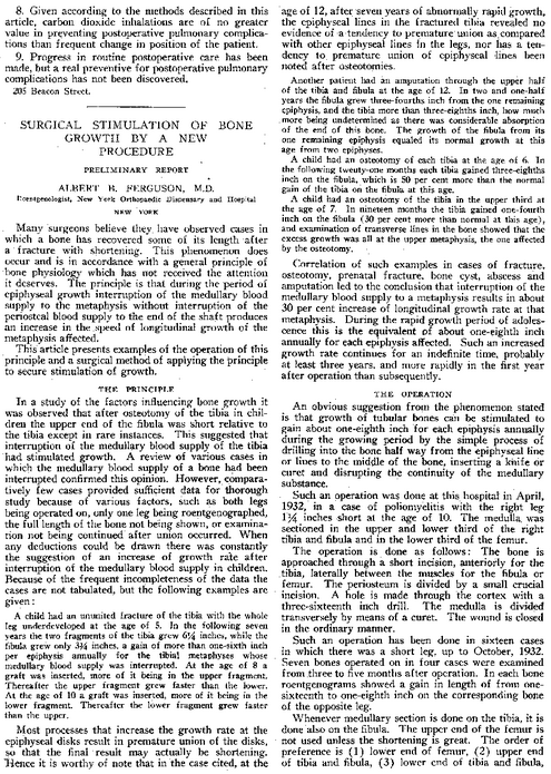
This article was written 80 years ago so it is very old but I wanted to try to take some idea or clue from it since the results we find from it are so unique and strange. This paper was cited by the study which I have looked at extensively ““A PROCEDURE FOR STIMULATION OF LONGITUDINAL GROWTH OF BONE, AN EXPERIMENTAL STUDY“. The experiment is entitled “SURGICAL STIMULATION OF BONE GROWTH BY A NEW PROCEDURE – PRELIMINARY REPORT” by ALBERT B. FERGUSON, M.D.
An clip of excerpt of the study is below.
Many surgeons believe they have observed cases in which a bone has recovered some of its length after a fracture with shortening. This phenomenon does occur and is in accordance with a general principle of bone physiology which has not received the attention it deserves. The principle is that during the period of epiphyseal growth interruption of the medullary blood supply to the metaphysis without interruption of the periosteal blood supply to the end of the shaft produces an increase in the speed of longitudinal growth of the metaphysis affected.
This article presents examples of the operation of this principle and a surgical method of applying the principle to secure stimulation of growth.
The article has very small text so is it is relatively hard to read but from what I have been able to gather, it seems that if you can disrupt the blod supply that is going through in the medullary cavity of children or people with still functional growth plates.
There is also another set of blood vessels that supply blood to the ends or the periosteal ends of the bones. Those will be kept intact. The disrupted metephyseal middle blood vessels would cause the metaphysis middle region to increase in its lengthening and growth.
Apparently even in the 1930s, 80 years ago this principle for increase bone lengthening was a well known physiological principle by surgeons.
The proposed idea was just to drill a small hole about 1/8th of an inch away from the growth plate in the metaphyseal middle region of any tubular (aka long) bone and then insert a knife or curret. Spin the knife/curret around and cut/disrupt all the blood vessels and supply from reaching the metaphyseal region of the lone bone.
For the two three cases that is cited in the one page that I can see, we find that if the tibia bone is cut (using osteotomy) the length increase we would see is about 30% faster than if the bone was growing naturally on average.
The authors do say that “during the rapid growth period of adolescent this is the equivalent of about 1/8th inch annually for each epiphysis affected.”
The most amazing thing that I can reveal in this post is that while most longitudinal increasing processes in the human body would only lead to the premature closure of the growth plates, like what we see in the increased estrogen level of girls earlier in age causing them to at least for a short time frame become taller than males but eventually often end up shorter and have their growth plates close earlier than the males, we don’t find it happening which this surgical method.
After a few years, each year having the patient go through with this, resulting in about 1/16th-1/8th extra tibia length increase, the growth plates under testing using roentgenogram showed that they were not closing at a faster rate as most endocrinologists might expect.
This idea for a minimal invasive surgical process that can be done in just a few hours and annually can to lead to a little bit of extra tibia lengthening is the first evidence that a minimal surgical way for leg bone lengthening is possible without any evidence that the growth plates would suffer the fate of early/premature closure.

