In one of the most pivotal posts I had written for the website entitled “The Connection Between Regenerating Deer Antlers and The PTHrP, PTH And IHH pathway for Cartilage Regulation, PTHrP Seems To Be The Answer (Big Breakthrough!)” I had looked at the connection between the IHH-PTHrP master regulator signaling pathway in the growth plates, and the principles behind how the antlers in deers continue to regenerate and grow longitudinally almost annually.
That post linked above is one of the biggest breakthroughs I have had in the research in being able to push the endeavor slightly closer.
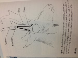
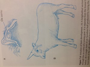
 I would now like to refer to the picture to the right which I took using my iPhone 4s of the medical research textbook “Bones & Cartilage, Developmental and Evolutionary Skeletal Biology” by Brain K. Hall. The book was found recently at the Health Sciences Library at the University of Washington.
Although the photo is crooked by 90 degrees, it is still very clearly visible that the way that the deer antlers seem to suggest that they grow is that there is a small pocket inside the relatively hard bone-like tissue where the mesenchymal stem cells or progenitor cells are located which will eventually turn into chondrocytes and then into osteocytes.
 This sort of suggests the idea that as long as we can get a sizable colony of mesenchyme into the human long bones, the mesenchyme might have the strength to push the bones apart and thus expand the bones longitudinally. Of course this idea must be checked and compared to the human body’s structures to see whether we can even take the principles of one biological system arrangement and use them in another.
The 2nd thing we must also consider is the fact that the way the antlers or horns are shaped suggest that there is a break in the bones so that the hard bone matrix does not completely surround the mesenchyme progenitor cells. Horns are actually different from antlers. Horns are made of a type of material that is not as strong as bones. From the picture of the horns in the 1st picture, we see that the horn material and the bones surround the mesenchyme. However, it seems that there is a break or a edge-edge boundary layer break between the horn material and the bone material which would suggest that the mesenchyme which will differentiate into chondrocytes use this boundary layer as the source where longitudinal lenghthening is even possible.
When we try to search further into this idea, we first have to do more research on the composition and structural arrangement of the deer antlers and ask whether the human long limbs have a similar structural arrangement.
From the Wikipedia article on the antler.…
While an antler is growing, it is covered with highly vascular skin called velvet, which supplies oxygen and nutrients to the growing bone. Antlers are considered one of the most exaggerated cases of male secondary sexual traits in the animal kingdom, and grow faster than any other mammal bone. Growth occurs at the tip, and is initially cartilage, which is later replaced by bone tissue. Once the antler has achieved its full size, the velvet is lost and the antler’s bone dies. This dead bone structure is the mature antler. In most cases, the bone at the base is destroyed by osteoclasts and the antlers fall off at some point.
Analysis:
It seems from the Wikipedia description that the tip is what grows taller being all cartilage, and the cover is the perichondrium. Once the mesenchyme runs out of progenitor stem cells to turn into chondrocytes, that is some type of molecular or genetic signaling pathway which gets triggered turning the velvet cover which is just the perichondrium hard into something like the analogue to the human’s long bone’s periosteum from vascularization and ossification. Then the antlers reach the final length and they get sheded. From one source, it says…
After the breeding season, cells start to de-mineralize the bone between the pedicle and antler, causing the antler’s connection with the skull to weaken and the antler to fall off
So that is the initial guess from me on what happens during the antler longitudinal growth.
However, is the deer antler made of the same type of composition as say the human long bones?
From the study “Mineral composition of antlers of three deer species reared in captivity” three species or types of deers antlers which were shed were taken and analyzed for their composition.
Dry matter, crude protein (31.99, 35.16 and 36.59%) and total ash (62.94, 62.54 and 60.27%) contents were similar among the three species, whereas the fat content was greater (P<0.05) for swamp deer (5.08%) compared to axis deer (2.31%) but compatible with hog deer (3.14%). Concentrations of calcium (23.44, 23.43 and 22.05%), magnesium (0.73, 0.80 and 0.74%), cobalt (9.01, 8.04 and 7.67 ppm) and zinc (33.50, 32.14 and 35.81 ppm) were not different among species…..Concentrations of the heavy metals, lead and cadmium, were similar among the three species. It was concluded that the concentrations of various minerals in the fallen hard antlers of the three species of deer were similar to those reported for bones of domestic animals.
This study sort of suggest that the antlers one might find shed in the woods have around the same composition as bones of farm animals that have been domesticated. But the key to ask is whether the matrix of the antler is as strong and resistive to tensile and compressive loads as the bones that is underneath it. My guess at this point is that the antler’s don’t have that high of a tensile strength.
Note: It is important right now to realize that there is a clear biological difference between the term horn and the term antler.
Let’s now look at the example of the elephant’s tusks, the rhinoceros’s horn, and the horns in other mammals like the medium sized whale like sea creature, the narwhal’s tusks. From the wikipedia articles on these mammals, the narwhal’s tusk seems to be an elongated canine teeth in the whale. For the rhino, it’s horns is made form keratin, which is the same type of basic element found in human hair and nails.
As for the elelphant’s tusks, they are supposed to be….”modified incisors in the upper jaw”. So the tusks do grow in length as the elephant is in it’s growing years.
From the article in Wikipedia about elephants…
“They replace deciduous milk teeth when the animal reaches 6–12 months of age and grow continuously at about 17 cm (7 in) a year. A newly developed tusk has a smooth enamel cap that eventually wears off. The dentine is known as ivory and its cross-section consists of crisscrossing line patterns, known as “engine turning”, which create diamond-shaped areas. As a piece of living tissue, a tusk is relatively soft; it is as hard as the mineral calcite. Much of the incisor can be seen externally, while the rest is fastened to a socket in the skull. At least one-third of the tusk contains the pulp and some have nerves stretching to the tip. As such it would be difficult to remove it without harming the animal. When removed, ivory begins to dry up and crack if not kept cool and moist.”
The whole point at this section of the post is to find other animals, hopefully mammals which have parts of their bodies that still grow while they are adults, similar in shape, structure, and strength to human bones.
We see two cases where the bone like material which grows in length is actually teeth in the animal. I would assume that if we cut off the tusks, then the bone like horn will not regrow back. In one case, with the rhino, the material of the horn is actually very weak, and made of the same type of compound as our hairs and nails. Elephant tusks are strong, but definitely not as strong as human bones. The ivory that human artisans which can carve into is weak if the bones become weak from drying. The ivory is just the elephant’s analogue to the dentin found in human teeth.
This short side look seems to suggest that the way and basic principles in which horns in animals grow in length can not really be used viably towards height increase. The material of horns are much weaker than human bones, so this might explain why lengthening is even possible when one tries to put a pocket of progenitor pluripotent cells inside to push the wall of organic matrix around it.
We have to look at antlers, not horns since antlers seem to be made of the the same type of components as human bones.
It seems that for antlers, the growth in length originates from the top, or tip of the appendage. For horns, the growth is from inside the horn, between the horn material and bone underneath. From the website for the National Park Service,….
“…Antlers, on members of the deer family, are grown as an extension of the animal’s skull. They are true bone and are a single structure They are generally found only on males. Antlers are shed and regrown each year.
Horns, found on pronghorn, bighorn sheep, bison, and many other bovine, are two-part structures. An interior of bone (also an extension of the skull) is covered by an exterior sheath grown by specialized hair follicles, as are your fingernails. In fact, your fingernails and the exterior sheath of horns are made of very similar materials. Horns are never shed and continue to grow throughout the animals life. The exception to this rule is the pronghorn which sheds and regrows its horn sheath each year.”
So it seems that antler’s are extensions of the skull of the animal, which is real bone. Antler’s can be shed (usually annually) while horns almost always continues to grow longer throughout life. As we remember from the previous post on the link between antler regeneration and the PTHrP-IHH main regulation pathway, for the longitudinal growth to happen in antlers, there must first be a wound, which must not be closed. The wound area must form a blastema, which will grow into the mesenchyme-bone arrangement seen in antler-based animals leading to longitudinal growth.
It might be that for us to be able to achieve longitudinal growth, we might have to inflict some type of bone disruption in the form of distraction ,incision, or cut which would give the bone wound area time to form a blastema, to push the separated bone areas apart from each other in the direction we desire.
If we are going to try to do that using the principles we learned from antler regeneration, we would also have to figure out how to get the progenitor cells which will development in the aftermath to stay on the path towards the chondrogenic lineage and not differentiate too quickly into the osteogenic lineage giving the cells enough time to enlarge/hypertrophize to push the area of incision apart.
{Tyler-Here’s a study I found recently on deer antler:
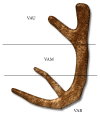
“The aim of this study was to compare the effectiveness of velvet antler (VA) from different sections for promoting longitudinal bone growth in growing rats. VA was divided into upper (VAU), middle (VAM), and basal sections (VAB). An in vivo study was performed to examine the effect on longitudinal bone growth in adolescent rats. In addition, in vitro osteogenic activities were examined using osteoblastic MG-63 cells. VA promoted longitudinal bone growth and height of the growth plate in adolescent rats. Bone morphogenetic protein-2 (BMP-2) in growth plate of VA group was highly expressed compared with control{If the growth is mainly due to BMP2 that does not show much promise for adult height increase as BMP2 is expressed in adults}. The anabolic effect of VA on bone was further supported by in vitro study. VA enhanced the proliferation, differentiation, and mineralization of MG-63 cells. The mRNA expressions of osteogenic genes such as collagen, alkaline phosphatase, and osteocalcin were increased by VA treatment. These effects of in vivo and in vitro study were decreased from upper to basal sections of VA. In conclusion, VA treatment promotes longitudinal bone growth in growing rats through enhanced BMP-2 expression, osteogenic activities, and bone matrix gene expressions{The non BMP2 effects have the most potential to taking effect in adults like the bone matrix effects/The bone matrix may actually be catabolic to growth by constraining so if VA can degrade the bone matrix that can stimulate growth}. In addition, present study provides evidence for the regional differences in the effectiveness of velvet antler for longitudinal bone growth.”
“Typically, the antler is cut off near the base after it is about two-thirds of its potential full size, between 55 and 65 days of growth, before any significant calcification occurs. “
“Longitudinal bone growth in control rat was 617.4 ± 34.2 μm/day, and administration of VAU significantly increased the longitudinal bone growth to 718.6 ± 48.6 μm/day”<-This is actually huge. 16% growth increase approximately. Assuming this works for all bones it would take a person from 69 to 80 inches or almost a foot taller. Note though that growth rate does not always correlate with total growth.
VAU(or upper region of the deer antler) was most anabolic to longitudinal bone growth. VAU has the most epiphysiseal content.
Deer Antler also stimulated osteoblastic growth which the scientists speculated is likely contribute to the longitudinal bone growth. However, this possibly could only increase growth rate but it could also increase total longitudinal bone growth by increasing the amount of growth per growth plate cell(more bone matrix, etc.)

Look at this http://www.ncbi.nlm.nih.gov/pubmed/22313625 ”It was further demonstrated that treatment with DAE (50 and 100μg/ml) significantly decreased the levels of MMP-2, MMP-9, TNF-α, and IL-6.” and this http://www.ncbi.nlm.nih.gov/pubmed/22951396
I would like I formation regarding deer antler extract and height .
Please reply to my email or call 912 660 9655
Pingback: Articular Cartilage At The End Of Epiphysis Do Growth Thicker Making Bones Longer (Big Breakthrough) | Natural Height Growth
Is it safe to take parathyroid related peptide as supplement?