The blood supply of the growth plate and the epiphysis: a comparative scanning electron microscopy and histological experimental study in growing sheep.
” The vascular supply of growth plate and epiphysis of the proximal tibia was reinvestigated using a modern technique, the Mercox-perfusion method, in six sheep aged 6-24 weeks. A comparison was made among pure perfusion specimens, the corrosion casts, and histological sections. The metaphyseal, epiphyseal, and perichondral blood supply systems were confirmed. However, there was evidence of regular transphyseal anastomoses{reconnection of two systems} between the metaphyseal and epiphyseal system. Based on the histological arrangement of the blood vessels, the arterial blood flow would appear to be from the metaphysis to the epiphysis. The existence of transphyseal arterial vessels originating metaphyseally and seen both in cast preparations and histological sections was added to the present description of the blood supply of the growth plate. Age-related differences in the vascularization of the growth plate were not found. ”
I couldn’t copy and paste from the full study at top.
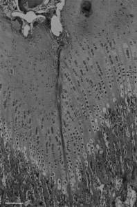
Some vessels do cross the growth plate.
Longitudinal bone growth may cease if blood supply is cut off.
Here’s an image of a growth plate artery:
Cartilage canals run parallel to growth plate columns.
These transphyseal vessels may only be present up to 24 weeks of age.

