“To investigate the effect of bone-marrow stimulation (BMS) on subchondral bone plate morphology and remodeling compared to untreated subchondral bone in a validated minipig model.
Three Göttingen minipigs received BMS with drilling as treatment for two chondral defects in each knee. The animals were euthanized after six months. Follow-up consisted of a histological semiquantitative evaluation using a novel subchondral bone scoring system and micro computed tomography (µCT) of the BMS subchondral bone. The histological and microstructural properties of the BMS-treated subchondral bone were compared to that of the adjacent healthy subchondral bone. The µCT analysis showed that subchondral bone treated with BMS had significantly higher connectivity density compared to adjacent untreated subchondral bone (26 1/mm3 vs. 21 1/mm3, P = 0.048). This was the only microstructural parameter showing a significant difference. The histological semiquantitative score differed significantly between the subchondral bone treated with BMS and the adjacent untreated subchondral (8.0 vs. 10 P = < 0.001). Surface irregularities were seen in 43% and bone overgrowth in 27% of the histological sections. Only sparse formation of bone cysts was detected (1%).BMS with drilling does not cause extensive changes to the subchondral bone microarchitecture. Furthermore, the morphology of BMS subchondral bone resembled that of untreated subchondral bone with almost no formation of bone cyst, but some surface irregularities and bone overgrowth(!).”
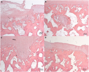
And the bone bone overgrowth can be seen to be length as seen in the histological slides.
“The objective of BMS is to create canals in the subchondral bone to the underlying bone marrow, either by use of a drill(subchondral drilling) or an awl (microfracture).”<- an awl is a tool for piercing holes. It’d be hard to mimic either a drill or awl safely using non-surgical methods but I don’t believe that it’s impossible.
The study mentions that other studies also showed bone overgrowth.
“Bone overgrowth was defined as areas where the subchondral bone plate had grown into the cartilage.”<-this could be due to endochondral ossification or longitudinal appositional growth.
“Nine out of the 12 samples contained bone overgrowth corresponding to 27% of the sections.”
“Hematoxylin eosin staining, scale bars: 200 μm. Defects treated with BMS. (A) Shows irregularity of the subchondral bone plate (top arrow marks the beginning of the defect). (B) Bone cyst marked by arrows. (C) Remaining drill hole in the subchondral bone plate. (D) Overgrowth in the subchondral bone plate marked by to arrows, top arrow marks the beginning of the defect site. BMS = bone marrow stimulation.”<-so the overgrowth is very minor but if there was some way to reproduce it, it could add up to height over time. The pigs used in the study were skeletally mature.
Here’s the study study cited in the above that also shows bone overgrowth:
“Trochlear cartilage defects debrided of the calcified layer were prepared bilaterally in 16 skeletally mature rabbits. Drill holes were made to a depth of 2 mm or 6 mm and microfracture holes to 2 mm. Animals were sacrificed 3 months postoperatively, and joints were scanned by micro–computed tomography before histoprocessing. Bone repair was assessed with a novel scoring system and by 3-dimentional micro–computed tomography and compared with intact controls. Correlation of subchon-dral bone features to cartilage repair outcome was performed.”
“Although surgical holes were partly repaired with mineralized tissue, atypical features such as residual holes, cysts, and bony overgrowth were frequently observed. For all treatment groups, repair led to an average bone volume density similar to that of the controls but the repair bone was more porous and branched as shown by significantly higher bone surface area density and connectivity density. Deeper versus shallower drilling induced a larger region of repairing and remodeling subchondral bone that positively correlated with improved cartilage repair.”
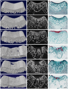
“Repair of subchondral bone 3 months after bone marrow stimulation does not result in complete regeneration of the normal bone structure as shown by 3-dimensional visual models (left column), 2-dimensional micro-CT images (middle column), and safranin O/fast green stain (right column). A through C, normal unoperated (control). D through F, a case of good bone repair with suboptimal lateral integration (sample from DRL2/GrpII). G through I, presence of bone overgrowth (sample from DRL6/GrpI). J through L, presence of residual holes (sample from DRL2/GrpI). M through O, presence of nonmineralized connective tissue in subchondral region—a subchondral cyst (sample from MFX2/GrpII). P through R, lack of integration to adjacent bone (sample from DRL6/GrpI). Bars = 1 mm.”
“drilling deeper to 6 mm versus 2 mm increased the volume of this repaired and remodeling subchondral bone region, resulting in greater cross-sectional area, as well as width and depth”
“Alterations of joint biomechanics and joint homeostasis caused by osteochondral defects may also contribute to the generation of bone over-growth.”<-so perhaps bone overgrowth can be generated by methods other than drilling.
The question is how can we induce something like a microfracture safely and without surgery?


