Monthly Archives: August 2022

Paper with interesting supplements that impact longitudinal bone growth

Novel Treatment Options in Childhood Bone Diseases

“Several novel treatment options have recently become available in childhood bone diseases. The purpose of this article is to provide an update on some of the therapeutic agents used in the treatment of pediatric osteoporosis, X-linked hypophosphatemic rickets, and achondroplasia (ACH){this is what we’re interested in as it’s related to height}Summary: Vitamin D3 and Ca supplementation remains the basis of childhood osteoporosis treatment. Bisphosphonate (BP) therapy is the main antiresorptive therapeutic option, while denosumab, a human monoclonal IgG2 antibody with high affinity and specificity for a primary regulator of bone resorption – RANKL, represents a possible alternative. Its potent inhibition of bone resorption and turnover process leads to continuous increase of bone mineral density throughout the treatment also in the pediatric population. With a half-life much shorter than BPs, its effects are rapidly reversible upon discontinuation. Safety and dosing concerns in children remain. Novel treatment options have recently become available in two rare bone diseases. Burosumab, a monoclonal antibody against FGF-23{FGF23 has an impact on height but there’s mixed evidence on whether it’s good or bad therefore Burosumab may have an impact on height too}, has been approved for the treatment of children with X-linked hypophosphatemic rickets older than 1 year. It presents an effective, more etiology-based treatment for rickets compared to conventional therapy, without the need for multiple daily oral phosphate supplementation. Its long-term efficacy and safety are currently being investigated. After years of anticipation, a novel treatment option for ACH has become available. C-type natriuretic peptide analog vosoritide effectively increases proportional growth and has a reasonable safety profile in children >2 years. Its effect on other features of the disease and the final height is yet to be determined.{studies show that vosoritide definitely increases growth rate but there’s yet a study that shows it’s impact on adult height; Micheal’s thoughts on vosoritide; I also speculate that CNP could help with longitudinal bone growth in adults if one as an adjunct to other methods or maybe to help grow via the cartilage in the spine or knee etc; CNP increases the proliferation of chondrocytes in general} Several other treatment options for ACH exploring different therapeutic approaches are currently being investigated. Key Messages: Denosumab is effective in the treatment of childhood-onset osteoporosis; however, further studies are necessary to determine the optimal treatment protocol. Burosumab is more etiology-based and convenient in comparison to conventional treatment of X-linked hypophospha­-temic rickets in children and adults. Vosoritide importantly changes the natural course of achondroplasia, at least in the short term.”

“Burosumab is the first etiologic treatment option that actively increases phosphate levels while also decreasing FGF-23 actions in XLH. It is a monoclonal IgG1 antibody that suppresses the actions of FGF-23. FGF-23 is the key phosphaturic hormone and acts as a regulator of phosphate homeostasis. It mediates its actions by binding to its cofactor alpha-Klotho and FGF-receptor 1 (FGFR1), through which it inhibits phosphate reabsorption in the kidney via downregulation of the sodium-dependent phosphate transporters (NaPi-2a and NaPi-2c) in proximal renal tubules. Additionally, it suppresses renal 1α-hydroxylase (CYP27B1) and activates 24-hydroxylase (CYP24A1), both of which contribute to lowering serum concentrations of 1,25-dihydroxy cholecalciferol and thus reduce intestinal uptake of phosphate. The elimination of burosumab follows the endogenous immunoglobulin degradation pathway”

“In another phase 3 study (study identifier NCT02915705) burosumab treatment was superior to conventional therapy regarding growth velocity and disease progression determined by RSS”<-burosumab’s impact on growth  velocity suggests that it may impact height.  IF you look at the study menionted figure 4,Height was increased by about 0.2cm.

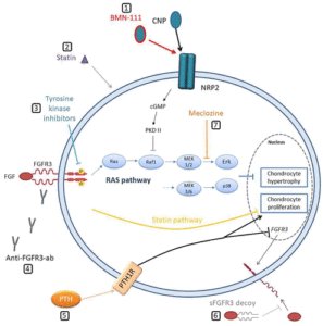
“Excessive FGFR3 activation results in downstream activation of multiple intracellular signaling pathways, leading to intensified inhibition of cartilage tissue formation at the level of chondrocyte proliferation (via STAT1), hypertrophy, differentiation, and synthesis of the extracellular matrix (via Erk-MAPK signaling pathway) “

“Growth hormone supplementation has not shown promising results and is not viewed as a standard treatment for ACH. The progress in the understanding of ACH pathogenesis has led to the development of many potential therapeutic strategies for modulating excessive FGFR3 activation. Approaches are varied and include inhibiting the tyrosine kinase activity of FGFR3 (infigratinib), producing artificial FGFR3 as a decoy for FGF ligand (recifercept), inhibition of FGFR3 downstream signaling pathways (meclizine, C-type natriuretic peptide [CNP] analogs), modulation of growth via natriuretic peptide receptor 2 (NPR2) receptor (CNP analogs) and use of aptamers or monoclonal antibodies to prevent binding of FGF to its receptor (aptamer RBM-007, vofatamab). The investigations into analogs of CNP, especially vosoritide, are currently the most advanced”

“Vosoritide is a recombinant CNP analog. Endogenous CNP and its action on the growth plate through NPR-B are recognized as one of the important regulating mechanisms of longitudinal bone growth. Coupled with NPR-B, CNP antagonizes downstream FGFR3 signaling by inhibiting the Erk-MAPK signaling pathway at the level of Raf. This leads to chondrocyte proliferation, differentiation and increases the extracellular matrix synthesis. CNP-targeted overexpression in the cartilage or its continuous delivery by intravenous infusion has shown normalization of the impaired bone growth in mouse models with ACH”

” In August 2021, results of the extension phase 3 clinical trial in children with ACH aged between 5 and 18, receiving vosoritide 15 μg/kg once daily in subcutaneous injection, were published. An increase in annualized growth velocity was observed, with 3.52 cm of height gain over a 2-year treatment period in comparison to untreated patients. In addition, improvement in the proportionality of body segments and no acceleration of the bone maturation process (determined by bone age assessment) was observed{this is a positive indicator that the treatment will increase adult height}

“Recent preclinical data in healthy cynomolgus monkeys showed that treatment with TransCon CNP subcutaneously once per week resulted in significant growth increases in body, tail, and long bones compared to controls. An increase in height was also more pronounced in comparison to the animals receiving a daily dose of CNP analog with the same amino acid sequence as vosoritide (5% vs. 3%, respectively), and no significant changes in bone quality were observed with both treatments. Moreover, sustained CNP release resulted in lower systemic CNP peak levels and has not been associated with adverse cardiovascular effects in monkeys treated with repeated weekly doses up to 100 μg/kg”

According to A long-acting C-natriuretic peptide for achondroplasia, “CNP-38 was slowly released into the systemic circulation and showed biphasic elimination pharmacokinetics with terminal half-lives of ∼200 and ∼600 h. Both preparations increased growth of mice comparable to or exceeding that produced by daily vosoritide.”

So both vosoritide and Transcon CNP increase height during development and I suspect may have some applications for adults as well.  Burosomab and the other FGFR3 inhibitors likely have impact on height as well.

{Note I accidentally made this post in Michael’s account}

Big Breakthrough: Vosoritide to grow taller

Update on Vosoritide:  New studies have come in about it that show very promising results and I am like 99% confident that it would work on children that do not have dwarfism because they also are impacted by CNP and have FGFR3 receptors.   Vosoritide is very promising and I think will eventually be used for children of normal growth velocity.

Vosoritide is basically a daily CNP injection.  It’s targeted for dwarfism but as everyone has FGFR3 receptors it can work normal children but testing would be needed.

<-From the video it seems that it’s progressing very slowly.  Which is unfortunate as it has potential to happen normal children and possibly even adults.  Unfortunately it doesn’t seem like they’r keen on testing Growth Hormone and CNP at the same time because Growth Hormone may only increase growth velocity but not final height but perhaps together.  They address other potential uses kindof at around the 25 minute mark.

Here’s more on CNP.<-“we developed transgenic mice with an elevated plasma concentration of CNP under the control of human serum amyloid P component promoter and exhibited that these mice showed prominent skeletal overgrowth phenotype”

CNP delays mineralization.->”The femur, skull, and spine (L2-4) measurements were longer than that of the wild-type littermates”  It could potentially affect adults via spinal height even if limbs do not increase.

CNP activates bone turnover and remodeling in vivo

More on CNP.<-also there’s a snippet from free patents online for CNP being used to increase height in people free of FGFR3 abnormalities meaning normal children.

Who knows if CNP could potentially increase height in adults until it is tested….

Apparently Biomarin did test this on adults but did the adults get taller?  Although the study was only for a short period of time approx two weeks which is not a lot of time to evaluate if the adults grew taller.

Note Meclozine has been associated with height growth too.

Note based on this image, barring other effects the benefit of CNP is limited based on how much growth inhibiting effects FGFR3 induces.  Though according to Dose dependent effect of C-type natriuretic peptide signaling in glycosaminoglycan synthesis during TGF-β1 induced chondrogenic differentiation of mesenchymal stem cells., CNP may induce differentiation of MSCs to chondrogenic lineage so it’s effects may not solely be limited based on how much FGFR3 there is to inhibit.

“A multinational study of 35 children (5–14 years of age) receiving daily subcutaneous vosoritide at a dose of 15 µg/kg demonstrated a sustained increase in the annualized growth velocity of approximately 1.5–2.0 cm/year over 42 months of treatment.” Let’s say that’s 1.5 inches over 3 years.  That’s pretty significant.  How long that can be sustained will be revealed with further testing.

Here’s another study on Vosorotide:

Once-daily, subcutaneous vosoritide therapy in children with achondroplasia: a randomised, double-blind, phase 3, placebo-controlled, multicentre trial

Methods: This randomised, double-blind, phase 3, placebo-controlled, multicentre trial compared once-daily subcutaneous administration of vosoritide with placebo in children with achondroplasia. The trial was done in hospitals at 24 sites in seven countries (Australia, Germany, Japan, Spain, Turkey, the USA, and the UK). Eligible patients had a clinical diagnosis of achondroplasia, were ambulatory, had participated for 6 months in a baseline growth study and were aged 5 to less than 18 years at enrolment. Randomisation was done by means of a voice or web-response system, stratified according to sex and Tanner stage. Participants, investigators, and trial sponsor were masked to group assignment. Participants received either vosoritide 15·0 μg/kg or placebo, as allocated, for the duration of the 52-week treatment period administered by daily subcutaneous injections in their homes by trained caregivers. The primary endpoint was change from baseline in mean annualised growth velocity at 52 weeks in treated patients as compared with controls. All randomly assigned patients were included in the efficacy analyses (n=121). All patients who received one dose of vosoritide or placebo (n=121) were included in the safety analyses. The trial is complete and is registered, with EudraCT, number, 2015-003836-11.

Findings: All participants were recruited from Dec 12, 2016, to Nov 7, 2018, with 60 assigned to receive vosoritide and 61 to receive placebo. Of 124 patients screened for eligibility, 121 patients were randomly assigned, and 119 patients completed the 52-week trial. The adjusted mean difference in annualised growth velocity between patients in the vosoritide group and placebo group was 1·57 cm/year in favour of vosoritide (95% CI [1·22-1·93]; two-sided p<0·0001). A total of 119 patients had at least one adverse event; vosoritide group, 59 (98%), and placebo group, 60 (98%). None of the serious adverse events were considered to be treatment related and no deaths occurred.”

1.57cm is small but significant.  And major if it can be maintained throughout development.

Here’s another study on Vosorotide:

Safe and persistent growth-promoting effects of vosoritide in children with achondroplasia: 2-year results from an open-label, phase 3 extension study

“Achondroplasia is caused by pathogenic variants in the fibroblast growth factor receptor 3 gene that lead to impaired endochondral ossification. Vosoritide, an analog of C-type natriuretic peptide, stimulates endochondral bone growth and is in development for the treatment of achondroplasia. This phase 3 extension study was conducted to document the efficacy and safety of continuous, daily vosoritide treatment in children with achondroplasia, and the two-year results are reported.

Methods

After completing at least six months of a baseline observational growth study, and 52 weeks in a double-blind, placebo-controlled study, participants were eligible to continue treatment in an open-label extension study, where all participants received vosoritide at a dose of 15.0 μg/kg/day.

Results

In children randomized to vosoritide, annualized growth velocity increased from 4.26 cm/year at baseline to 5.39 cm/year at 52 weeks and 5.52 cm/year at week 104. In children who crossed over from placebo to vosoritide in the extension study, annualized growth velocity increased from 3.81 cm/year at week 52 to 5.43 cm/year at week 104. No new adverse effects of vosoritide were detected.”

Now growth velocity does not always coincide with final height.

“Due to the inherent variability of growth and the lesser magnitude of the pubertal growth spurt in children with achondroplaisa, these long-term effects will only be known once these children reach final adult height”

Efficacy of vosoritide in the treatment of achondroplasia

“Achondroplasia is the commonest form of dwarfism and results from a mutation in the fibroblast growth factor receptor 3 (FGFR3) gene on chromosome 4p16.3. The mutation is at nucleotide 1138 resulting in a G-to-A transition (134934.0001). This condition is characterized by full penetration meaning that everyone with this genetic mutation will exhibit the phenotypic characteristics of achondroplasia. It is a gain-of-function mutation that causes increased inhibition of cartilage formation. C-type natriuretic peptide (CNP) acts on the growth plate through the natriuretic peptide receptor-B (NPR-B) causing the transformation of guanosine 5′-triphosphate into cyclic guanosine monophosphate. However, CNP cannot be used in the treatment of achondroplasia because it is rapidly degraded by neutral endopeptidase. Vosoritide is a modified recombinant human CNP and has a half-life 10 times that of CNP. Clinical trials have demonstrated that vosoritide is effective in significantly increasing the annualized growth velocity in children with achondroplasia before the fusion of the epiphyses.”

<-couldn’t get this full study.

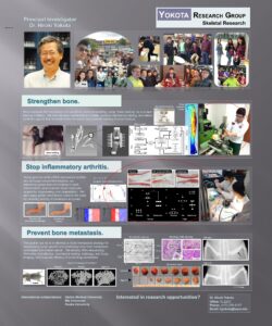
Yokota has possibly looked at a chisel and hammer method in the past

You can see in the circled image a student using a hammer and chisel on an object.  I’m currently trying the hammer method with a ball pein hammer.  Unlike suggested in the post I am currently tapping the epiphysis of the bone and not just the diaphysis and I am not using a chisel because I think it is too cumbersome.  Now it’s possible that the hammer here is being used to cause a fracture rather than as a bone stimulatory agent, I’m just suggesting that using a lateral impact force is not unheard of.  Also, the method alluded to in the poster would have the issue that the hammer would puncture the skin(hence why I’m trying a ball pein hammer).

Also of note in the poster(and circled) Is that salubrinal and joint loading is used on articular cartilage and it seems to be noticeably thicker than even the control group.  Also if you look at the rat foot on the same line.  the salubrinal(and joint loaded) treated group looks longer than the osteoarthritis group but that could be an optical illusion.

Study shows the presence of “microgrowth plates”

Here are David Burr’s thoughts on the study.  I have spoken to David Burr before as he’s add that microdamage in bone does not heal by endochondral ossification.  He’s said:  “Diffuse damage(and linear microcracks that are are not fully fractures) don’t heal by endochondral ossification.”  “Large cracks always heal by remodeling, but full fractures, trabecular or otherwise, can heal by endochondral ossification.”  I pointed out this study to him given that he’s made the above statements.  Here’s what he had to say: “The endochondral ossification and chondrocytes that are present here are in woven bone.  That is not unusual.  There is none of that in the pre-existing bone.  Several things here are remarkable (though not that cartilage can appear in woven bone).  One question is why is there woven bone when the strains are only 640 microstrain (not high, as suggested by the authors).  This suggests there was some independent effect of muscle stimulation on bone, beyond the level of strain.  This could be due to the manner of stimulation, or to the fact that these were quite young rats – not adult – started at 8 weeks, or that the muscle stimulation occurred consistently over a long period of time. At some point in the very distant past, we showed that high strains (which were not present here) in dogs can cause woven bone formation without fracture.  It is simply a response either to a need to adapt quickly, or to damage that is not evidenced in a fracture.  So, I don’t find anything particularly novel about this, and some fundamental flaws in the model and analysis.”<-I think Burr is arguing that endochondral ossification is only occurring in immature bone that is not under repair.

I suggested that the independent effect of muscle stimulation could be fluid flow.  Here’s what he had to say: “Strain is still too low – but it depends on the strain rate (ie, strain x frequency).  But, if the frequency is too high, then fluid can’t relax and so the effect is blunted”

I talked to Dan Huey in the past about the possibility of endochondral ossification within the bone.

“”While MSCs derived from bone marrow have shown the ability to differentiate down the chondrocytic lineage both in vitro and in vivo the efficiency and completeness of this process hinders the formation of stable hyaline tissue. Ectopic differentiation of MSCs into chondrocytes does not occur in the marrow cavity due to a lack of the appropriate signals (both mechanical and biochemical)These MSCs are tuned by their environment to contribute to the natural bone remodeling process. However, even when these cells are introduced into a cartilage defect via microfracture, complete chondrocyte differentiation does not occur, as evidenced by the formation of fibrous tissue. For these cell to undergo complete chondrogenesis the proper combination of mechanical and biochemical cues must be provided. As the clot formed in microfracture is quite soft the cells within the clot will not receive the appropriate level of mechanical forces for chondrogenesis. With regards to the biochemical signals, a cartilage stimulating growth factor analagous to BMP’s effect for osteogenic differenetiation has not been identified. With respect to the term microfracture, in cartilage and bone it means two different things. For cartilage microfracture is a surgical procedure that involves creating holes in the bone underlying a cartilage defect to allow stem cells to enact a healing response. With regards to bone, microfractures are the very small breaks in bone that occur during strenuous activity.  does not occur in cartilage as it does in bone. In bone, microfracture occurs during strenuous activity and heals.”<-it’s possible that muscular contraction along with other mechanical stimuli could help induce the signals needed for chondrogenic differentiation.

The below study shows the presence of endochondral ossification in non-growth plate regions within bone itself.  The problem is that at least according to this stimulus it is not in a longitudinal direction.  But this is a huge breakthrough study as it shows that chondrogenic regions can occur outside of the growth albeit in developing rats.

Morphological and histological adaptation of muscle and bone to loading induced by repetitive activation of muscle

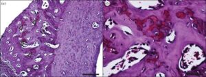
“Muscular contraction plays a pivotal role in the mechanical environment of bone, but controlled muscular contractions are rarely used to study the response of bone to mechanical stimuli. Here, we use implantable stimulators to elicit programmed contractions of the rat tibialis anterior (TA) muscle. Miniature stimulators were implanted in Wistar rats (n = 9) to induce contraction of the left TA every 30 s for 28 days. The right limb was used as a contralateral control. Hindlimbs were imaged using microCT. Image data were used for bone measurements, and to construct a finite-element (FE) model simulation of TA forces propagating through the bone. This simulation was used to target subsequent bone histology and measurement of micromechanical properties to areas of high strain. FE mapping of simulated strains revealed peak values in the anterodistal region of the tibia (640 µε ± 30.4 µε). This region showed significant increases in cross-sectional area (28.61%, p < 0.05) and bone volume (30.29%, p < 0.05) in the stimulated limb. Histology revealed a large region of new bone, containing clusters of chondrocytes, indicative of endochondral ossification. The new bone region had a lower elastic modulus (8.8 ± 2.2 GPa) when compared with established bone (20 ± 1.4 GPa). Our study provides compelling new evidence of the interplay between muscle and bone.”

“eight-week-old Wistar rats (weights 228–282 g) underwent surgical implantation of miniature neuromuscular stimulators”

“Stimulators delivered 0.2 ms pulses at 100 Hz for 200 ms every 30 s, resulting in a total of 9.6 min of stimulation per day. Each 200 ms burst of nerve stimulation at 100 Hz caused a very brief but fused (tetanic, near maximum force) contraction.”<-it would be very hard to induce a maximum force contraction.  But muscle stimulation increase fluid forces in bone.  So it is not only possible for muscle contraction to stimulate new endochondral ossification within bone but any method that stimulates fluid forces.

“The volume of the stimulated muscles, TA and EDL, showed a significant decrease of volume of, on average, 19% and 16% (p < 0.05), respectively, when compared with the contralateral control limb”<-It’s interesting that the muscle actually decreased in size.

“Histological sectioning targeted to this distal region revealed that the cross-sectional geometry of the contralateral control and the stimulated tibias were markedly different (figure 8a). The stimulated bone shows a large region of primary osteon formation (figure 9). Safranin O staining revealed the presence of clusters of chondrocytes within the region of primary osteon formation!!!!!! (figure 10).”

“Bone has a highly ordered structure, and mature bone cells are highly differentiated, so the deposition of bone is an iterative surface-bound (and often time-consuming) process.”
” In rats, the surface deposition of bone by osteoblastic apposition has a maximal rate in the region of 3.5 µm a day. By contrast, cartilage is able to dramatically increase its volume, as its cells are capable of division and hypertrophy across its volume. This capability can be seen in the growth plates, for which the growth rate can be up to 125 µm per day in rats. The chondrocytes then undergo cell death, and are replaced by bone cells. This mechanism provides a mechanically sound structure capable of faster growth than direct growth of bone. The presence of chondrocytes within the region of new bone reported here implies that the adaptive response of the tibia to the muscle load was initially a rapid phase of cartilage growth followed by endochondral ossification. Investigation of our EMS model at earlier time points will help reveal the exact nature of the response.”
” Here, we used approximately 2880 cycles per day, resulting in a total of 9.6 min of 10 N loading per day and provoking a large region of bone growth. In Turner et al.‘s experiments, it was found that the lower limit for bone formation was 40 N; however, the loading protocol involved only 36 cycles per day, totalling 10.8 min of 40 N of loading per day. So, while the overall loading time is fairly similar, applying loads more frequently (and possibly at a higher rate) appears to have a greater effect than simply increasing the magnitude of the load, assuming the nature of the adaptive response was similar to the apparent endochondral process reported for the first time here.”<-this is the possible reasoning behind tapping as a stimulus.

Grant with potential implications on growth plate regeneration

Here’s the grant Cartilage progenitor cells for growth plate regeneration

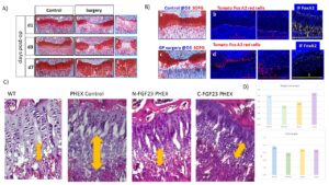
“Growth plate (GP) injuries result in growth arrest, formation of a “bony bar”{a technique that gets rid of the bony bar could potentially be used to make a new growth plate in adults} and angular limb deformities in children. Novel therapeutic approaches directed towards prevention of bone formation and growth arrest have to integrate cellular grafts, biomaterials and growth factors with the ultimate goal of recapitulating the complex zonal organization of the growth plate. One endogenous source of cartilage progenitor cells is thought to be the resting zone of the growth plate. Until now, the lack of specific marker(s) for the resting zone restricted the examination of this population. Currently, the evaluation of potential strategies for growth and cartilage disorders can mainly be achieved in vivo, therefore we proposed to use genetic modified mice to characterize the GP population and to characterize its development. Mice are the most appropriate model to use for several reasons: 1) GP is a tissue hardly approachable in vitro 2) we aim to characterize the GP population, and GP dynamics during the process of growing for which there is no optimal in vitro assays 3) we chose PHEX hemizygous mice for the study of GP dynamics because it is a well stablished model of X-linked hypophosphatemia (XLH){also called vitamin D resistant rickets}, which has being used for decades to study growth plate ossification. An other thing to keep in mind is that the standard treatments for XLH patients do not completely rescue the rickets and bone deformities. Serious side effects such as nephrocalcinosis and hyperparathyroidism have also been observed. Antagonizing FGF23 activity with antibodyes (Burosumab treatment){here’s some papers that indicate that FGF23 inhibition may potentially be part of a growth plate reactivation treatment.  FGF23 impacts bone mineralization so that may be why FGF23 inhibition could potentially help with growth plate restoration } is a recent and very promising therapy. However, this treatment requires at least a monthly infusion, is very costly and alleviates symptoms in many patients but not all. Hence, it is of the utmost necessity to identify more affordable strategies for therapy. Dr. Santos’ Laboratory demonstrated that inhibiting the MAPK pathway (FGF23 downstream pathway) in PHEX mice partially rescues growth impairment by normalizing the GP structure, specifically in the hypertropy zone. Nevertheless, it is not completely understood how FGF23 inhibition affects GP dynamics or how this is translated into a growth rescue and whether this treatment would be suitable for paediatric patients. Consequently, we will utilize the PHEX mice and FGF23 to 1) gain a better understanding of GP development and 2) look for alternative therapies to antagonize FGF23 activity.”

“FoxA2+ cells exhibit high clonogenicity and longevity. Moreover, FoxA2+ cell number expand in response to trauma and the data suggest that these cells participate in the production of hyaline cartilage, allowing for successful cartilage regeneration”

“Unlike other cartilage regions such as articular cartilage, GP has the ability to regenerate.”

Since Wild Type was the longest we don’t know it this treatment would have any impact in healthy developing individuals.