There are studies in mice/rats that show that swimming increases bone length. But the kind of swimming that rats due is different than the kind of swimming that humans do.
Effects of swimming training on bone mass and the GH/IGF-1 axis in diabetic rats
“The aim of this study was to examine the influence of moderate swimming training on the GH/IGF-1 growth axis and tibial mass in diabetic rats. Male Wistar rats were allocated to one of four groups: sedentary control (SC), trained control (TC), sedentary diabetic (SD) and trained diabetic (TD). Diabetes was induced with alloxan (35 mg/kg b.w.). The training program consisted of a 1 h swimming session/day with a load corresponding to 5% of the b.w., five days/week for six weeks. At the end of the training period, the rats were sacrificed and blood was collected for quantification of the serum glucose, insulin, GH, and IGF-1 concentrations. Samples of skeletal muscle were used to quantify the IGF-1 peptide content. The tibias were collected to determine their total area, length and bone mineral content. The results were analyzed by ANOVA with P < 0.05 indicating significance. Diabetes decreased the serum levels of GH and IGF-1, as well as the tibial length, total area and bone mineral content in the SD group (P < 0.05). Physical training increased the serum IGF-1 level in the TC and TD groups when compared to the sedentary groups (SC and SD), and the tibial length, total area and bone mineral content were higher in the TD group than in the SD group (P < 0.05). Exercise did not alter the level of IGF-1 in gastrocnemius muscle in nondiabetic rats, but the muscle IGF-1 content was higher in the TD group than in the SD group. These results indicate that swimming training stimulates bone mass and the GH/IGF-1 axis in diabetic rats.”
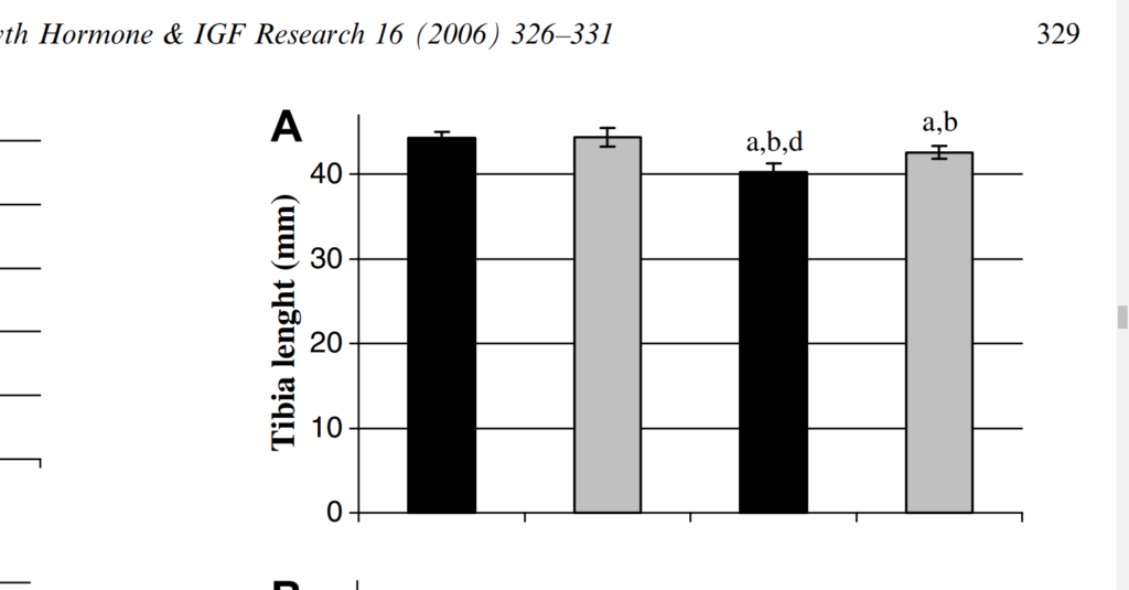
Columns: SC – sedentary control, TC – trained control, SD – sedentary diabetic, TD – trained diabetic. So the swimming groups had non-statistically significant increases in bone length between sedentary and trained control and statistically significant between sedentary diabetic and trained diabetic.
Here’s another study that finds an increase in bone length:
The Effect of Swimming on Bone Modeling and Composition in Young Adult Rats
“The purpose of this study was to investigate the adaptability of long bones of young adult rats to the stress of chronic aquatic exercise. Twenty-eight female Sabra rats (12 weeks old) were randomly assigned to two groups and treatments: exercise (14 rats) and sedentary control (14 rats) matched for age and weight. Exercised animals were trained to swim in a water bath (35°±1°C, 1 hour daily 5 times a week) for 12 weeks loaded with lead weights on their tails (2% of their body weight) (BW). At the end of the training period following blood sampling for alkaline phosphatase, all rats were sacrificed and the humeri and tibiae bones were removed for the following measurements: bone morphometry, bone water compartmentalization, bone density (BD), bone mineral content (BMC), and bone ions content (Ca, Pi, Mg, Zn). The results indicate that exercise did not significantly affect the animals’ body weight, bone volume, or length and diameters. However, bone hydration properties, BD, bone mass, and mineralization revealed significant differences between swim-trained rats and controls (P<0.05). Longitudinal (R1) measurement was higher by 43% for both humerus and tibia, and Transverse (R2) relaxation rates of hydrogen proton were higher by 117 and 76% for humerus and tibia, respectively; fraction of bound water was higher by 36 and 46% for humerus and tibia, respectively. BD, bone weight, and ash were higher by 13%. BMC and bone ions content were higher by 10%, and alkaline phosphatase was higher by 67%. These results indicate that long bones of young adult rats after the age of rapid growth can adapt positively to nonweight-bearing aquatic exercise. This adaptation is evident by an increase in bone mass, density, mineralization, and hydration properties.”
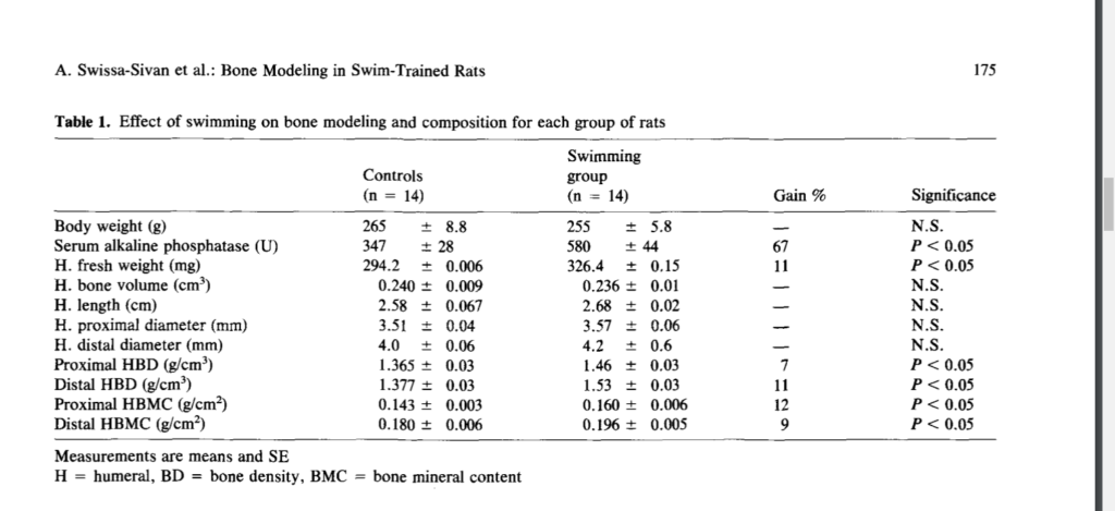
So perhaps upright doggy paddling is a method to induce torsional strain on relevant bones and increase height.
Histomorphometry of long bone growth plate in swimming rats
“In exercises involving running, muscle power and gravitational forces act together to affect bone mass in accordance with Wolff’s law. However, the direct effect of muscle activity on bones in non-weight-bearing activities, such as swimming, has not been explored. Previous data indicate that swimming exerts a positive effect on bone growth and development in young rats. We performed a histomorphometric study on the effect of swimming on the growth plate and subepiphyseal area of young adult rats. The experiments were carried out on 28 12-week-old albino Sabra rats. One group of 14 rats was trained to swim 1 hour/day, 5 days a week, for 12 weeks. Another group of 14 rats served as controls. The proximal femur and humerus of each animal were examined histomorphometrically. There was an increase in the subepiphyseal cancellous bone trabecullae of the femur. In the growth plate there was an increase in the number of column cells and proliferative cells. These changes were more pronounced in the femur than the humerus. We conclude that swimming induces an increase in subepiphyseal cancellous bone in young adult rats by enhancing growth plate activity.”
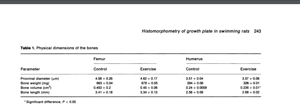
Humerus length increased but femoral length decreased.
Articular cartilage height increased in both groups which is very promising:
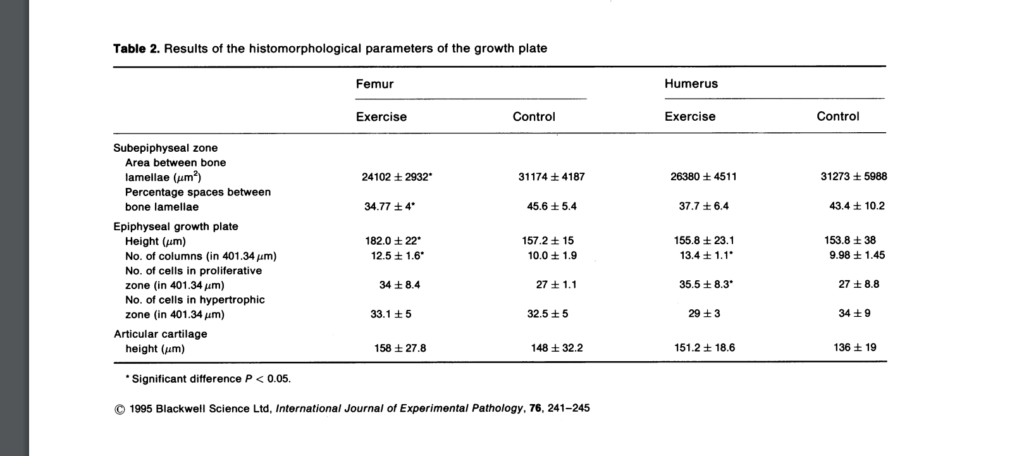
while the forelegs move in a small circular, balancing fashion. Therefore the effort of the hind legs is greater than that of the forelegs, explaining the differences in the muscle stresses of the humerus and femur and the consequent histomorphological changes in the bones.”
Swimming Enhances Bone Mass Acquisition in Growing Female Rats
“Growing bones are most responsive to mechanical loading. We investigated bone mass acquisition patterns following a swimming or running exercise intervention of equal duration, in growing rats. We compared changes in bone mineral properties in female Sprague Dawley rats that were divided into three groups: sedentary controls (n = 10), runners (n = 8) and swimmers (n = 11). Runners and swimmers underwent a six week intervention, exercising five days per week, 30min per day. Running rats ran on an inclined treadmill at 0.33 m.s−1, while swimming rats swam in 250C water. Dual energy X-ray absorptiometry scans measuring bone mineral content (BMC), bone mineral density (BMD) and bone area at the femur, lumbar spine and whole body were recorded for all rats before and after the six week intervention. Bone and serum calcium and plasma parathyroid hormone (PTH) concentrations were measured at the end of the 6 weeks. Swimming rats had greater BMC and bone area changes at the femur and lumbar spine (p < 0.05) than the running rats and a greater whole body BMC and bone area to that of control rats (p < 0.05). There were no differences in bone gain between running and sedentary control rats. There was no significant difference in serum or bone calcium or PTH concentrations between the groups of rats. A swimming intervention is able to produce greater beneficial effects on the rat skeleton than no exercise at all, suggesting that the strains associated with swimming may engender a unique mechanical load on the bone.”
“There was no significant difference for femur length between the runners (34.55 ± 1.40mm), swimmers (33.50 ± 0.70mm) or sedentary control rats (34.21 ± 1.99mm, F=1.34, p = 0.28).”<-another study that found shorter femur length for swimming rats. It could be biomechanically upright swimming results in compressive deformation.
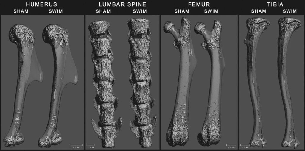
Reduced Bone Mass Accrual in Swim-Trained Prepubertal Mice

“Prepubertal female mice underwent a 16-wk training program, in which they swam for progressively increasing durations up to 55 min for 5 d·wk−1. A sham group was subjected to the water, but they did not perform the swimming exercise. Skeletal sites that were assessed included the proximal humerus, lumbar spine, midshaft and distal femur, proximal tibia, and the skull.”
This study found a decrease in length.
Here are the bone images:
The swimming studies that resulted in growth seem to be swimming studies with attached weight. That may be the key/weight equals more torsion.

Can this benefit people with closed growth plates? Does it induce microfractures?