Last night while I was laying in bed the subject of the phenomena where certain females noticed that they have grown taller during pregnancy reappeared in my mind. I couldn’t stop thinking about the issue and tried to make a somewhat educated guess on what exactly is happening.
Why is it that there have been so many (maybe a dozen on online forums and discussion boards so far which I have found) females who report that they have noticed that they have become taller from pregnancy?
I thought that it was from the dramtic, accelerated increase in progesterone in the post Analysis On The Possible Cause For Height Increase During Pregnancy, but that can’t be since progesterone is supposed to be able to increase bone mineral density, which means that the bones should be getting harder, and less resistant to bone remodeling.
The other idea I wrote about was that the hormone Follicle-Stimulating Hormone (FSH) which is involved in Klinefelter Syndrome, was the growth hormone that was being overstimulated. FSH comes from the same place in the anterior pituitary gland as somatotropins. They are stimulated by gonadotrophs. FSH is over stimulated during certain phases of pregnancy.
However, the main problem was alway over how could the pregnant female’s body ever increase in height, especially with a fetus in their uterus? Which bones exactly would be a stretched out for it?
Then I remembered the fact that multiple pediatricians have warned pregnant women that they might develop osteoporosis because of the transfer of calcium from the mother’s body to the fetus’. This is what I am guessing is the critical process I was missing.
The Process
1. The development of the fetus from just a embryo to a organic being which required calcium minerals to start being formed in its developing body means that the calcium level in the mother’s entire body is dropped.
How much is the drop in calcium level?
From the study Calcium and Bone Metabolism in Pregnancy and Lactation*
“…calcium and bone metabolism is substantially altered during the normal reproductive periods of pregnancy and lactation, and bone density can drop and regain 3–10% in the span of a few months in normal, healthy women.”
So the drop can be as much as 10% of the calcium phosphatase crystals.
2. Because of the position of where the uterus is located, the calcium will be drawn more from the upper part of the body, the torso, than the legs, since the bones in the leg are consistently being remodeled and made thicker and stronger from the effect of body weight loading.
3. If the calcium from the torso is being removed from the mother, then her bones would become weaker. Assuming the normal care of a pregnant women, she would start to spend less time on her feet, and more time lying on her back or in a fetal position in bed.
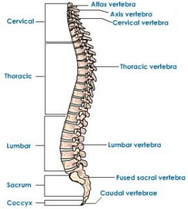
 4. The vertebrate curvature is reduced as she is lying in bed more. Her intervertebral discs are decompressed completely.
Her vertebrate bones after a 10% decrease in calcium mineral loss has become weak enough to realign themselves to be much more straight. It is in contrast to the normal curvature we find in most vertebrate of people.
The thoracic and lumbar region of most people’s vertebrate has a natural curvature, which gets straightened out by the way the pregnant women lies on the bed.
 I refer both the lying on the back position and the fetal position.
Notice the picture of the ballerina on the right. Notice how the fetal position some of us get into for sleep is actually a sort of body contorsion which allows for the dorsal side of the vertebrate to be stretched and decompressed.
With the addition of a baby that is uniquely positioned in the lower stomach area, the lumbar vertebrate are the bones that are specifically realigned and positioned as the hardness of the bones are decreased.
What I am proposing is that from the combination of 7 main factors, the pregnant women being more bed ridden would experience height growth.
1. The decrease in calcium levels in her own body from the transfer to her baby by upwards of 10%. This makes the bones more malleable.
2. The fact that the pregnant female is more likely bed ridden causing her to lie on her back more. This slightly helps in the vertebrate realignment idea.
3. The only other bed position is the fetal position which means that her vertebrate is naturally becoming decompressed and stretched out. obviously a pregnant women can’t lie on a bed face down, or that would crush her womb.
4. The position of where the womb is means that more structurally strain is placed in the lower back area, so if the women is in the fetal position in bed, the weight of the womb and baby contributes in the vertebrate realignment.
5. The fact that the irregular bones in the feet of the females get slightly bigger in width contributes. This is from the phenomena of where a large percentage of females have shown that their feet have grown longer and wider from pregnancy, most probably from periosteal appositional growth. I proposed that besides just length and width of the irregular bones in the bones that have increased, the height of the irregular bones in the female have also increased. This will contribute slightly to the height increase.
6. Tyler’s proposal of the onset of relaxin means that the ligaments holding bones in tight structural alignement is slightly loosened for bone realignment.
7. The release of the Follicle Stimulating Hormone by the gonatotrophs in the anterior pituitary gland means that it might act like a growth hormone for the women who is in a position with weak enough bones, where they can be ‘stretched out’
All of these 7 factors causes what I propose is the lumbar and thoracic vertebrate bones to become weaker from a significant drop in calcium levels losing the high compressive strength, have looser ligaments holding them together, and being in positions in bed for a long time which would allow for the natural curvature of the vertebrate to be decreased from a realignment of the individual irregular vertebrate bones.
However, there are also multiple cases of women who have experienced height loss from pregnancy due to their bones becoming too weak and developing fractures. The truth is that height loss from pregnancy makes much more anatomical sense than height increase.
When you are standing up when pregnant, the extra weight of the baby and womb would cause the lower back to become more curved, not less. The bones being weaker in the compressive strength (NOT tensile strength) should mean that when standing, the pregnant women would have their upper body weight loading the vertebrate and bones causing the body to become shorter.
Like always, this is just a theory I am proposing. Something to maybe reference in future posts to either disprove or validate new research, and idea we might have.

Micheal i recently found out that nerves usually heal about 1mm per day is this another reason for the 1mm per day LL or does it not matter
With Lsjl people grow in width in the exact place they clamp right? so if you did some sort of axial loading would this be beneficial? Just a thought.