Author Archives: Tyler

BREAKTHROUGH! Sea cucumber may boost longitudinal bone growth


Sea cucumber is available for sale but it is very expensive.  I found some sea cucumber tablets that were cheap however it’s not for certain that the tablets have all the benefits of eating sea cucumbers directly.

Novel Peptides from Sea Cucumber Intestine Hydrolysates Promotes Longitudinal Bone Growth in Adolescence Mice through Accelerating cell cycle progress by Regulating Glutamine Metabolism

“Sea cucumber intestines are recognized as a major by-product in sea cucumber processing industry and have been shown to have antioxidant properties. However, whether the sea cucumber intestine is beneficial to osteogenic remains unknown. In this study, low molecular weight rich in glutamate/glutamine peptides were obtained from sea cucumber intestines (SCIP) by enzymatic hydrolysis, and orally administered to adolescent mice to investigate the effects on longitudinal bone growth. The results showed that the SCIP supplement significantly increased the femur length and new bone formation rate by 9.6% and 56.3%, and elevated the serum levels of osteogenic markers ALP, Collagen I and OCN{this is pretty nuts.  I couldn’t find studies directly measuring what effect glutamine has on longitudinal bone growth directly}. Notably, H&E staining showed that SCIP significantly increased the height of the growth plate, in which the height of the proliferation zone was elevated by 97.4%. Glutamine is a major determinant of bone growth. We found that the SCIP supplement significantly increased glutamine levels in the growth plate by 44.2% and elevated the expression of glutamine metabolism-related enzymes Gls1 and GLUD1 in the growth plate{so it’s possible that sea cucumbers could have a benefit on glutamine in the growth plate due to some other ingredient in the cucumber other than the glutamine itself}. Further, SCIP upregulated growth plate acetyl coenzyme A levels to promote histone acetylation and accelerated cell cycle progression by upregulating Sox9 expression, thereby contributing to the rapid chondrocyte proliferation. To our best knowledge, this is the first report that SCIP could enhance longitudinal bone growth via promoting growth plate chondrocyte proliferation. Our present study will provide new ideas and a theoretical basis for high-value utilization of sea cucumber intestine.”

“the peptide from Euphausia superba promoted longitudinal bone growth in adolescent mice.  supplement with enzymatic hydrolysate of Stichopus japonicus could promote bone formation in vivo and in vitro.”

“the avascular nature of the growth plate makes glutamine a potential factor for regulating longitudinal bone growth”

“the SCIP supplement significantly increased femur length by 6.8% (SCIP-L{LOW DOSE}) and 9.6% (SCIP-H{High dose}) compared with the normal group”<-so we don’t know if even more is better than the doses mentioned.  The effect of growth plate height, proliferative height, and hypertrophic chondrocytes was also greater based on dose.

“the serum glutamine levels were gradually increased to a maximum between 0.25 h and 0.5 h and returned to baseline after 2 h. Conversely, as serum glutamine levels fell back, glutamine levels in muscle and femur peaked sequentially at 2 h and 3 h.”<-theoretically it could be worthwhile to take glutamine every 2 hours which would be very difficult to do.

“glutamine metabolism has been shown to upregulate chondrocyte proliferation genes (especially Sox9) by epigenetic modification”<-obviously epigenetic modification is very powerful.

“glutamine promoted the growth plate chondrocyte proliferation during endochondral ossification through acetylation modification of the histone 3K9”

So they did have a glutamine only group.  And the sea cucumber growth increased growth plate height further than that.  Also if you look at the growth plates, it’s a pretty dramatic difference.

Based on the information in this paper, it may be worthwhile for everyone growing to take a low cost sea cucumber supplement(i’d say actual sea cucumber is too expensive).  Not only for the growth plate but also joint cartilage.

This study Stichopus chloronotus aqueous extract as a chondroprotective agent for human chondrocytes isolated from osteoarthitis articular cartilage in vitro finds that sea cucumber has anabolic effects on articular cartilage.

Need help evaluating x-rays

I happened to get x-rays a year ago.  I was performing bone tapping at the time and was unable to get a copy of them because I couldn’t find any computer that would play a CD.  I found something that maybe an abnormality and evidence that bone tapping can induce changes in bone morphology but since I don’t have before and after I can’t be sure.  Like I said I just happened to have had xrays ordered.

Here’s the image of what may be an abnormality(if requested I can post full versions of all three xrays I have):

Here’s another tibia x-ray:

Above is a normal tibial x-ray.  Also here the x-rays of someone who performed LSJL.  Essentially the area circled in red looks clearer(less dense) than normal.  Is that indicative of neo-bone formation?  I can’t be sure.  So if someone could find an expert that would be helpful.  I can also provide copies of all the x-rays.  Please comment or email if you can help.

Study shows proof of concept that microfractures can increase bone length

“To investigate the effect of bone-marrow stimulation (BMS) on subchondral bone plate morphology and remodeling compared to untreated subchondral bone in a validated minipig model.
Three Göttingen minipigs received BMS with drilling as treatment for two chondral defects in each knee. The animals were euthanized after six months. Follow-up consisted of a histological semiquantitative evaluation using a novel subchondral bone scoring system and micro computed tomography (µCT) of the BMS subchondral bone. The histological and microstructural properties of the BMS-treated subchondral bone were compared to that of the adjacent healthy subchondral bone.  The µCT analysis showed that subchondral bone treated with BMS had significantly higher connectivity density compared to adjacent untreated subchondral bone (26 1/mm3 vs. 21 1/mm3, P = 0.048). This was the only microstructural parameter showing a significant difference. The histological semiquantitative score differed significantly between the subchondral bone treated with BMS and the adjacent untreated subchondral (8.0 vs. 10 P = < 0.001). Surface irregularities were seen in 43% and bone overgrowth in 27% of the histological sections. Only sparse formation of bone cysts was detected (1%).BMS with drilling does not cause extensive changes to the subchondral bone microarchitecture. Furthermore, the morphology of BMS subchondral bone resembled that of untreated subchondral bone with almost no formation of bone cyst, but some surface irregularities and bone overgrowth(!).”
And the bone bone overgrowth can be seen to be length as seen in the histological slides.
“The objective of BMS is to create canals in the subchondral bone to the underlying bone marrow, either by use of a drill(subchondral drilling) or an awl (microfracture).”<- an awl is a tool for piercing holes.  It’d be hard to mimic either a drill or awl safely using non-surgical methods but I don’t believe that it’s impossible.
The study mentions that other studies also showed bone overgrowth.
Bone overgrowth was defined as areas where the subchondral bone plate had grown into the cartilage.”<-this could be due to endochondral ossification or longitudinal appositional growth.
“Nine out of the 12 samples contained bone overgrowth corresponding to 27% of the sections.”
“Hematoxylin eosin staining, scale bars: 200 μm. Defects treated with BMS. (A) Shows irregularity of the subchondral bone plate (top arrow marks the beginning of the defect). (B) Bone cyst marked by arrows. (C) Remaining drill hole in the subchondral bone plate. (D) Overgrowth in the subchondral bone plate marked by to arrows, top arrow marks the beginning of the defect site. BMS = bone marrow stimulation.”<-so the overgrowth is very minor but if there was some way to reproduce it, it could add up to height over time.  The pigs used in the study were skeletally mature.

Joint loading promotes stem cell migration

New study related to lateral synovial joint loading:

Knee Loading Enhances the Migration of Adipose‑Derived Stem Cells to the Osteoarthritic Sites Through the SDF‑1/CXCR4 Regulatory Axis

“Osteoarthritis (OA) is a whole joint disorder that is characterized by cartilage damage and abnormal remodeling of subchondral bone. Injecting adipose-derived stem cells (ASCs) into the knee joint cavity can assist in repairing osteoarthritic joints, but their ability to migrate to the damaged site is limited. Our previous studies have shown that knee loading can improve the symptoms of OA, but the effect and mechanism of knee loading on the migration of ASCs in OA remain unclear. We employed a mouse model of OA in the knee and applied knee loading (1 N at 5 Hz for 6 min/day for 2 weeks) after the intraarticular injection of ASCs. The cartilage and subchondral bone repair were assessed by histopathological analysis. Immunofuorescence assays were also used to analyze the migration of ASCs. Using cell cultures, we evaluated the migration of ASCs using the transwell migration and wound healing assays. In vivo experiments showed that knee loading promoted the migration of ASCs, increased the local SDF-1 level, and accelerated the repair of the OA-damaged sites. Mechanistically, the observed effects were blocked by the SDF-1/CXCR4[SDF-1/CXCR4 promotes neovascularization which is great] inhibitor. The in vitro results further revealed that knee loading promoted the migration of ASCs and the inhibition of SDF-1/CXCR4 significantly suppressed the beneficial loading effect. The results herein suggested that the migration of ASCs was enhanced by knee loading through the SDF-1/CXCR4 regulatory axis, and mechanical loading promoted the joint-protective effect of ASCs”

Now we don’t know how much more lateral knee loading stimulates neo-vascularization etc. than regular exercise.  Nor do we know if knee loading promotes subchondral bone repair independently of adipose derived stem cells.  But neovascularization and new cartilage formation are very important findings that could potentially be linked to what’s needed to grow taller.

“A variety of physical rehabilitation regimens can afect the remodeling process of articular cartilage and subchondral bone and alter the symptoms of OA, such as whole-body vibration, axial loading, and bending loading. The knee loading modality, developed by our laboratory, is a mild non-invasive physical therapy, which applies transverse loading to a synovial joint, such as the elbow, knee, and ankle. Knee loading has been shown as an effective loading option for improving OA symptoms. Previous studies showed that knee loading reduced MMP13 activity and prevented OA-induced cartilage degeneration through cross-talk of the cartilage with subchondral bone. Mechanical loading also mitigated OA symptoms by regulating the stress to the endoplasmic reticulum and autophagy. Further, knee loading facilitated repairing osteoporotic OA by relieving the abnormal remodeling of the subchondral bone via Wnt/β-catenin signaling. It contributed to promoting the diferentiation of MSCs into osteogenic and chondrogenic fates”

“Stromal cell-derived factor 1 (SDF-1), a chemokine of a CXC family, and its receptor CXC chemokine receptor 4 (CXCR4) exist widely in the body, and they regulate a variety of physiological processes, including cell proliferation, migration, adhesion, differentiation, and wound healing ”

“A total of 108 female C57BL/6 mice (14 weeks old, Animal Center of Academy of Military Medical Sciences, China) were used.”<-not quite as old as we would like.

“After the induction of OA, mice were injected with GFP-ASCs, followed by daily knee loading for 2 weeks. The injected ASCs in the knee joints of all groups were identified by immunofuorescence staining with antibodies against GFP. In vivo studies showed that GFP-positive cells were found in the OA cartilage 2 weeks after their injections”

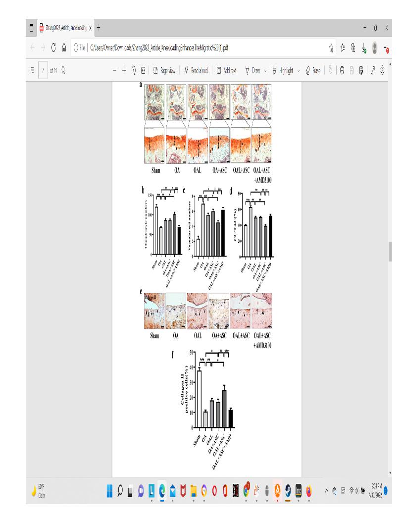
” The number of chondrocytes and vacuolar cells was counted. The number of chondrocytes in the OA group was significantly decreased compared to the sham-treated control group, but the number was restored by knee loading or the injection of ASCs”<-it’s very promising that the chondrocyte number was restored be knee loading independently of stem cell injection but again these mice are not as old as we would like.

” IHC analysis showed that collagen II-positive cells in the OA group were significantly decreased. However, the numbers in the OAL group and OA+ASCs group were increased.”<-again very promising that collagen II positive cells were increasing independently of ASC injection

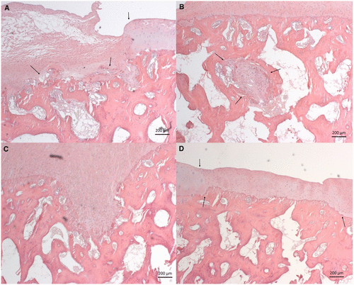
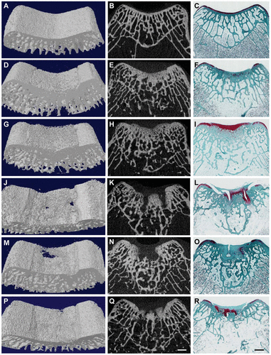
In the top images you can see that the mice still have their growth plate.  It’d be hard to distinguish between changes in the bone caused by Osteoarthritis and those caused by loading.  But if you look at the loaded groups(Loading with and without ASC) you can see in red cartilage in places that did not have cartilage in either the control or the osteoarthritis group at the longitudinal ends of the bone.  And you don’t really see that in the OA+ASC group.  I could be reading it wrong though.

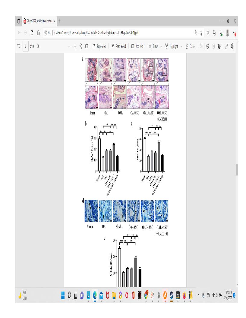
Again here you see cartilage at the sides of the longtiduinal ends of the bone that you don’t really see in the non-loaded groups.

Distraction Histogenesis: Arteries

In limb lengthening surgery, there is concern to how the soft tissue will adapt to the growing bone. Here is a paper that provides evidence that the arteries do adapt after limb lengthening surgery:

Effect of mechanical tension stress on arterial vessels after limb osteotomy in rabbits

<-osteomy is bone cutting an important stage in limb lengthening surgery

Limb lengthening by tensile stress{limb lengthening surgery is primarily an osteomy plus tensile stress} has become an effective measure in the treatment of fracture and limb amputation, but the underlying mechanism of how mechanical tensile stress affects limb arteries and vessels has not been clarified.  
To investigate the effect of slow steady unidirectional mechanical tension stress on limb artery.
 Totally 75 adult New Zealand rabbits were randomly divided into model control group (the hind limb was amputated and not prolonged), experimental group (the hind limb was amputated and lengthened) and blank group (no limb treatment), with 25 rabbits in each group. The prolonged speed of the experimental group was 0.5 mm/time, twice a day, for 10 days. Saphenous artery specimens were collected at 8, 10, 12, 16, and 19 days (i.e., 3, 5, 7, 11, and 14 days from the start of distraction osteogenesis) after Ilizarov external fixation. The length of saphenous artery was observed and compared. At the same time, X-ray examination was conducted to detect the osteotomy of the affected limb. Arterial structure and inflammatory response were observed by hematoxylin-eosin staining.  
(1) In terms of vascular length: Since the 5th day of Ilizarov external fixation stent pulling, the length of saphenous artery in the experimental group was significantly increased compared with the model control group (P < 0.01). (2) In terms of X-ray examination, at 16 days after osteotomy (i.e., 1 day after the extension ends), the end of osteotomy in the experimental group was extended by about 10 mm, and no callus was formed in the gap. (3) Hematoxylin-eosin staining showed that at 8, 10, 12, 16, and 19 days after external fixation, the arterial tissue structure of the experimental group was intact, without intima vascular injury, smooth muscle cell necrosis or inflammatory cell infiltration. (4) These results indicate that slow and steady unidirectional mechanical stretch stress can prolong the limb arteries and keep the arterial vascular structure stable while lengthening the length of tibial osteotomy end, suggesting that the potential mechanism of stretch stress promoting limb lengthening lies in the stimulation of arterial vascular growth.

Unfortunately, I could not get this full study.

New LSJL Study with histological slides

New LSJL study was published that has some good stuff.  Maybe the paper will finally give the insight on how to use mechanical loading to gain height.

Loading-driven PI3K/Akt signaling and erythropoiesis enhanced angiogenesis and osteogenesis in a postmenopausal osteoporosis mouse model

“Bone vasculature influences osteogenesis[any type of new bone formation and haematopoiesis[production of blood cells in the bone marrow] in the bone microenviroment. Mechanical loading has been shown to stimulate the formation of osteogenesis-related type H vessels in an ovariectomy (OVX)- induced osteoporosis mouse model. To determine the loading-driven mechanism of angiogenesis and the formation of type H vessels in bone, we evaluated the roles of PI3K/Akt signaling and erythropoiesis in the bone marrow. The daily application of mechanical loading (1 N at 5 Hz for 6 min/day) for 2 weeks on OVX mice inhibited osteoclast activity{although osteoclast activity is good for our purposes as degradation of bone is needed to theoretically create a new growth plate}, associated with an increase in the number of osteoblasts and trabecular volume ratio. Mechanical loading enhanced bone vasculature and vessel formation, as well as PI3K/Akt phosphorylation[Akt phosphorylation indicates general anabolism] and erythropoiesis[production of red bood clells] in the bone marrow. Notably, LY294002, an inhibitor of PI3K signaling, blocked the tube formation by endothelial progenitor cells, as well as their migration and wound healing. The conditioned medium, derived from erythroblasts, also promoted the function of HUVECs with elevated levels of VEGF, CD31, and Emcn. Collectively, this study demonstrates that mechanical loading prevents osteoporotic bone loss by promoting angiogenesis and type H vessel formation. This load-driven preventing effect is in part mediated by PI3K/Akt signaling and erythropoiesis in the bone marrow”

“A specific vessel subtype called a type H vessel, which is positive for CD31 and endomucin (CD31hiEmcnhi), has been reported to link angiogenesis with osteogenesis”

“Vascular-forming endothelial cells provide a framework of bone homeostasis and metabolism, acting as a cellular highway for blood cells, leukocytes, and other types of cells throughout the body ”

“Erythropoiesis is characterized by the movement of lineage-committed cells with progenitors, precursors, and mature red blood cell (RBC) compartments, which are located in the fetal liver and the postnatal bone marrow”

“Molecular signals by mechanosensing and transduction, leading to a series of cellular responses. The phosphatidylinositol 3-kinase (PI3K)/Akt signaling pathway has been shown to play a critical role in the regulation of diverse cellular functions and is also involved in angiogenesis and erythropoiesis. joint loading increases the expression of Akt, which may promote bone formation and homeostasis. The effect of PI3K signaling on endothelial progenitor cells (EPCs) in the bone marrow of OVX mice, however, has not yet been demonstrated in response to joint loading. ”

“we hypothesized that the reduction in type H vessels and the hematopoietic function in OVX mice is suppressed by mechanical loading through PI3K/Akt signaling, which stimulates erythroid differentiation of hematopoietic stem cells (HSC) and promotes vessel formation”<-so joint loading may not suppress osteoclast activity in normal bone as the OVX environment is different.

“Ninety female C57BL/6 mice (~16 weeks of age, Animal Center of Academy of Military Medical Sciences, China) were used. Mice were randomly divided into three groups: the sham control group (Sham, n = 30), the ovariectomized group (OVX, n = 30), and the loading-treated ovariectomized group (OVXL, n = 30).”<-unfortunately no loaded control group.

Above is histological slides.  Can’t really see anything abnormal.  There may be something atypical going on in F where the added arrow is pointing.

According to figure 2G PI3K, AKT, and VEGF activity was greatly enhanced by joint loading even over controls.  But according to 2H, I, J it did not increase the ratios of phosphorylated PI3K and AKT to total PI3K and AKT and VEGF relative to Beta-Actin.

“The formation of type H vessels was correlated to the stimulation of osteogenesis, and the result suggests that the promotion of type H formation and angiogenesis are important in response to load-driven suppression of OVX-induced bone loss. ”

“knee loading stimulated PI3K/Akt phosphorylation in the bone, and this stimulation can be linked to loading-driven bone metabolism”<-although this alone is definitely not sufficient to induce new longitudinal bone growth.

“Knee loading enhanced cell migration and wound healing”

“in OVX mice, load-driven angiogenesis via erythropoiesis contributed to suppressing the loss of osteoporotic bone. “<-erythropoiesis is the creation of new red blood cells.  Erythropoiesis is thought to not typically occur in adult bones so if joint loading can induce new erythropoiesis that is a big deal.

The mice here were subjected to 1n loading at 5Hz for 6 min/day.  This is actually higher than that performed on the original bone lengthening LSJL study 5-min bouts at 0.5 N[at 5Hz).

Here’s a graphical representation in the study at how mechanical loading might stimulate bone:

The estimated increase in type H vessels is dramatic.  Osteoclast activity as depicted is way done too.

And here’s the description of type H vessels “Type H vessels are noted most abundantly in young, growing animals, adjacent to the growth plate. The elongation of long bones by the process of endochondral ossification occurs at the growth plate and involves continuous replacement of cartilage matrix by calcified bone to acquire their typical shapes through bone modeling.”

Could type H vessels be the key for new longitudinal bone growth here’s a study explaining type H vessels.

Type H blood vessels in bone modeling and remodeling

“In the mammalian skeletal system, osteogenesis and angiogenesis are intimately linked during bone growth and regeneration in bone modeling and during bone homeostasis in bone remodeling. Recent studies have expanded our knowledge about the molecular and cellular mechanisms responsible for coupling angiogenesis and bone formation. Type H vessels, termed such because of high expression of Endomucin (Emcn) and CD31, have recently been identified and have the ability to induce bone formation. Factors including platelet-derived growth factor type BB (PDGF-BB), slit guidance ligand 3 (SLIT3), hypoxia-inducible factor 1-alpha (HIF-1α), Notch, and vascular endothelial growth factor (VEGF) are involved in the coupling of angiogenesis and osteogenesis.”

“in bone modeling associated with endochondral ossification, hypertrophic chondrocytes express high levels of vascular endothelial growth factor (VEGF), which promotes the vascular invasion of cartilage, recruits chondroclasts to resorb hypertrophic cartilage and osteoblasts to build the bone matrix”

“In bone modeling in response to mechanical loading, macrophage/non-resorbing osteoclast lineage cells secrete platelet-derived growth factor type BB (PDGF-BB) to recruit both endothelial and osteoblast precursor cells to couple angiogenesis with osteogenesis”

“activation of mechanistic target of rapamycin complex (mTORC) in articular chondrocytes promoted VEGF secretion into subchondral bone and stimulated type H vessel formation.”

“type H vessels were reported to digest the cartilage matrix, which may be another mechanism of cartilage degeneration in OA.”

In fracture healing, type H vessels appear during the stage of fibrocartilaginous callus formation. Type H vessels form in the fracture callus and induce bone formation. Factors, such as SLIT3, can promote fracture healing by augmentation of type H vessel induced osteogenesis.”<-Limb lengthening basically if LSJL can induce type H vessels and if type H vessels are important in neocartilage formation than that would be a big find.  But I can only find that Type H vessels are important in absorbing the cartilage matrix not in creating the cartilage matrix.  Although increased Type H Vessels as shown in this study could be indicative of enhanced and maybe even new growth plate activity would would be massive.  IF the mice got new Type H blood vessels past skeletal maturity that would be a huge find 16 week of the type used in this study are pretty far along skeletal wise so new type H vessels is still a big deal but they are still not quite old enough.

Here’s the supplementary data attached

This for the sham and the joint loaded control no osteoarthritis.  The bones look significantly different too me with the loaded bone significantly bigger and and maybe slightly longer(hard to tell). I’d say the width difference is striking and huge. The growth plate is clearly open.  Bone parameters are bigger in the loaded group despite not being “statistically significant”.