Author Archives: Tyler

Interesting study about Epiphyseal Growth Plate Fusion

Understanding Growth Plate Fusion(and growth plate senescence) will help us understand if these processes can be reversed.  And just as a note I am still working on devices and methodology to grow taller.

A Computed Microtomography Method for Understanding Epiphyseal Growth Plate Fusion.

“The epiphyseal growth plate is a developmental region responsible for linear bone growth, in which chondrocytes undertake a tightly regulated series of biological processes. Concomitant with the cessation of growth and sexual maturation, the human growth plate undergoes progressive narrowing, and ultimately disappears. Despite the crucial role of this growth plate fusion “bridging” event, the precise mechanisms by which it is governed are complex and yet to be established. Progress is hindered by the current methods for growth plate visualization; these are invasive and largely rely on histological procedures. Here, we describe our non-invasive method utilizing synchrotron X-ray computed microtomography for the examination of growth plate bridging, which ultimately leads to its closure coincident with termination of further longitudinal bone growth. We then apply this method to a dataset obtained from a benchtop micro computed tomography scanner to highlight its potential for wide usage. Furthermore, we conduct finite element modeling at the micron-scale to reveal the effects of growth plate bridging on local tissue mechanics. Employment of these 3D analyses of growth plate bone bridging is likely to advance our understanding of the physiological mechanisms that control growth plate fusion.”

“It is the terminally differentiated hypertrophic chondrocyte, which mineralizes its surrounding extracellular matrix. This process, thought to involve membrane-limited matrix vesicles, is biphasic and tightly regulated by a number of enzymes and factors including alkaline phosphatase (Alpl), PHOSPHO1, the ankylosis protein (Ank), ecto-nucleotide pyrophosphatase/phosphodiesterase-1 (Enpp1) ”

“as growth slows, the human growth plate undergoes progressive narrowing as bony bridges form and span its width. This ultimately leads to complete growth plate closure and the cessation of human growth. These bone bridges are also known to form upon growth plate injury, thought to be through an intramembranous ossification mechanism”

“in two genetic mutations resulting in estrogen deficiency (in the estrogen-receptor gene, and in the CYP19 gene), the growth plate fails to fuse and growth persists, albeit rather slowly, into adulthood”

“Evidence from studies in both humans and rats revealed the cessation of growth long before any histological evidence of growth plate fusion, suggesting that epiphyseal fusion is a marker of growth cessation and not its cause “<-Thus it may be possible to renew growth plate growth by reversing cessation and not fusion.


“young (8 weeks old) CBA wild-type mice, growth plate bridging is associated with locations that contain high local von Mises stresses. Moreover, we reveal that with aging an increased number and density of growth plate bridges is observed”

” in wild-type mice, increased growth plate bridging translates into increased stresses in the bone directly beneath the growth plate.”<-So maybe these stresses contribute to growth plate closure.

“At 8 weeks, few bridges are detected and overall the growth plate is squeezed in a “sandwich” configuration. This suggests that compressive hydrostatic stresses are engendered across major volumes and that higher shear stresses are generated only at the peripheral edges of the growth plate. Yet, the results of numerous mechanobiological models support that growth and ossification is accelerated by tensile strain (or shear stresses) and that cartilage tends to be maintained by hydrostatic compressive stress“<-so for a longer growth period we need to encourage hydrostatic compressive stress.


Switch in stem cell renewal over time in epiphyseal growth plate

A radical switch in clonality reveals a stem cell niche in the epiphyseal growth plate.

“Longitudinal bone growth in children is sustained by growth plates, narrow discs of cartilage that provide a continuous supply of chondrocytes for endochondral ossification. However, it remains unknown how this supply is maintained throughout childhood growth. Chondroprogenitors in the resting zone are thought to be gradually consumed as they supply cells for longitudinal growth, but this model has never been proved. Here, using clonal genetic tracing with multicolour reporters and functional perturbations, we demonstrate that longitudinal growth during the fetal and neonatal periods involves depletion of chondroprogenitors, whereas later in life, coinciding with the formation of the secondary ossification centre, chondroprogenitors acquire the capacity for self-renewal, resulting in the formation of large, stable monoclonal columns of chondrocytes{if these chondroprogenitors never become senescent then we can grow forever}. Simultaneously, chondroprogenitors begin to express stem cell markers and undergo symmetric cell division. Regulation of the pool of self-renewing progenitors involves the hedgehog and mammalian target of rapamycin complex 1 (mTORC1) signalling pathways. Our findings indicate that a stem cell niche develops postnatally in the epiphyseal growth plate, which provides a continuous supply of chondrocytes over a prolonged period.”

“CD73 (encoded by Nt5e) was among the most upregulated stem cell surface markers (4.9 ± 1.2-fold; P28 versus P2; P = 0.018) and immunohistochemical analysis confirmed de novo expression of CD73 at P28 (markers for cell proliferation (Ki67) and differentiation (MEF2C) were controls for developmental changes”

“Cells in the growth plate might also orient as a result of migration or stacking due to tissue polarization, which is reflected by the orientation of primary cilia. Cilia on flat chondrocytes, but not on chondroprogenitors, were polarized (with 61.3 ± 4.8% and 32.8 ± 2.2% of cilia, respectively, oriented longitudinally, P = 0.005, n = 3;”

3j is supposed to summarize it

Martial Arts Bone Conditioning and LSJL

LSJL is lateral loading of the synovial joints or possible the epiphysis of the bone to stimulate fluid flow to stimulate degradation(remodeling) of the cortical bone and creation of mesenchymal stem cell chondrogenesis through a favorable microenvironment.

Martial arts bone condition is another form of this as impact is a form of loading.  However impact loading would not stimulate the soft tissues surrounding the bone which are all connected to the bone this is an important distinction.  Axial loading does not really drive fluid flow so that elements punching as a studiable stimulus.  However, several martial artists do tapping of the bones of the leg and arm.

The different between this and an LSJL style tapping would be that the tapping would not necessarily be at the longitudinal ends of the bone so the bone would be more innervated and there would be less of a fluid pressure gradient than at the longitudinal ends of the bone.  The goal of martial arts bone conditioning is described as something like calcification or bone thickening which is not exactly what we want.

The other difference between LSJL and martial arts bone conditioning is the intensity of the load.  Short intense bursts is more osteogenic whereas longer less intense bursts of loading are more chondrogenic.  This is primarily because osteocytes respond to sudden changes in fluid flow but osteocytes(probably) cannot make you taller as osteocytes produce bone and bone is what blocks you from growing taller!  Soft tissue is what allows for interstitial growth.  So for LSJL style loading you would tap the longitudinal ends of the bone with more frequency and less intensity.

If the bones weren’t so innervated would you be able to grow via martial arts style loading?  Probably not by rolling a bottle up and down your leg.  But we know that martial artists fracture legs during kicks.

And fractures definitely result in longitudinal bone growth because the bone clot results in a lot of fluid stimulus and it removes the cortical bone impediment.

We also know that there’s a phenomenon known as stress fractures where bones can develop fracture over time to non-overly traumatic loads just as the bone is no longer able to recover.

But we want to know if it’s possible to induce the longitudinal bone growth stimulus without fracturing a bone.  And could it be possible with a tapping stimulus?  The key is to target a less innervated part of the bone which happens to be the epiphysis although there are some epiphysis where regions are fairly innervated.  Tapping the epiphysis also generates a fluid pressure gradient.

With these fluid pressures we want to stimulate bone degradation via osteoclasts(remodeling)[this process could be accelerated via HGH] but we don’t want bone deposition via osteocytes and osteoblasts we want cartilage deposition although it’s feasible that other fibrocartilage tissues could work so that’s why we want sustained loading pressures over time so consistent taps.  The stress fracture phenomenon is also beneficial for our purposes.  Microcracks are good at stimulating fluid forces.

Nerve issues are probably unavoidable but we want to choose the least nerved location.

Does anyone have any anecdotal evidence of martial artist effects on bone?

I know there’s not a lot of solid proof of anything here but basically I’m testing epiphyseal tapping over prolonged periods to generate bone degradation and creating a favorable microenvironment for replacement via soft tissues.  I wanted people to know what I’m working on as I haven’t posted here in a while.  I’m solely dependent on generating proof at this point so if anyone has any evidence of martial artists generating any kind of visible bone adaptation whatsoever that would be extremely helpful.


p-Coumaric acid<- potential height increasing supplement

According to the study it only increases IGF-1, but it may have other effects that are not known about.  Red Clover flowers contain p-Coumaric Acid.  I could only find p-coumaric acid available for scientific use.

p-Coumaric acid stimulates longitudinal bone growth through increasing the serum production and expression levels of insulin-like growth factor 1 in rats.

“The aim of the present study was to examine the effects of p-coumaric acid on the longitudinal growth of the long bone in adolescent male rats. Teatment with p-coumaric acid significantly increased the tibial length and the height of each growth plate zone and the ratio of 5-bromo-2′-deoxyuridine-positive cells relative to total proliferative cells. Expression of insulin-like growth factor 1 and its receptor in the proliferative and hypertrophic zones, and serum levels of growth hormone and insulin-like growth factor 1 were significantly increased as well in the p-coumaric acid-treated group. Via increasing both the serum level of insulin-like growth factor 1 and its expression, p-coumaric acid could promote cell proliferation in growth plate zones. These results suggest that p-coumaric acid has the potential to increase height and may be a feasible alternative to growth hormone therapy.“<-the statement that this may be a feasible alternative to growth hormone therapy makes this seem more powerful than just your typical supplement.

The study also mentions other supplements that have beneficial effects on chondrogenesis.  If you look at figure 1 and 2 you can see that there are differences between p-Coumaric Acid and Growth Hormone growth plates.  The scientists suggest that p-Coumaric acid upregulates IGF-1 and IGF-1R.

Ligaments constrain growth

The reason that the jaw can grow via stretching by forwarding positioning and bite-jumping appliances is that it is not as constrained by ligaments as other bones.  It’s possible that other movable joint regions like the wrists, fingers, and toes would also be stretchable.  The wrists would come under extreme stress due to the farmers walk so if this true there should be anecdotal evidence of longer arms due to the farmers walk.  It is possible that a new height increase method could be invented that causes enough tensile strain in the articular cartilage to activate endochondral ossification in that region.  The ligaments holding the bone together would constrain this.  This would also help explain why people with Marfan’s become taller as they have more flexible joints and therefore their growth is less constrained by ligaments.   Here’s another connective tissue disorder that may affect longitudinal bone growth.  It’s also possible that differences between condylar cartilage and articular cartilage allow this to happen in the mandible but not in other joints.

So if anyone could look for instances of longer arms due to heavy farmers walk.  This would be much easier to do than LSJL.  So any research on this would be a great boon to further the cause of height increase.

The adaptive remodeling of condylar cartilage—a transition from chondrogenesis to osteogenesis.

“Mandibular condylar cartilage is categorized as articular cartilage but markedly distinguishes itself in many biological aspects, such as its embryonic origin, ontogenetic development, post-natal growth mode, and histological structures. The most marked uniqueness of condylar cartilage lies in its capability of adaptive remodeling in response to external stimuli during or after natural growth. The adaptation of condylar cartilage to mandibular forward positioning{basically bringing your jaw forward} constitutes the fundamental rationale for orthodontic functional therapy, which partially contributes to the correction of jaw discrepancies by achieving mandibular growth modification. The adaptive remodeling of condylar cartilage proceeds with the biomolecular pathway initiating from chondrogenesis and finalizing with osteogenesis{so basically by stretching the articular cartilage you activate enchondral ossification enabling you to grow on the longitudinal ends of the bone}. During condylar adaptation, chondrogenesis is activated when the external stimuli, e.g., condylar repositioning, generate the differentiation of mesenchymal cells in the articular layer of cartilage into chondrocytes, which proliferate and then progressively mature into hypertrophic cells. The expression of regulatory growth factors, which govern and control phenotypic conversions of chondrocytes during chondrogenesis, increases during adaptive remodeling to enhance the transition from chondrogenesis into osteogenesis, a process in which hypertrophic chondrocytes and matrices degrade and are replaced by bone. The transition is also sustained by increased neovascularization, which brings in osteoblasts that finally result in new bone formation beneath the degraded cartilage.  The repositioning of the mandibular condyle in adult rats led to a reactivation of chondrogenesis in condylar cartilage which otherwise is at resting status, and finally results in increased bone formation{this is huge if as it means a bigger stronger jaw}

“chondrogenic activity of BMP-2 in vitro involves the action of the cell-cell adhesion protein, N-cadherin, which functionally complexes with beta-catenin the change of condyle position relative to the glenoid fossa constitutes an important trigger for [the endochondral ossification related adaptation of the mandible].”

The deviation of the condyle from the glenoid fossa by mandibular forward translation is the basis for orthodontic functional therapy, which aims to enhance condylar growth and therefore to eliminate the discrepancy between upper and lower jaws.

a decrease in compressive loading enhances condylar growth, whereas an increase in loading inhibits growth”

Note that they do say that condylar cartilage is distinct from articular cartilage.

Here’s another study:

Murine TMJ loading causes increased proliferation and chondrocyte maturation.

“The purpose of this study was to examine the effects of forced mouth opening on murine mandibular condylar head remodeling. We hypothesized that forced mouth opening would cause an anabolic response in the mandibular condylar cartilage. Six-week-old female C57BL/6 mice were divided into 3 groups: (1) control, (2) 0.25 N, and (3) 0.50 N of forced mouth opening. Gene expression, micro-CT, and proliferation were analyzed. 0.5 N of forced mouth opening caused a significant increase in mRNA expression of Pthrp, Sox9, and Collagen2a1, a significant increase in proliferation{These alone will not increase height except maybe the increase in proliferation}, and a significant increase in trabecular spacing in the subchondral bone, whereas 0.25 N of forced mouth opening did not cause any significant changes in any of the parameters examined. Forced mouth opening causes an increase in the expression of chondrocyte maturation markers and an increase in subchondral trabecular spacing.”

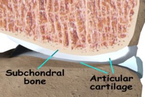
“The articulating surfaces of the temporomandibular joint (TMJ) are covered by fibrocartilage, which is composed of chondrocytes at various stages of maturation that are organized into four zones: (1) superior articular zone—cells in this zone express proteoglycan 4 (Prg4); (2) polymorphic zone—cells in this zone express SRY-box 9 (Sox9) and parathyroid-hormone-related protein (Pthrp); (3) flattened zone—the cartilage cells in this layer express collagen type II (Col 2a1); and (4) hypertrophic zone—the chondrocytes express collagen type X (Col 10a1)”

“Growth in the condylar cartilage proceeds from cells exiting the proliferative pool in the polymorphic zone and undergoing endochondral ossification in the inferior cartilage zones. The mandibular condylar cartilage is able to remodel its structure in response to mechanical strains

F and G are both forced mouth opening with F and G to a higher degree.  C and D have longer bones but there is less separation between the two bones with C and D.  The fear with articular cartilage endochondral ossification is that you’ll decrease the joint space.

“Functional appliance therapy is believed to place the mandible in a protrusive position, causing the condyle to be unseated caudally and ventrally from the articular fossa, while forced mouth opening is believed to cause a compressive force between the articular eminence and the mandibular condyle”

“A possible explanation for both functional appliance therapy and forced mouth opening causing similar effects in the mandibular condylar cartilage is that the mandibular condylar cartilage responds similarly to different types of loading compression for forced mouth opening and tension for functional appliance therapy{another possibility is that both activate the lateral pterygoid muscle and that’s what pulls on the cartilage}. Another possible explanation is that forced mouth opening and functional appliance therapy influence the masticatory muscles in such a way that the actual deformational strains within the mandibular condylar cartilage are similar for both.”

Forward mandibular positioning enhances the expression of Ang-1 and Ang-2 in rabbit condylar chondrocytes

“Functional appliances correct dental malocclusion, partly by exerting an indirect mechanical stimulus on the condylar cartilage, initiating novel bone formation in the condyle. Angiopoietin is involved in the angiogenesis associated with novel bone formation. This study aimed to determine the expression of angiopoietin (Ang)‑1 and ‑2 following forward mandibular positioning (FMP) in the condylar chondrocytes of rabbits. Sixty rabbits (age, 8 weeks) were randomly allocated to the experimental and control groups (n=30 per group). In the experimental group, FMP was induced by a functional appliance. Five rabbits from the experimental group and the control group were sacrificed following 3 days and 1, 2, 4, 8 and 12 weeks, respectively. The right temporomandibular joints (TMJs) were collected and the expression of Ang‑1 and -2 was evaluated by immunohistochemical staining. The expression of Ang-1 increased at day 3 and reached a peak at 2 weeks, whereas Ang‑2 reached maximal expression 4 weeks after FMP. Subsequently, the expression of Ang‑1 and ‑2 gradually decreased. Thus, FMP enhanced the expression of Ang‑1 and Ang‑2 in condylar cartilage, which is related to angiogenesis in the process of endochondral ossification.”

“Autocrine Ang-1/Tie-2 modulates blood vessel plasticity and contributes to vascular maintenance. In addition, Ang-1 enhances survival, migration and network formation of endothelial cells in vitro, and induces neovascularization in vivo. Ang-2 is a naturally occurring antagonist of Ang-1 that inhibits Ang-1-induced activation of Tie2. Ang-1 and -2 are located at sites of endochondral bone formation in the growing skeleton”

“The appliances were worn for 24 h to produce a continuous forward and downward positioning of the mandible.”

Look at all the cartilage growth.  There seems to be some endochondral ossification going on in the longitudinal ends of bone which is very good for height growth.

Here’s a normal jointNotice how they are connected strongly by a ligament(Li)

Here’s Mandibular Condylar Cartilage as a Juvenile and as an adult:

Microcracks effect on fluid flow. Long term loading may be key for LSJL.

This study is important because it indicates that microcracks may be bad for fluid flow which stimulates bone growth but good for cartilage growth as if fluid is not flowing than it is building up pressure and pressure is more conducive to chondrogenesis.  This indicates that a method to induce fluid flow such as  clamping/tapping should be higher duration and more “fatigue loading” based to insure the induction of microcracks.  Fatigue loading is the act of inducing bone damage not by a sudden large damage but by sustained bouts of loading over time.

Influence of interstitial bone microcracks on strain-induced fluid flow.

“microcracks act as a stimulus for bone remodelling, initiating resorption by osteoclasts and new bone formation by osteoblasts.  Microcracks alter the fluid flow and convective transport through the bone tissue. [We evaluate] the strain-induced interstitial fluid velocities developing in osteons in presence of a microcrack in the interstitial bone tissue. Based on Biot theory in the low-frequency range, a poroelastic model is carried out to study the hydro-mechanical behaviour of cracked osteonal tissue. the presence of a microcrack in the interstitial osteonal tissue may drastically reduce the fluid velocity inside the neighbouring osteons{So maybe microcracks will increase hydrostatic pressure as hydrostatic pressure is the pressure exterted by a fluid at rest}. This fluid inactive zone inside osteons can cover up to 10% of their surface. Consequently, the fluid environment of bone mechano-sensitive cells is locally modified.”

“Cortical bone constitutes the outer shell of long bones. This live entity is continuously renewed by bone cells in response due to the loading generated by daily activity”

“microdamage occurring inside the osteonal volume may generate a cell-transducing mechanism based on ruptured osteocyte processes. Concomitantly, microcracks are likely to alter the fluid flow and convective transport through the bone tissue and thus modify the hydraulic vicinity of the sensitive cells”

“the drag force caused by the pericellular fibres is thought to activate the cellular biochemical response through the interactions with the cytoskeleton”

“the pressure inversely increases from its Haversian reference to reach its maximum in the interstitial tissues.”

“the presence of the microcrack strongly modifies the fluid flow velocities in the osteons located in the immediate vicinity of the damage. It may generate an “inactive zone” inside the osteon wherein the fluid velocities are relatively low and thus the osteocytes stimulation too”<-But even though fluid flow may be low hydrostatic pressure may be high which may be better for chondrogenesis.