Author Archives: Tyler

Negative Pressure Cupping any link to height increase?

Negative pressure is a potential novel technique that can be used to manipulate height growth but there is nothing in the evidence to suggest that it will increase height growth.  The main selling point behind it is that it’s novel and is inversely related to the concept of LSJL(which wants to increase pressure).  The inverse relation may actually eventually prove useful as you could do rapid sunctioning and unsunctioning to build pressure or you could cup every area other than the epiphyseal region of the bone(But the blood just seems to rise to the area of sunction not to other areas).

Here’s a cupping device:

cupping

Cupping essentially manipulates fluid and blood flow.  So is there a way to use such a cupping device on the bone or maybe the cartilage?

The device is not expensive:

Rather than cupping the back, you would cup the synovial joints or epiphyseal region if this had any potential to work.  The bruising redness often seen with cupping is reported to be caused by a discharge of blood from the vessels but can this be used to induce height growth.

First, this would have to manipulate the blood within the bone itself and it’s possible that it does because blood vessels are interconnected but in addition it would have to manipulate blood flow to increase hydrostatic pressure in the epiphyseal region.  You would either have to put the cup on the target area or everywhere but the target area.  If cupping makes the blood flow it has to go somewhere and unfortunately it seems to head into the red spots seen post cupping.

One thing that could be done is to rapidly cup and uncup a region creating a pressure gradient.

I think cups like this with might work better for that:

According to this cupping website, cupping can activate the secretion of synovial fluids but I’m not sure that can cause height growth unless their are nutrients in the synovial fluid that can stimulate endochondral chondrogenesis.

Cupping is basically the inverse LSJL.  LSJL involves lateral tissue compression whereas cupping is negative pressure.

According to Effect of Negative Pressure on Human Bone Marrow Mesenchymal Stem Cells In Vitro:

“The aim of this study was to determine how low-intensity intermittent negative pressure affects the differentiation and proliferation of human mesenchymal stem cells (MSCs), as well of OPG and OPGL mRNA expression in MSCs. MSCs were isolated from adult marrow using the density gradient separation method, passaged for three generations, and divided into the vacuum group, which was administrated at pressure of −50 kPa{So LSJL involves positive pressure and this involves negative pressure}, for 30 min at a frequency of 2/d, and a control group. The differentiation of MSCs was examined through inverted phase contrast microscopy, measurement of alkaline phosphatase activity, alizarin-red staining, and immunohistochemistry for type I collagen, hypoxia-inducible factor-1α (HIF-1a), and vascular endothelial growth factor (VEGF). The MTT assay and flow cytometry were used to measure proliferation and apoptosis. Real-time PCR detected the expression of mRNA from OPG/OPGL. Compared to the control group, there was a decrease in the proliferation of cells in the vacuum group. The number of cells in S phase was reduced by 62.4%, while the rate of apoptosis, the activity of ALP, and calcium release all increased under vacuum conditions. Calcium nodes could be observed through alizarin-red staining, and the expression of collagen type I, VEGF, and HIF-1a were increased significantly. Expression of OPG mRNA was increased and the expression of OPGL mRNA decreased in the vacuum group relative to the control group. In conclusion, low-intensity intermittent negative pressure can inhibit the proliferation of human MSCs, induce differentiation to bone cells, promote the OPG mRNA expression, and reduce OPGL mRNA expression.

This sounds like something unhelpful for height growth unless you make MSCs undergo a chondrogenic versus an osteogenic lineage but you never know.

Finger progress/methodology update

I found an interesting observation while evaluating my finger progress.  I realize that a lot of this may be confusing to understand as I’m not explaining it very well.  But the core idea is that when clamping you tend to clamp the same way every time and that may be supoptimal to growth.  That may foster angular growth which may slow the overall longitudinal bone growth process so if I find a way to balance the clamping I can grow straighter/faster.  I’m also trying this on the bigger bones in the legs and arms too.  It’s just a lot easier to monitor progress in the fingers and I don’t have to worry about clamping force being a limiting factor.  The entirety of my body can generate enough force to clamp the finger.

If you don’t understand don’t worry about it.  I’m just letting you that I had a methodology epiphony and am still working on finding a way to grow taller.

The InfoVisual.info site uses images to explain objects.


20160804_123712

You can see that the right pinky finger is slightly crooked.

20160804_124101

You can see that the pinky is crooked in solitude as well.  This is likely due to the fact that the way I’ve been clamping has resulted in the bone tilting in a certain direction.  If only one side of the bone has been growing that won’t result in as much longitudinal growth as could be as it will be weighed down by the shorter side.  Naturally you tend to clamp the same way every time so I’m adjusting it to try to see if I can fix the angular growth.

20160804_123815

So for instance rather than clamping like this.  I’m going to be feeling what parts of the bone are underclamped and try to focus on clamping at that angle.  Underclamped regions of bone don’t feel as thick and tend to feel to smoother than the other regions of bone.  For instance like:

20160804_123801

I’m clamping the right hand pinky phalanges on the epiphysis of the distal phalange on the inner side but the middle phalange on the outer side this could result in crooked growth.  For the lower areas it’s inner medial/outer proximal and inner proximal/outer metarcapal.  It wasn’t intentional but I tended to clamp the same style every time.  Actually when I was clamping my thumb I was clamping around the proximal phalanx on both sides and I do not see signs of curved growth but I will try clamping more on the distal phalanx to see if that can inspire more growth

I realize this may be hard to understand but I’d rather focus on doing it rather than trying to draw some diagrams.  It’s possible that I will go back in the future.  So now what I’m going to do is invert it.  Clamp the middle phalange on the inner side and the proximal phalange on the outer side.  It’ll be very difficult to do this on the proximal/metacarpal intersection this way due to the web intersection.

If inbalanced growth is an issue and this new hand clamping strategy corrects it then this will be the LSJL proof I’ve been looking for as angular growth can severely reduce overall growth.  If only one side of the bone is growing you’re going to get angular changes but not much longitudinal bone growth until both are growing so if I manage to correct this it will be a huge improvement.

Whether you be a skeptic or a believer regardless I’m going to try to change my clamping technique and I’ll either have strong evidence of LSJL or not but adjusting clamping this way already feels different as one part of the epiphysis feels a lot more underdeveloped than the other.

Finger/Toe Clamping Update

Here is the last toe/finger clamping progress post.  Essentially, I’m using finger and toe clamping as a proof of concept with LSJL without being limited by force.  I’m still doing LSJL on ankles, knees, wrist, and elbows but also on these smaller joints as well.  Last time someone mentioned hiking as increasing shoe size by two.  Pregnancy also tends to increase shoe size and that could be linked to hydrostatic pressure so that’s why I’m attempting toe clamping.

toe before

Here’s the before pic.  It’s not optimal but it’s something.  I’ve been hand clamping my right toe at least 5 minutes a day.  I took this picture on May 14th.  So now it’s been about a month.

June foot comparison

I realized that keeping my heel on the floor would make things more level but if I get more significant results I can always go back to the first image.  The right toe looks bigger in the after picture but it also looks bigger than the left in the before picture too.  I’ll have to keep clamping and see if there are further developments.  One thing to note is that shoes feel tighter around my right foot so that’s a positive indication.  Hopefully I’ll keep clamping and will eventually outgrow a shoe.

Here’s the finger clamping progress:

thumb compare

Yeah I know it’s slanted but so was the eariler thumb pic shown in the link above.  It used to be that the left thumb was longer than the right but now they appear more equal in length.  Not much right now but enough for me to try to clamp harder to try to squeeze out more results.

finger compare

The most promising clamping.  I could always get an xray to validate but with all the other stuff I’m clamping I haven’t been clamping as intently as I could be.

So basically I don’t have enough yet but I have enough indications that I’m starting to get some results so I should try clamping harder/more intently until I have something more concrete.

For fingers I always have the hand xrays I got before so I can get new x-rays and compare.  For toes I don’t have x-rays however if I no longer fit into the same shoe that’s a strong result.


Bone Deformation

Understanding deformation will understand how to help grow taller as bone lengthening is a form of bone deformation.  The problem with tensile(stretching) loading of bone is that it’s plastic so it’ll bounce back to it’s original length.  It’s also hard to generate enough force to stretch bone.

Material heterogeneity in cancellous bone promotes deformation recovery after mechanical failure.

“Many natural structures use a foam core and solid outer shell to achieve high strength and stiffness with relatively small amounts of mass. Biological foams, however, must also resist crack growth. The process of crack propagation within the struts of a foam is not well understood and is complicated by the foam microstructure.  in cancellous bone, the foam-like component of whole bones, damage propagation during cyclic loading is dictated not by local tissue stresses but by heterogeneity of material properties associated with increased ductility of strut surfaces. The increase in surface ductility is unexpected because it is the opposite pattern generated by surface treatments to increase fatigue life in man-made materials, which often result in reduced surface ductility. the more ductile surfaces of cancellous bone are a result of reduced accumulation of advanced glycation end products compared with the strut interior. Damage is therefore likely to accumulate in strut centers making cancellous bone more tolerant of stress concentrations at strut surfaces. Hence, the structure is able to recover more deformation after failure and return to a closer approximation of its original shape{we would not want this in terms lf lengthening bone, we don’t bone to return to it’s original shape we want it to be longer}. Increased recovery of deformation is a passive mechanism seen in biology for setting a broken bone that allows for a better approximation of initial shape during healing processes and is likely the most important mechanical function. Our findings suggest a previously unidentified biomimetic design strategy in which tissue level material heterogeneity in foams can be used to improve deformation recovery after failure.”

“Whole bones consist of a dense shell of cortical bone surrounding a foam-like tissue called cancellous bone. Bone tissue itself is a hierarchical composite consisting of a mineral component (primarily impure hydroxyapatite) and an organic polymer component (primarily type I collagen).”

“Completed remodeling sites have highly mineralized boundaries known as cement lines that contribute to crack deflection, thereby increasing tissue toughness”

Here’s an image showing what microcracks look like in trabecular bone although to get bone length you’d to cause microcracks in cortical bone:

microcracks

“the presence of a more ductile strut surface forces tissue damage and associated permanent deformations into strut centers.”

“On unloading, struts that accumulate tissue damage in the center (where stresses are lower) will recover more deformation from bending and torsion than struts accumulating damage at surfaces.”

Here’s a paper that shows plastic deformation:

Acute plastic deformation of the ulna in a skeletally mature individual

“Acute plastic deformation of a bone refers to traumatic bending or bowing without a detectable cortical defect{we don’t want bending or bowing we want longitudinal stretching but this shows that plastic deformation is possible}. We present a case that is unusual in that bowing of the ulna occurred in a skeletally mature individual and was associated with injury to the distal radioulnar joint. In this patient, the symptoms were severe enough to warrant an ulnar osteotomy. The patient regained satisfactory function. Acute plastic deformity should be suspected whenerver abnormal curvature of a long bone is noted, even in adults. If the distal radioulnar joint is dislocated, the deformation should be corrected as soon as possible to avoid permanent loss of forearm rotation.”

Couldn’t get the full study but just the abstract is pretty much a breakthrough because it proves that plastic deformation is possible in a skeletally mature individual.  But longitudinal plastic deformation is very difficult.

LSJL Adjunct Studies 1: Muscle Stimulation to stimulate bone adaptation

These papers will cover the potential to use muscle stimulation to induce stimulation in the bone.  Most of the papers will not address longitudinal growth directly but they will cover additional ways to stimulate the bone which possibly could stimulate length growth.  Could occlusion bands be helpful in increasing intramedullary pressure in the bone?

Dynamic skeletal muscle stimulation and its potential in bone adaptation

“Intramedullary pressure (ImP) and low-level bone strain [is] induced by muscle stimulation (MS)”

“Because bone is biomechanically linked to muscle and bones adapt to their mechanical environment, which includes muscle forces during development, it is assumed that muscle and bone grow in proportion to one another.”<-Why then would bodybuilders not get taller?  Presumably because they are not stimulating the muscle in the right way.

“Pressure waves from muscle pump contractions, aided by increased blood pressure during exercise and coupled with temporary occlusion of arteries and veins leading to and from bone, increase hydraulic pressure in cortical bone capillaries, thus amplifying capillary filtration”<-Although capillary filtration is not going to make you taller after skeletal maturity.  It could make you taller pre-maturity by increasing growth plate nutrition.

“The results of paired comparisons, involving experimental and control tibia, showed an increase in venous pressure, and an increase in periosteal new bone formation on the side of increased venous pressure.”

“Two disposable needle-sized electrodes were inserted into the quadriceps, about ~5 mm anterior to the femur. The electrodes were then connected to a 100MHz arbitrary waveform generator which applied MS at various frequencies (1, 2.5, 5, 10, 15 20, 30, 40, 50, 60 and 100 Hz) to induce MS. Stimulation was induced at 2V with 1ms square pulse for one second, followed by a rest of 4 seconds. Three signals (ImP, bone strain, load feedback) were collected simultaneously using a strain gauge amplifier with a 160 Hz low-pass filter and A/D conversion at 1000 Hz with 16-bit resolution.”

“Normal heart beat generated approximately 5 mmHg of ImP in the femur at a frequency of 5.37±0.35 Hz. The ImP value (peak-peak) was increased significantly by dynamic MS at 5, 10, 15, 20, 30, and 40 Hz. The response trend of the ImP against frequency was nonlinear; the ImP reached a maximum value of 45±9.3 mmHg (peak-peak) at 20 Hz, although there was no significant difference between 10, 20, and 30 Hz. The MS generated ImP in the marrow cavity with values of 17.4±6.2, 24±5.4, 37.5±11.0, 26.3±11.1, and 3.7±1.5 mmHg at frequencies of 1, 5, 10, 40, and 100 Hz, respectively.”

“These results suggest that muscle force alone, if applied at a low rate, such as resistant weigh lifting with high intensity, would not be able to generate sufficient strain and fluid pressure in bone. [Muscular stimulation] with relatively high rate and small magnitude, however, can trigger significant fluid pressure in the skeleton.”<-So this could be why bodybuilders do not grow taller.  So you would perhaps do bicep curls very rapidly with low weights?  Although it is likely that some weightlifters do this, it is not nearly as novel to do this as it is to do lateral loads.

“Both ImP and matrix strain have indicated a nonlinear response in the MS spectrum between 1 Hz and 100 Hz, though peaked differently at 20 Hz (ImP) and 10 Hz (strain).”  Hz is cycles per second so theoretically you’d have to 20 bicep curls a second?   It’d be much easier to charge the muscle with Electrical Muscle Stimulation at that rate.

But it’s not necessarily the exercise that effects strain cycles per second.  And throughout the exercise different muscles can get used.  I don’t know if you can make a direct correlation between reps and frequency.  You’d have to do a study per exercise.  I think rapid, limited range of motion exercise would be the best way to stimulate high frequency.

Study that shows that Fluid flow stimulation like induced by LSJL stimulates MSCs

This study is focused mainly on bone formation but it looks at MSCs in general and stimulating stem cells would be an important step in growing taller.

Dynamic Fluid Flow Mechanical Stimulation Modulates Bone Marrow Mesenchymal Stem Cells.

“Osteoblasts are derived from mesenchymal stem cells (MSCs){Also chondrocytes are derived from MSCs}, which initiate and regulate bone formation. New strategies for osteoporosis treatments have aimed to control the fate of MSCs. While functional disuse decreases MSC growth and osteogenic potentials, mechanical signals enhance MSC quantity and bias their differentiation toward osteoblastogenesis. Through a non-invasive dynamic hydraulic stimulation (DHS), we have found that DHS can mitigate trabecular bone loss in a functional disuse model via rat hindlimb suspension (HLS). To further elucidate the downstream cellular effect of DHS and its potential mechanism underlying the bone quality enhancement, a longitudinal in vivo study was designed to evaluate the MSC populations in response to DHS over 3, 7, 14, and 21 days. Five-month old female Sprague Dawley rats were divided into three groups for each time point: age-matched control, HLS, and HLS+DHS. DHS was delivered to the right mid-tibiae with a daily “10 min on-5 min off-10 min on” loading regime for five days/week. At each sacrifice time point, bone marrow MSCs of the stimulated and control tibiae were isolated through specific cell surface markers and quantified by flow cytometry analysis. A strong time-dependent manner of bone marrow MSC induction was observed in response to DHS, which peaked on day 14. After 21 days, this effect of DHS was diminished. the MSC pool is positively influenced by the mechanical signals driven by DHS. Coinciding with our previous findings of mitigation of disuse bone loss, DHS induced changes in MSC number may bias the differentiation of the MSC population towards osteoblastogenesis, thereby promoting bone formation under disuse conditions. the mechanism of MSC induction in response to mechanical loading [is time sensitive], and for the optimal design of osteoporosis treatments. ”

Whether MSCs are driven towards osteo or chondrogenesis could be related to the microenvironment.  So how strong the existing bone is could be a key as to whether the MSCs are driven torwards chondro or osteogenesis.

DHS was delivered through a custom-made inflatable cuff placed around the right hind limb above the tibia.{so maybe occlusion bands could help?} An oscillatory actuator-driven syringe, a force-controlled syringe and a pressure sensor, were connected to the stimulation cuff. The actuator-driven syringe was controlled by a programmable 100 MHz waveform/signal generator. The hydraulic pressure was monitored by the pressure sensor throughout the entire treatment. With a stimulation frequency of 2 Hz, the pressure stimulation magnitudes were 30 mmHg static pressure + 30 mmHg (peak-to-peak) dynamic pressure. Daily stimulation of the “10 min on-5 min off-10 min on” loading regime was applied to each stimulated animal, while under anesthesia (isoflurane inhalation), for five days/week.”<-This is less than 0.01MPa well below the 0.1 to 10MPa to induce chondrogenesis.

Hydraulic Stimulation increased the number of bone marrow by 50%.

Dynamic fluid flow induced mechanobiological modulation of in situ osteocyte calcium oscillations.

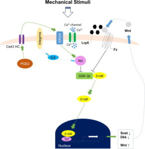
“Distribution of intramedullary pressure (ImP) induced bone fluid flow has been suggested to influence the magnitude of mechanotransductory signals within bone. As osteocytes have been suggested as major mechanosensors in bone network, it is still unclear how osteocytes embedded within a mineralized bone matrix respond to the external mechanical stimuli derived from direct coupling of dynamic fluid flow stimulation (DFFS). While in vitro osteocytes show unique Ca(2+) oscillations to fluid shear, the objective of this study was to use a confocal imaging technique to visualize and quantify Ca(2+) responses in osteocytes in situ under DFFS into the marrow cavity of an intact ex vivo mouse femur. This study provided significant technical development for evaluating mechanotransduction mechanism in bone cell response by separation of mechanical strain and fluid flow factors using ImP stimulation, giving the ability for true real-time imaging and monitoring of bone cell activities during the stimulation. Loading frequency dependent Ca(2+) oscillations in osteocytes indicated the optimized loading at 10Hz, where such induced response was significantly diminished via blockage of the Wnt/β-catenin signaling pathway. The results provided a pilot finding of the potential crosstalk or interaction between Wnt/β-catenin signaling and Ca(2+) influx signaling of in situ osteocytes in response to mechanical signals. Findings from the present study make a valuable tool to investigate how in situ osteocytes respond and transduce mechanical signals, e.g. DFFS, as a central mechanosensor.”<-Our main concern isn’t Ca2+ oscillations as those are not likely to induce chondrogenesis directly although it’s still possible that it may have an impact or that Ca2+ could be complementary.

Changes in the pressure or velocity of bone fluid flow (BFF) act as a communication medium that connects external loading signals and internal cellular activities in bone, which ultimately regulate the bone remodeling process”<-can we manipulate this loading signals and cellular activities in such a way that we grow taller?

“DHS[Dynamic Hydraulic Stimulation] generated local ImP that acted independently from simultaneous bone strain.”

Here’s a diagram of how Calcium signaling may alter a cell.  So Ca2+ is anabolic but it doesn’t really seem to have a direct impact on what lineage a cell traverses(osteo- versus chondro-).

fluid flow stimuli

Interrelation between external oscillatory muscle coupling amplitude and in vivo intramedullary pressure related bone adaptation.

“Interstitial bone fluid flow (IBFF) is suggested as a communication medium that bridges external physical signals and internal cellular activities in the bone, which thus regulates bone remodeling. Intramedullary pressure (ImP) is one main regulatory factor of IBFF and bone adaptation related mechanotransduction. Our group has recently observed that dynamic hydraulic stimulation (DHS), as an external oscillatory muscle coupling, was able to induce local ImP with minimal bone strain as well as to mitigate disuse bone loss. The current study aimed to evaluate the dose dependent relationship between DHS’s amplitude, i.e., 15 and 30mmHg, and in vivo ImP induction, as well as this correlation on bone’s phenotypic change. Simultaneous measurements of ImP and DHS cuff pressures were obtained from rats under DHS with various magnitudes and a constant frequency of 2Hz. ImP inductions and cuff pressures upon DHS loading showed a positively proportional response over the amplitude sweep. The relationship between ImP and DHS cuff pressure was evaluated and shown to be proportional, in which ImP was raised with increases of DHS cuff pressure amplitudes. A 4-week in vivo experiment using a rat hindlimb suspension model demonstrated that the mitigation effect of DHS on disuse trabecular bone was highly dose dependent and related to DHS’s amplitude, where a higher ImP led to a higher bone volume. This study suggested that sufficient physiological DHS is needed to generate ImP. Oscillatory DHS, potentially induces local fluid flow, has shown dose dependence in attenuation of disuse osteopenia. ”

“Direct ImP measurements in rats under DHS over a frequency spectrum indicated that its optimal loading at 2Hz can generate maximum ImP of 14.48±3.10mmHg. On the other hand, dynamic components with large loading amplitudes typically result in pronounced osteogenic responses”

“a micro-cardiovascular pressure transducer was carefully inserted into the tibial marrow cavity through a 1mm drilled hole, and was then tightly sealed within the drilled hole”

” the observed ImP inductions were found to be positively proportional to DHS cuff pressure, namely, which indicates promising potential of DHS loading dose dependency on ImP related bone adaptation.”

“DHS loading at 2Hz between 28.41±10.50 mmHg and 33.75±10.69 mmHg cuff pressures can induce significant ImP increases. Coincided with these observations, our 4-week in vivo study showed that DHS loading at 2Hz with 15 mmHg and 30 mmHg dynamic pressure was able to mitigate disuse trabecular bone loss in a rat functional disuse model.”

“At baseline, normal heart beats generated approximately 1mmHg tibial ImP within the marrow cavity.”

Dynamic hydraulic fluid stimulation regulated intramedullary pressure.

“Physical signals within the bone, i.e. generated from mechanical loading, have the potential to initiate skeletal adaptation. Strong evidence has pointed to bone fluid flow (BFF) as a media between an external load and the bone cells, in which altered velocity and pressure can ultimately initiate the mechanotransduction and the remodeling process within the bone. Load-induced BFF can be altered by factors such as intramedullary pressure (ImP) and/or bone matrix strain, mediating bone adaptation. Previous studies have shown that BFF induced by ImP alone, with minimum bone strain, can initiate bone remodeling. However, identifying induced ImP dynamics and bone strain factor in vivo using a non-invasive method still remains challenging. To apply ImP as a means for alteration of BFF, it was hypothesized that non-invasive dynamic hydraulic stimulation (DHS) can induce local ImP with minimal bone strain to potentially elicit osteogenic adaptive responses via bone-muscle coupling. The goal of this study was to evaluate the immediate effects on local and distant ImP and strain in response to a range of loading frequencies using DHS. Simultaneous femoral and tibial ImP and bone strain values were measured in three 15-month-old female Sprague Dawley rats during DHS loading on the tibia with frequencies of 1Hz to 10Hz. DHS showed noticeable effects on ImP induction in the stimulated tibia in a nonlinear fashion in response to DHS over the range of loading frequencies, where they peaked at 2Hz. DHS at various loading frequencies generated minimal bone strain in the tibiae. Maximal bone strain measured at all loading frequencies was less than 8με. No detectable induction of ImP or bone strain was observed in the femur. This study suggested that oscillatory DHS may regulate the local fluid dynamics with minimal mechanical strain in the bone, which serves critically in bone adaptation. These results clearly implied DHS’s potential as an effective, non-invasive intervention for osteopenia and osteoporosis treatments.”

” bone fluid flow (BFF) with altered velocity or pressure acts as a communication media between an external load and the bone cells, which then regulate bone remodeling. In converse, discontinuous BFF can initiate bone turnover and result in osteopenia”<-Initiating bone turnover could be a good thing if bone structure inhibits anabolism so lowering the frequency may be key.

“DHS was achieved through a costume-made inflatable cuff placed around the right tibia”

” the inflation and deflation of the cuff was driven by a syringe pump with the loading magnitudes and frequencies delivered by a programmable waveform/signal generator”

” 1mmHg tibial ImP and 5mmHg femoral ImP were generated by normal heart beat within the marrow cavities.”

” muscle contraction compresses the blood vessels in muscle, which generates an arteriovenous pressure gradient that further increases the hydraulic pressure in skeletal nutrient vessels and amplifies the capillary filtration in bone”

” using oscillatory muscle stimulation (MS), in which oscillatory MS induced maximal ImP at 20Hz. Oscillatory MS was achieved via two disposable needle-sized electrodes inserted into the quadriceps of the stimulated rats. The electrodes were then connected to the waveform generator with 2V supplies to induce muscle contraction”

“Due to the different physical orientations of how oscillatory MS and DHS contact the loaded tissue, as well as the different material densities and viscosities within hard and soft tissues, maximal DHS-induced ImP may result at relatively lower frequency compared to MS.  Direct hydraulic coupling may influence bone adaptation in a more physiological frequency range, where normal heart rate is 360 times per minute.”

“Changes of MSC number in response to DHS bias their differentiation towards osteoblastogenesis, leading to bone formation even under disuse conditions”

“increased bone fluid pressure and bone fluid flow regulation are strongly correlated, our current results of DHS-driven ImP induction suggest a possible mechanism that the induced ImP may subsequently enhance bone fluid flow.”