Author Archives: Tyler

Quantifying bone adaptations then and now: Breakthrough from 1920 study(paget’s disease)

Is some of the recent scientific advancements hiding key features that may give the secrets to growth?  Compare this recent study to a 1920s study that provides an insight about adult longitudinal bone growth related to bone softening.

Development of a protocol to quantify local bone adaptation over space and time: Quantification of reproducibility.

In vivo micro-computed tomography (µCT) scanning of small rodents is a powerful method for longitudinal monitoring of bone adaptation{May be helpful to measure LSJL?}. However, the life-time bone growth in small rodents makes it a challenge to quantify local bone adaptation. Therefore, the aim of this study was to develop a protocol, which can take into account large bone growth, to quantify local bone adaptations over space and time. The entire right tibiae of eight 14-week-old C57BL/6J female mice were consecutively scanned four times in an in vivo µCT scanner using a nominal isotropic image voxel size of 10.4µm. The repeated scan image datasets were aligned to the corresponding baseline (first) scan image dataset using rigid registration. 80% of tibia length (starting from the endpoint of the proximal growth plate) was selected as the volume of interest and partitioned into 40 regions along the tibial long axis (10 divisions) and in the cross-section (4 sectors). The bone mineral content (BMC) was used to quantify bone adaptation and was calculated in each region. All local BMCs have precision errors (PE%CV) of less than 3.5% (24 out of 40 regions have PE%CV of less than 2%), least significant changes (LSCs) of less than 3.8%, and 38 out of 40 regions have intraclass correlation coefficients (ICCs) of over 0.8. The proposed protocol allows to quantify local bone adaptations over an entire tibia in longitudinal studies, with a high reproducibility, an essential requirement to reduce the number of animals to achieve the necessary statistical power. ”

“in rodents like mouse, bone growth spans across the animal׳s life time”

Here’s a 1926 study regarding the regeneration of bone:

THE REGENERATION OF BONE

An early theory of the bone: “the “juice” of bone is released by a fracture,
fills the gap between the ends and there solidifies.”

“The excision of a necrotic bone (the modern sequestrectomy) was more than once reported with complete reconstitution of the affected bone.”

“bone regeneration depends upon the periosteum.  the thickening of this membrane and its later consolidation into bone in experimental fractures and drill holes.”

The four main elements of adult bone: bone cells; periosteal cells; endosteal cells; calcified matrix.

The marrow cavity of a bone(that which contains the juice) is broken up by the trabeculae of spongy bone.

“the bony shell or cortex, we find a definite architecture. On the inner and outer surfaces, the matrix appears in layers lying parallel to the surface. Further in, these layers lose the parallel arrangement and are there grouped in a series of concentric circles. In the center of each of these circles is an opening, the haversian canal, containing a blood vessel and lined with a membrane.  Throughout the substance of the matrix between these canals and in the parallel layers are numerous smaller openings, the lacunae, in which lie the bone cells.”

“Enveloping the outer aspect of the bone is the periosteum, a membrane consisting of several layers of cells resembling connective tissue cells, with an elongated nucleus and abundant fibrillar intercellular substance.”

“solid bone will not grow in length from within by expansion. The calcified matrix cannot expand. Whatever growth takes place must take place by accretion.”<-it’s possible to grow by accretion on the longitudinal ends.

“Completely calcified bone will not grow interstitially but soft bone will grow interstitially. The experiment just described, if done on a young bone not yet hard, will result in separation of the nails. This is an observation which has a curious reference to adult pathology. Bones in certain pathologic states that render them soft, will occasionally increase in length long after the epiphyseal cartilages are united. This growth is undoubtedly a genuine interstitial addition of new bone, a true expansion of the matrix exactly comparable to such growth in young bones. The commonest conditions in which this phenomenon is encountered are osteomyelitis, Paget’s disease of bone (osteitis deformans) and Recklinghausen’s disease (neurofibromatosis).
all of which result in a certain degree of decalcification, that is a softening of the bone”

“bones assume a shape adapted to the function they are to perform.”

“Suppose a small drill passed through the cortex of a bone into the marrow cavity. The first effect, of course, is hemorrhage. On withdrawing the drill, the defect fills with blood. Within a few hours this clot begins to be replaced by a plug of fibrin containing polymorphonuclear leucocytes. Fibrin is the culture medium, so to speak, of young tissue. Then appears a proliferation of the periosteal and endosteal cells, the production of periosteal and endosteal fibroblasts which differ from connective tissue fibroblasts only in their inherent capability of producing bone. These proliferating cells heap up at the edges of the defect, producing a macroscopic thickening of the periosteum and endosteum, and then grow down and up respectively into the fibrin that fills the defect, until it is completely replaced. The periosteal and endosteal reaction extends on the surfaces of the bone over a considerable distance from the point of injury.”

“Before the process of fibrosis is complete, ossification begins at the points where the periosteal and endosteal proliferation began, namely over the surfaces of the bone at the edges of the defect.”

“when a foreign body is brought into contact with bone, one must anticipate that the regenerative processes will be diminished and retarded and that actual bone destruction may occur.”

A discussion attached to the end of the paper suggests that periosteums function merely is a membrane that constrains the bone and that it’s the fluid within the bone that results in bone regeneration.  The periosteum also serves as a mechanism of communication with other tissues.  The commenter stated that the periosteum only appears after the bone has taken place.

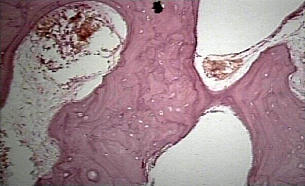
Here’s the bone histology for Paget’s disease:

paget's disease bone histology

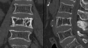
Here’s Paget’s disease in an x-ray of the spine:
pagets disease spine

Here’s someone with paget’s disease in the arm:
pagets disease arm

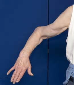
Paget’s disease can make bones longer but not in a real symmetrical way so it’s not really appealing.

Paget’s disease is a disease where bone undergoes break down and rebuilding much faster than normal(so basically faster bone remodeling cycle) and this results in weaker bones as bone does not have time to develop strength.  If there was a way that Paget’s disease was found to increase height we could mimic the effects somewhat by increasing the amount of bone remodeling that occurs.

Here’s more info on paget’s disease:

Clinical manifestations and diagnosis of Paget disease of bone

“Paget disease of bone (PDB), also known historically as osteitis deformans, is a focal disorder of bone metabolism that occurs in the aging skeleton; it is characterized by an accelerated rate of bone remodeling, resulting in overgrowth of bone at single (monostotic PDB) or multiple (polyostotic PDB) sites and impaired integrity of affected bone. Commonly affected areas include the skull, spine, pelvis, and long bones of the lower extremity.”

” Its onset is typically after age 55″<-so we could theoretically apply whatever is causing paget’s disease to normal healthy bone to make it grow longitudinally.

“Paget disease of bone (PDB), which is characterized by an accelerated rate of bone remodeling resulting in overgrowth of bone at selected sites and impaired integrity of affected bone, is believed to be a disease of the osteoclast. Osteoclasts are the only cell known to resorb bone. Osteoclast differentiation requires pathways involving receptor activator for nuclear factor kappa-B ligand (RANKL) and macrophage colony-stimulating factor (MCSF), which are both necessary for osteoclast activation. RANKL binds to its receptor, RANK, on osteoclast precursors to promote osteoclast differentiation and activation via activation of nuclear factor kappa-B (NFkB)-dependent pathways. Osteoclast differentiation is inhibited by osteoprotegerin (OPG; a soluble decoy receptor for RANKL) and further modulated by cytokines and hormones. It is not known why some areas of bone are affected while others are not.”<-osteoclasts are the key to growing taller as you need to break down bone in order for new growth to occur as bone inhibits growth.

“The osteoclasts in pagetic bone demonstrate an unusual appearance, with a disproportionate number of osteoclasts with too many nuclei.”

“When symptoms [from Paget’s disease] do arise, these are usually due to overgrowth, deformity, or pathologic fracture of the affected bone at a given skeletal location.”

“Enlargement of the skull, which can occur after many years of disease”<-the skull can make you taller if it grows in the right way.

“Long bones – Bowing deformities in the long bones and early arthritis of affected joints are common, with a heightened risk of fracture over the lifetime of the individual. The deformities are caused by enlarging and abnormally contoured bones, which result in bowing”<-bowing makes you shorter not taller.

Osteomylitis seems caused by infection which would be hard to manage.

Neurofibromatosis can cause overgrowth of specific extremeties related to NF1 and NF2 mutations.

New study shows LSJL induces Bone Deformation

Bone Deformation is change in the bone shape or structure.  This deformation can be compression of various cavities, stretching of the bone, twisting, and so son.  This is important as bone deformation is one way to increase hydrostatic pressure by decreasing the cavity size.  Hydrostatic pressure is the pressure exerted by a fluid at rest.   Compressing the bone laterally inhibits the fluids ability to move thus increasing hydrostatic pressure.   If there is more fluid within a smaller space than it follows that hydrostatic pressure increases. Hydrostatic pressure has been consistently shown to induce chondrogenic differentiation.  Chondrogenic tissue is the key for longitudinal bone growth as traditionally chondrogenic tissue is capable of interstitial(growth from within) whereas bone is not.  Only interstitial bone growth has been shown traditionally to induce significant longitudinal bone growth but there are potentially other ways to stimulate longitudinal bone growth.

Knee loading inhibits osteoclast lineage in a mouse model of osteoarthritis.

“Osteoarthritis (OA) is a whole joint disorder that involves cartilage degradation and periarticular bone response. Changes of cartilage and subchondral bone are associated with development and activity of osteoclasts from subchondral bone{Since osteoarthritis does affect the subchondral bone that does affect our ability to say that LSJL affects bone deformation in a normal bone but there is no reason why it shouldn’t}. Knee loading promotes bone formation. Knee loading regulates subchondral bone remodeling by suppressing osteoclast development, and prevents degradation of cartilage through crosstalk of bone-cartilage in osteoarthritic mice{This “crosstalk” may stimulate chondral tissue within the bone as well}. Surgery-induced mouse model of OA was used. Two weeks application of daily dynamic knee loading significantly reduced OARSI scores and CC/TAC (calcified cartilage to total articular cartilage), but increased SBP (subchondral bone plate) and B.Ar/T.Ar (trabecular bone area to total tissue area). Bone resorption of osteoclasts from subchondral bone and the differentiation of osteoclasts from bone marrow-derived cells were completely suppressed by knee loading{Knee loading affects the differentiation of bone marrow-derived cells which is the first step in proving that it causes chondrogenic differentiation}. The osteoclast activity was positively correlated with OARSI scores and negatively correlated with SBP and B.Ar/T.Ar. Furthermore, knee loading exerted protective effects by suppressing osteoclastogenesis through Wnt signaling. Overall, osteoclast lineage is the hyper responsiveness of knee loading in osteoarthritic mice. Mechanical stimulation prevents OA-induced cartilage degeneration through crosstalk with subchondral bone. Knee loading might be a new potential therapy for osteoarthritis patients.”

“Daily dynamic knee loading was applied at 1 N, 5 Hz, 5 min/day for 2 weeks”

joint loading on subchondral bone

Compare the OA+ loading to the control bone.  The subchondral bone plate looks much more dynamic.  There are three bone marrow regions rather than two(bone marrow is the blue dots).

You’ll also note that loading+OA increased the ratio of calcified cartilage out of total articular cartilage(but not above statistic significance.  It did not fully restore the thickness of the bone plate.  Alendronate is an anti reobsorption agent.  Given that the ALN and loading group is different we can say that change in subchondral bone shape is likely not related to inhibiting osteoclast activity and is something unique to the loading group.

loading affect on subchondral bone2

Compare the joint capsule region of control and OA+loading group.  The Joint Capsule is the region that’s not inside the bone.  The cells are a lot more spread out.  There’s a dense redness in the control group which is not present in the OA+Loading group

We can see that the loaded group again has distinct characteristics and we can also see the growth plate.  The growth plate of the LSJL group is distinct and there does seem to be signs of cellular migration.  I’ll have to blow it up.

cell migration

Circled is the region of possible cell migration.

LSJL-growth-plates

Similar signs of migration in earlier in LSJL studies(above taken from Lengthening of Mouse Hindlimbs with Joint Loading).

“the expression of Wnt3a was significantly increased by knee loading. However, the protein and mRNA levels of NFATc1, RANKL, TNF-α, and Cathepsin K were significantly suppressed by knee loading”

“Female C57BL/6 mice (~14 weeks of age)”

If you look at this image of bone marrow derived cells extracted from the loaded group and the other groups you can see that the cells are more condensed and condensation is a prerequisite for chondrogenic differentiation,

Loaded cells are more condensed

This is an image of what mesenchymal condensation looks like:

mesenchymal condensation

Finger clamping update

So I’ve been hand clamping the proximal part of my finger and it looks like there may be some significant results.  I’d have to validate with x-rays.  Here’s the last finger clamping results.

So now I’m going to try a few things.  i’m going to clamp the proximal part of my finger once more.  And I’m going to start clamping my thumbs.  The left thumbs is a little bit longer than the right due to previous LSJL experimentation so I’m going to start clamping my right to see if I can get my right thumb equal and perhaps surpass my right.

I’ve also been hand clamping on my wrists, elbows, knees, and ankles.  There may be possible results or it could be mind trickery.  I’ll keep trying.  Hand clamping does seem to be working better than quick grip clamping possibly due to the fact it is more precise and you can feel the deformation of bone.

Below is comparison images of my right(hand clamped) versus left index finger.  They are aligned based on the middle lines of the finger due to the fact that it is much harder to align on the base of the finger.  Also is the before image of the thumbs.  I’ll start by clamping the medial joint of the right thumb.

20160419_155041

20160419_155106

Finger pulling/clamping progress update

Here is the last finger clamping results.  In that I was clamping the distal point of the right pinky finger and now I am clamping the medial region.  I have abondoned the pulling motion as it seemed to be more the hand clamping that was generating my results.  Now I’m still doing LSJL using the Irwin Quick Grip clamp and one of the areas I clamp includes my left index finger with the Irwin Quick Grip.  I have not seen the changes with the quick grip clamping that I have with hand clamping.

A number of people doing LSJL reported that they felt it was more effective to use your hands to manually generate pressure.  This may indicate that there may be a deficiency in the Irwin Quick Grip and that an alternative clamping method may be needed.  That will be something that I am exploring.

Right now I’m going to clamp the base of the proximal end of the finger like so:

proximal finger pinch

Then I’ll see if I can pull out a little more growth.  There does seem to be some kind of conditioning effect where the body becomes more resistant to clamping.

Below is the progress pic.  I try to align based on the bottom based on the middle part of the finger because it’s extremely difficult to align based on where the proximal finger begins.  Now this image isn’t going to prove anything.  I’m going to need x-rays or a lot more significant growth.  I’ll see how the finger base clamping goes.

pinkyfinger growth progress


Axial loading devices at physiological loads can be helpful with the right stimulus

Axial loading can help with growth if the right stimulus is in place namely existing remodeling conditions.

Effects of mechanical loading on cortical defect repair using a novel mechanobiological model of bone healing.

Mechanical loading is an important aspect of post-surgical fracture care. The timing of load application relative to the injury event may differentially regulate repair depending on the stage of healing. Here, we used a novel mechanobiological model of cortical defect repair that offers several advantages including its technical simplicity and spatially confined repair program, making effects of both physical and biological interventions more easily assessed. Using this model, we showed that daily loading (5N peak load, 2Hz, 60 cycles, 4 consecutive days) during hematoma consolidation and inflammation disrupted the injury site and activated cartilage formation on the periosteal surface adjacent to the defect. We also showed that daily loading during the matrix deposition phase enhanced both bone and cartilage formation at the defect site, while loading during the remodeling phase resulted in an enlarged woven bone regenerate. All loading regimens resulted in abundant cellular proliferation throughout the regenerate and fibrous tissue formation directly above the defect demonstrating that all phases of cortical defect healing are sensitive to physical stimulation. Stress was concentrated at the edges of the defect during exogenous loading, and finite element (FE)-modeled longitudinal strain (εzz) values along the anterior and posterior borders of the defect (~2200με) was an order of magnitude larger than strain values on the proximal and distal borders (~50-100με){2000 is within physiological microstrain}. It is concluded that loading during the early stages of repair may impede stabilization of the injury site important for early bone matrix deposition, whereas loading while matrix deposition and remodeling are ongoing may enhance stabilization through the formation of additional cartilage and bone.”

“Compressive axial loading (100 cycles/day, 1 Hz, 5 days per week for 2 weeks at 0.5 N, 1 N,
and 2 N peak load) was applied across the flexed knee and ankle immediately after fracture
or after a 4-day delay, which coincided with the hematoma and inflammation stages”<-This is axial loading in contrast to lateral loading.

” femoral segmental defects subjected to daily cyclic bending (900 cycles, 1Hz, 15 min/day for 5 consecutive days per week for 1, 2 or 4 weeks) beginning on post-surgical day 10, which coincided with a provisional matrix scaffold, led to formation of pseudarthrosis with enhanced cartilage formation”<-pseudoarthrosis is a fracture that won’t heal properly.

“In sum, loading produces a strain field around the defect that is high on the anterior and posterior borders and low on the proximal and distal borders”

” Daily loading during the inflammatory phase (PSD 2 to 5) delays hematoma clearance and bone matrix deposition, stimulates cellular proliferation and osteoclast activity, and promotes cartilage formation.”

” Proliferating cells were observed within the defect at all time points post-loading and within the elevated periosteum and surrounding cartilage nodules suggesting that loading activated proliferation even when strains were relatively low (50-100με). ”

“low stress and strain lead to direct intramembranous bone formation, compressive stress and
strain lead to chondrogenesis, and high tensile strain leads to fibrous tissue formation”

Functional in situ assessment of human articular cartilage using MRI: a whole-knee joint loading device.

“The response to loading of human articular cartilage as assessed by magnetic resonance imaging (MRI) . An MRI-compatible whole-knee joint loading device for the functional in situ assessment of cartilage was developed and validated in this study. A formalin fixed human knee was scanned by computed tomography in its native configuration and digitally processed to create femoral and tibial bone models. The bone models were covered by artificial femoral and tibial articular cartilage layers in their native configuration using cartilage-mimicking polyvinyl siloxane. A standardized defect of 8 mm diameter was created within the artificial cartilage layer at the central medial femoral condyle, into which native cartilage samples of similar dimensions were placed.  After describing its design and specifications, the comprehensive validation of the device was performed using a hydraulic force gauge and digital electronic pressure-sensitive sensors. Displacement controlled quasi-static uniaxial loading to 2.5 mm (δ2.5) and 5.0 mm (δ5.0) of the mobile tibia versus the immobile femur resulted in forces of 141±8N(δ2.5) and 906±38 N (δ5.0) (on the entire joint)and local pressures of 0.680±0.088MPa (δ2.5) and 1.050±0.100 MPa (δ5.0) (at the site of the cartilage sample). Upon confirming the MRI compatibility of the set-up, the response to loading of macroscopically intact human articular cartilage samples (n = 5) was assessed on a clinical 3.0-T MR imaging system using clinical standard proton-density turbo-spin echo sequences and T2-weighted multi-spinecho sequences. Serial imaging was performed at the unloaded state (δ0) and at consecutive loading positions (i.e. at δ2.5 and δ5.0). Biomechanical unconfined compression testing (Young’s modulus) and histological assessment. All samples were histologically intact(Mankinscore,1.8±1.3)and biomechanically reasonably homogeneous (Young’s modulus, 0.42 ± 0.14 MPa). They could be visualized in their entirety by MRI and significant decreases in sample height [δ0:2 .86±0.25mm; δ2.5:2 .56±0.25mm; δ5.0:2 .02±0.16mm; p < 0.001 (repeated-measures ANOVA)] as well as pronounced T2 signal decay indicative of tissue pressurization were found as a function of compressive loading. In conclusion, our compression device has been validated for the noninvasive response-to-loading assessment of human articular cartilage by MRI in a close-to-physiological experimental setting. Thus, in a basic research context cartilage may be functionally evaluated beyond mere static analysis and in reference to histology and biomechanics”

“In terms of hydration, compressive loading most likely induced considerable water redistribution within and possibly out of the tissue.”

Breakthrough-Genetically Engineered Tomatos for Height Increase

This study provides evidence that supplementation of Quercetin, Kaempferol, and Rutin may affect height during development but you can’t be sure until it’s tested.  The method by which it induces height growth is by increasing the expansion of the bone lacunae per hypertrophic chondrocyte generating more bang for the buck.

Genetically engineered flavonol enriched tomato fruit modulates chondrogenesis to increase bone length in growing animals.

“Externally visible body and longitudinal bone growth is a result of proliferation of chondrocytes[not necessarily; chondrocyte hypertrophy plays a large role]. In growth disorder, there is delay in the age associated increase in height[not always]. The present study evaluates the effect of extract from transgenic tomato fruit expressing AtMYB12 transcription factor on bone health including longitudinal growth. Constitutive expression of AtMYB12 in tomato led to a significantly enhanced biosynthesis of flavonoids in general and the flavonol biosynthesis in particular. Pre-pubertal ovary intact BALB/c mice received daily oral administration of vehicle and ethanolic extract of wild type (WT-TOM) and transgenic AtMYB12-tomato (MYB12-TOM) fruits for six weeks. Animal fed with MYB12-TOM showed no inflammation in hepatic tissues and normal sinusoidal Kupffer cell morphology. MYB12-TOM extract significantly increased tibial and femoral growth and subsequently improved the bone length as compared to vehicle and WT-TOM. Histomorphometry exhibited significantly wider distal femoral and proximal tibial growth plate, increased number and size of hypertrophic chondrocytes in MYB12-TOM which corroborated with micro-CT and expression of BMP-2 and COL-10, marker genes for hypertrophic cells. We conclude that metabolic reprogramming of tomato by AtMYB12 has the potential to improve longitudinal bone growth thus helping in achievement of greater peak bone mass during adolescence.”

What’s significant is that this improves bone growth in normal individuals.

“the proliferative zone contains replicate chondrocytes arranged in columns parallel to the long axis of the bone”

“the proliferative chondrocytes located farthest from the resting zone stop replicating and enlarge to become hypertrophic chondrocytes which subsequently form bone. these cells also maintain the columnar alignment in the hypertrophic zone.”

“growing female mice fed with extract from MYB12-TOM affected formation, quality and length of the bone. Extract from MYB12-TOM significantly increased the length by interstitial growth of the epiphyseal plate of bones by the expansion of the lacunae in the hypertrophic cells in pre-pubertal stage.”

“genetically engineered transgenic tomatoes (MYB12-TOM) expressing a flavonol specific transcription factor from Arabidopsis, AtMYB1214. The fruits of MYB12-TOM accumulated significantly higher amount of flavonols as compared to wild type tomatoes (WT-TOM).”

“expansion of the lacunae in the hypertrophic cells leads to the increased longitudinal growth”

The genetically altered tomatoes had much higher levels of Quercetin, Kaempferol, and Rutin than normal,

“Extracts from MYB12-TOM increased the femur length significantly by 6.1%as compared to WT-TOM. In case of tibia, WT-TOM and MYB12-TOM increased the bone length by 5.54% and 9.98% as compared to control group respectively. Further, comparisons within experimental groups show that MYB12-TOM increased tibial length significantly by 4.2% as compared to WT-TOM group”

So it may be possible that Quercetin, Kaempferol, and Rutin supplements to have a significant impact on height during development.

“transgenic MYB12-TOM group exhibited significantly higher COL10a expression from control group  and WT-TOM”