Author Archives: Tyler

New Experiment-Finger Pulling

toddler hands

Correlation does not imply causation.  Just because the growth plates disappear correlates with the near cessation of longitudinal bone growth does not mean that the disappearance of growth plates causes the cessation of longitudinal bone growth.  Maybe a uniform factor causes both the disappearance of growth plates and cessation of growth.  Maybe the cessation of growth causes the disappearance of growth plates.  May another factor ceases both.

I’m testing the hypothesis that maybe it’s the joints themselves that place the constraint on longitudinal bone growth.  If you look at the above image of a toddlers hands you notice the near absence of joints/knuckles.  One of the main difference between adult and childs bones is that adults have much prominent epiphysis.  LSJL attempts to stimulate that epiphysis in one way. What if the epiphysis itself constrains growth. If you look at the image below by pulling my finger that I’m sort of creating the knuckle gap that’s like the child hand.

20160104_101759

So the test is to see if by pulling the finger that we can alleviate the potential constraints on growth.  I can’t do it 24/7 and if it does constrained growth there should only be a need for certain periods of time where growth is unconstrained by the epiphysis.  So I’m going to be pulling my right pinky finger for 15 minutes.  I need time to perform LSJL too.  All my left fingers are longer than my right and there are some images below so if my right pinky becomes longer than my left.

We can only really draw conclusions if this works.  If this works then we can see that it is the epiphysis itself that is the sole constraint on longitudinal bone growth and that by pulling the finger for enough time during the day we allow for growth to occur.  If it doesn’t work then we can’t really say anything.  Maybe I didn’t do it for enough time or maybe you need to stimulate neo-growth plate formation in addition to preventing the epiphysis’ from constraining growth.

If this works it also bodes well for growing taller via spinal height as you can stretch those as well.  However, usually when you stretch in one way you usually compress another and any activity involving the spine is more dangerous due it’s important bodily role.

This may sound crazy.  How can it possibly work?  I just saw a toddlers hands and noticed that gap where joints could be.  Then I pulled my finger and saw the gap sort of made my hand more toddler like.  The logical stretch is that how can you possibly grow without stimulating growth plates.  Well, every single field of science is understudied.  Correlation does not apply causation.  The hypothesis being tested here is:

The epiphysis puts a constraining factor on longitudinal bone growth by pulling the finger apart you allow for release from that factor temporarily allowing for possible new longitudinal bone growth to occur.

20160104_101624 20160104_101713

LSJL Update-12/10/15

I recent did some LSJL measurements and I didn’t have any significant increase in length.  I think the reason for this is that I wasn’t clamping long enough.  During the last few months I’ve been clamping hard so that I stop clamping sooner due to pain and can get it over with.  However, I don’t think that is enough time to build up hydrostatic pressure.  This is contrast to the method I post recently that had a very long clamping time.  I think that my attempt to shortcut the process by getting into more intense clamping faster denied me of results.  Slower clamping also allows for the surrounding muscles and tendons to adapt to the clamping meaning an eventual more intense clamping force before they start resisting.

Given that mesenchymal stem cells can differentiate into chondrocytes via hydrostatic pressure it’s very likely that their is an epithelial intermediary to allow for chondrogenic differentiation and epithelial cells are important to form the resting zone of the growth plate.  LSJL tries to increase hydrostatic pressure.  Although response to hydrostatic pressure may be dependent on cell matrix interactions.

I do have strong faith in LSJL given wingspan increase results and possible finger length increase.  I performed LSJL on my right finger.  My right finger appeared longer than my left.  I got X-rays and after exhaustive analysis all my left finger bones were in fact longer except for my right metacarpal.  And upon observation most of my left hand bones do appear to be longer than my right.  Thus, the difference in size between the right and left metacarpal is very likely due to LSJL.

So now I will be performing LSJL on my left index finger to try to get the left metacarpal longer than my right and I will be ramping up the clamping of other joints that I perform LSJL on more slowly and longer duration.

Factors that alter the bone microenvironment

Here’s a review paper on factors that alter the bone microenvironment.  There’s a lot of stuff here.  More than is mentioned.

 Biomechanical and biophysical environment of bone from the macroscopic to the pericellular and molecular level.

” Bones with complicated hierarchical configuration and microstructures constitute the load-bearing system. Mechanical loading plays an essential role in maintaining bone health and regulating bone mechanical adaptation (modeling and remodeling). The whole-bone or sub-region (macroscopic) mechanical signals, including locomotion-induced loading and external actuator-generated vibration, ultrasound, oscillatory skeletal muscle stimulation, etc., give rise to sophisticated and distinct biomechanical and biophysical environments at the pericellular (microscopic) and collagen/mineral molecular (nanoscopic) levels, which are the direct stimulations that positively influence bone adaptation. While under microgravity, the stimulations decrease or even disappear, which exerts a negative influence on bone adaptation. A full understanding of the biomechanical and biophysical environment at different levels is necessary for exploring bone biomechanical properties and mechanical adaptation. In this review, the mechanical transferring theories from the macroscopic to the microscopic and nanoscopic levels are elucidated. First, detailed information of the hierarchical structures and biochemical composition of bone, which are the foundations for mechanical signal propagation, are presented. Second, the deformation feature of load-bearing bone during locomotion is clarified as a combination of bending and torsion rather than simplex bending. The bone matrix strains at microscopic and nanoscopic levels directly induced by bone deformation are critically discussed, and the strain concentration mechanism due to the complicated microstructures is highlighted. Third, the biomechanical and biophysical environments at microscopic and nanoscopic levels positively generated during bone matrix deformation or by dynamic mechanical loadings induced by external actuators, as well as those negatively affected under microgravity, are systematically discussed, including the interstitial fluid flow (IFF) within the lacunar-canalicular system and at the endosteum, the piezoelectricity at the deformed bone surface, and the streaming potential accompanying the IFF. Their generation mechanisms and the regulation effect on bone adaptation are presented. The IFF-induced chemotransport effect, shear stress, and fluid drag on the pericellular matrix are meaningful and noteworthy. Furthermore, we firmly believe that bone adaptation is regulated by the combination of bone biomechanical and biophysical environment, not only the commonly considered matrix strain, fluid shear stress, and hydrostatic pressure, but also the piezoelectricity and streaming potential. Especially, it is necessary to incorporate bone matrix piezoelectricity and streaming potential to explain how osteoblasts (bone formation cells) and osteoclasts (bone resorption cells) can differentiate among different types of loads. Specifically, the regulation effects and the related mechanisms of the biomechanical and biophysical environments on bone need further exploration, and the incorporation of experimental research with theoretical simulations is essential.”

“Osteocyte processes are attached to the canalicular wall via transverse tethering elements
(the average spacing between the elements is 32 ± 14 nm) and only directly contact the canalicular wall at specialized collagen fibril protrusions ”

“In trabecular bone, the trabecular rods and plates form three-dimensional structures, and within the trabeculae are less regular arranged lamellae and osteocytes”

“The organic material, which is primarily type I collagen (90%) and nonfibrillar organic matrix
(osteocalcin and osteopontin present in a large proportion) gives the bone toughness. The mineral salts, mainly nanocrystallite apatite materials, permeate the organic matrix and provide the characteristic rigidity and strength of bone. Common belief holds that the apatite materials are hydroxyapatite [Ca10(PO4)6(OH)2] (HAP). In fact, there is no OH-band in bone mineral detected in a Raman spectrum, therefore the bone apatite is not hydroxyapatite ”

“in two adults tibiae during comfortable walking and strenuous running; the results suggested that the compressive strain ranged from approximately 400 to 1300 µε, and the tensile strain ranged from approximately 380 to 750 µε ”

“The pericellular space in the lacunar-canalicular system (LCS) of cortical bone and the porous
structures within trabecular bone are filled with interstitial fluid. Due to the low permeability of mineralized bone matrix, interstitial fluid flow (IFF) is principally generated during alteration of intramedullary pressurization (ImP) and bone matrix deformation

“Pulsatile extravascular pressure resulting from blood circulation can also drive IFF, but it is much smaller than that induced by ImP and bone deformation “<- so maybe muscular contraction is not as important as thought and that’s why bodybuilders have not been shown to grow taller as a result of bodybuilding.

“The IFF generated from ImP usually follows two spatially distinct flow profiles: IFF within the
LCS and at the endosteal surface.  Uniform pressurization [such as that due to decreased intramedullary cavity resulting from elevated bone marrow lipids induced by high level of corticosteroid administration will generate radial flow from the intramedullary compartment to the endosteal surface and into the LCS due to the pressure gradient from the marrow cavity to the bone matrix. Non-uniform pressure gradients within the intramedullary cavity [such as those due to local heterogeneous permeability or fluid displacement changes in the intramedullary compartment from the interaction between mechanical loading/oscillatory muscle stimulation and capillary filtration in bone tissue will cause tangential fluid flow to the endosteal surface ”

“Because the mineralized matrix is nearly impermeable{This is partially why it is so hard to grow taller} and the pericellular annular fluid space surrounding the osteocyte cell body and processes is narrow (gap < 1 µm) , the transportation of nutrients (e.g., glucose), oxygen, waste products (e.g., lactic acid), and regulatory signals (e.g., sex hormones, nitric oxide, prostaglandins, cytokines, and growth factors) by simple diffusion is not sufficient to maintain cell viability and function IFF within the LCS serves as the primary transport mechanism between the blood supply and osteocytes”

“hydrostatic pressure is induced by the bone compression that results from physiological loading. Based on the data that walking at a frequency of 1 Hz induces bone strain of 1000 µε and 0–18 MPa cyclic stress{this is surprisingly high},   the maximum hydrostatic pressure in LCS is approximately 0.27 MPa{this is actually within the chondrogenic range of 0.1-10MPa; there must be additional factors going on}, this pressure is estimated to be 12% of the applied axial stress and 40 times higher than that in the vasculature”

” the compressed areas develop negative potentials, whereas the tensioned areas develop positive potentials. These changes are known as the direct piezoelectric effect ”

“The researchers subjected demineralized type I collagen to bending deformation and bathed the collagen in continually flowing physiologic fluid over a period of weeks. Interestingly, the compressed internal surface (negative) of the collagen attracted calcium ions (positive), leading to the subsequent nucleation and crystallization of apatite and an increase in precipitate. In contrast, the tensioned portion (positive) of the collagen showed no difference in precipitate compared with that in the undeformed collagen. ”

“Apatite is piezoelectric by nature ”

“dry collagen is piezoelectric, but fully hydrated collagen is not piezoelectric because the structured water may increase the symmetry of the collagen molecule”

” In the presence of apatite, access of water to parts of the collagen fibril and the collagen swelling to its fully hydrated form can be prevented, thus the collagen can show some piezoelectricity even when the bone is fully hydrated ”

” the negative and positive potentials caused by polarization can be stored in apatite for years ”

“osteons are irregular cylinders always with complex branching and interconnections”

“The strain-induced movement of interstitial fluid through bone porosities can induce another
bioelectrical potential in addition to piezoelectricity; this potential is called the streaming potential ”

“At the interface of the bone matrix and interstitial fluid, negative charges accumulate on the bone surface due to the electronegativity of collagen; these charges attract positive ions in the liquid, and the positive ions concentrate near the interface, forming an electrical double layer.  The electric potential in the interfacial double layer relative to the interstitial fluid away from the interface is the zeta potential (ζ), and when interstitial fluid flows tangentially along the interface under pressure, a streaming potential appears along the flow direction. The zeta potential is mainly generated by deformed collagen and corresponds to the streaming potential ”

” the body fluid shifts[during space flight], and perfusion is diminished in the lower limbs and increased in the upper limbs and head ”

“ultrasound may primarily exert an effect on increasing blood flow and producing micromechanical strains; the actual acoustic intensity experienced by the bone cells at the target site might be lower due to the possible attenuation caused by the overlying soft tissue ”

“[Oscillatory muscle stimulation] at 20 Hz generated a maximum ImP and a relative high bone strain”

Understanding Mesenchymal to Epithelial cell transition may be key for neo-growth plates

This study is huge because we can get adult epithelial cells to potentially form new growth plates.  Since the resting zone is composed of mainly endothelial-like cells and the resting zone is the foundation of the growth plate.  Understanding how to transition adult mesenchymal stem cells into endothelial cells may be the basis for forming a new growth plate.

Epithelial-mesenchymal transition and mesenchymal-epithelial transition response during differentiation of growth-plate chondrocytes in endochondral ossification.

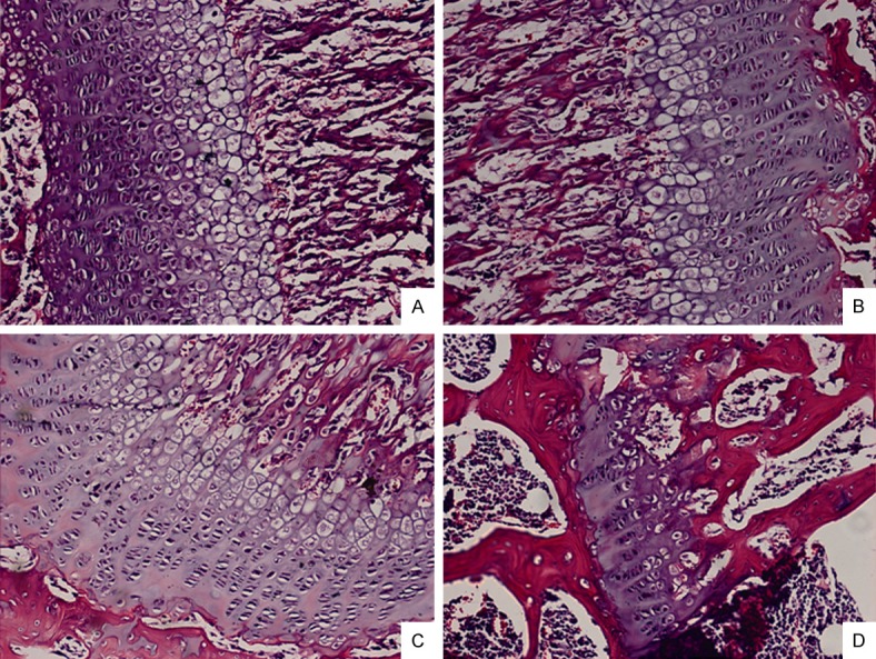
“For linear longitudinal bone elongation, the stem-like progenitor chondrocytes distributed in resting zone (RZ) of growth plate have a capacity to differentiate towards the spindle chondrocytes in proliferative zone (PZ), then towards the columnar and tightly adjacent chondrocytes in hypertrophic zone (HZ). We hypothesized this process of endochondral ossification with cells morphological change was occurred along with the inter-conversion between epithelial to mesenchymal cell types. Consistent with this hypothesis, the chondrocytes highly expressed mesenchymal-like biomarkers and loss of epithelial surface markers in PZ, while converse in RZ and HZ of the growth plate in mice distal tibia in vivo.  The 4-week old male and female mice were treated with estradiol cypionate or oxandrolone, then investigated the response of epithelial- and mesenchymal biomarkers, and demonstrated that estrogen blocked the EMT process from RZ to PZ while androgen promoted MET from PZ to HZ. Our observations supported the hypotheses that the growth plate firstly go through EMT from RZ to PZ, then MET process from PZ to HZ during the epiphyseal fusion. Our results could interpret the different roles of estrogen and androgen in growth plate cartilage [undergoing] endochondral ossification.”

In Epiphelial to Mesenchymal Cell Transition, cells lose cell-cell adhesion and cell polarity properties to become more migratory mesenchymal cells.  Since the growth plate firstly grows via Epipthelial to Mesenchymal Cell Transitiation at the RZ to PZ that is the most important stage for us to focus on as that causes the formation of the growth plate and the formation of the growth plate would be a great help in growing taller.  However, the cell condensation stage to establish the resting cell zone should first require epithelial cell types as cell to cell adhesion would be needed.

“Multiple tissues differentiation and organs formation in embryonic development arise from a
series of conversion from epithelial to mesenchymal cells, through epithelial to mesenchymal
transition (EMT) or mesenchymal to epithelial transition (MET). In primary EMT process,
the primitive epithelia lose their characterization of rounded shape, sequential arrangement and compact junctions to convert a population of spindle, loosely organized but motile mesenchymal cells{hydrostatic pressure may help organize the stem cells and change the structure of these cells} for gastrulation formation and neural crest migration. Then, after a transient epithelial structure condensation through MET, these population in notochord, somites, somatopleure and splanchnopleure derived from mesoderm generate mesenchymal cells which have ability to differentiate into specific cells types of diverse tissues via the secondary EMT

” the neural crest cells migrate to somites of mesoderm following stereotyped pathways and undergo a secondary EMT to generate mesenchymal condensation.  These mesenchymal cells differentiate into osteoprogenitors for intramembranous ossification and chondrocytes for endochondral ossification. ”

” Pluripotent stem cells exhibit epithelial characteristics, down-regulate the epithelial markers such as Cdh1, Cldn6, Epcam and enhance the mesenchymal markers including Snai1/2, Zeb1,
CtnnbIP1″

” estrogen administration maintains the epithelial type genes expression in growth plate particularly in RZ implies that estrogen appears to block EMT process.”

“Not like human or rabbit, the expression of estrogen receptors within HZ of growth plate in mice and rat was extremely low until at the last time point prior to epiphyseal fusion, which also
reflects the less effect of estradiol cypionate to in the HZ in our study. Conversely, Androgen
effectively promotes EMT for  chondrocytes differentiation”

” estrogen may interdict TGF-beta, then further repress Smad3 expression, so that
postpone chondrocytes differentiation via EMT blocking. ”

“Androgen is determined to promote EMT for differentiation. However, androgen improves Smad3 expression but appears to have no response to SIS3, which indicates that androgen may participate in other pathways rather than TGF-beta/Smad3.”

” A notable presence of growth plate was observed in distal tibia of the 4-week but not in 16-week old mice”<-Note that LSJL has worked in 16-week old mice.  Although in the LSJL study they used Sprauge -Dawley rats in contrast to outbred ICR mice.

” The mRNA level of epithelial markers including Cdh1, Cldn6, Col4a1, Krt19, Lamc1 expressed in RZ and HZ were significantly higher than in PZ while the mesenchymal markers such as Acta2, Ctnnb1, Smad3, displayed the converse tendency. The results suggested that a process of EMT occurred in the programming of RZ towards PZ and MET in PZ towards HZ.”

Here’s the 16-week old growth plate, still present but weak:

16-week old mice growth plate

Epithelial cells may already exist in adult bone marrow.

Epithelial cells in bone marrow: do they matter?

“epithelial-like cells can be detected in the bone marrow of many patients not known to have cancer. ”

Inducing a mesenchymal to epithelial transition would be difficult as I haven’t found any studies of it occuring due to physiological stimuli.  Another possibility though would be to have a growth plate without a resting zone as the proliferative zone consists mainly of mesenchymal cells.  The viability of this depends on the viability of a growth plate without a resting zone.  The resting zone may play a role in growth plate orientation which makes sense as epithelial cells tend to be involved in cell polarity and cell adhesion.

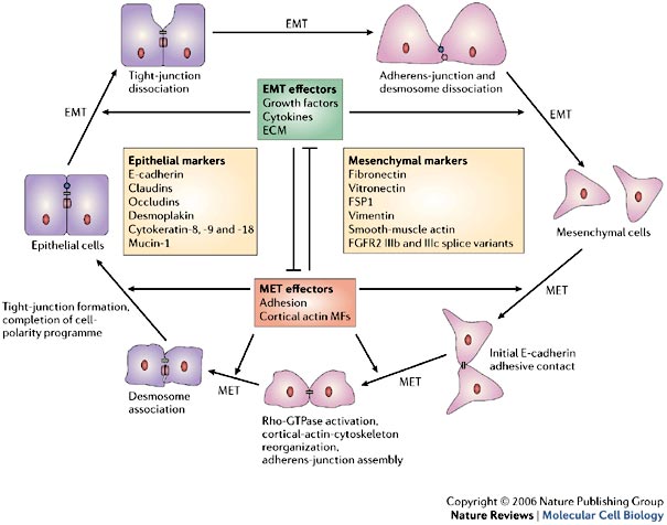
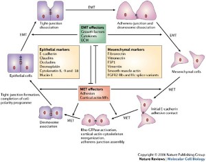
Here’s a diagram of the mesenchymal-epithelial transition:

mesenchymal-epithelial transition

LSJL upregulates Pcdhb2(protocadherin beta 2), Cdh13, Ctnna3, Fat1(a cell adhesion model).  It downs regulates protocadherin subfamily A, 4(Pcdhga4), AK002616(a miscellaneous Cadherin related protein), Celsr2, Cdh15, Cdh11.  So LSJL has the definite potential to affect the mesenchymal-epithelial transition although how isn’t clear as it affects a lot of related genes but not in a clear pattern

For the other parts LSJL downregulates Cldn13, Dsp(Desmoplakin isoform 1, the downregulation of this gene suggests that LSJL likely encourages the epithelial to mesenchymal transition but that doesn’t mean that it doesn’t encourage the mesenchymal to epithelial transition as well), and upregulates a gene related to Cldn19, Muc3.  It also downregulates an anti-mucin gene.

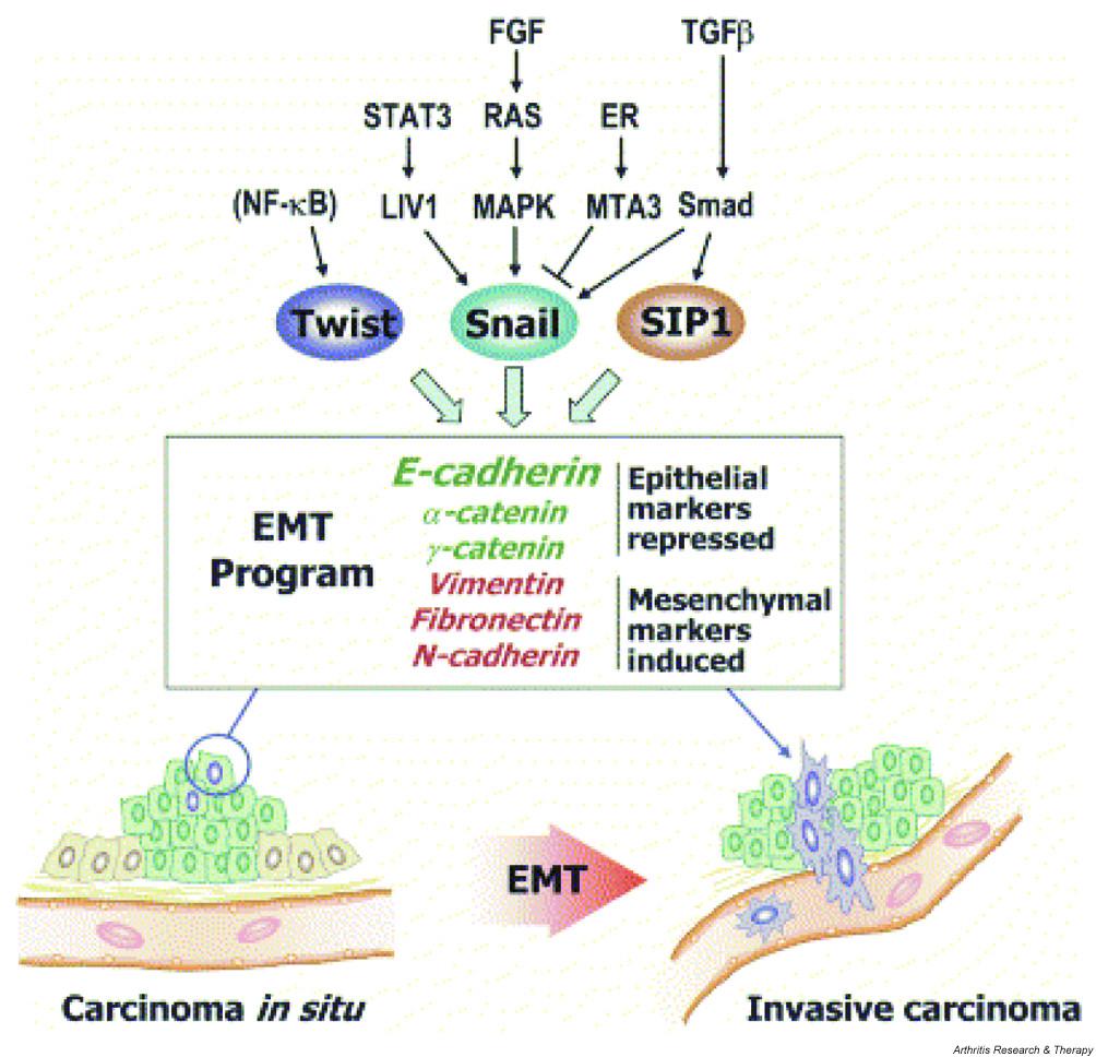
This diagram mentions the reverse markers:
EMT

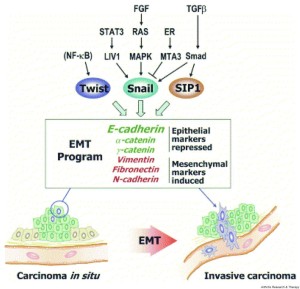
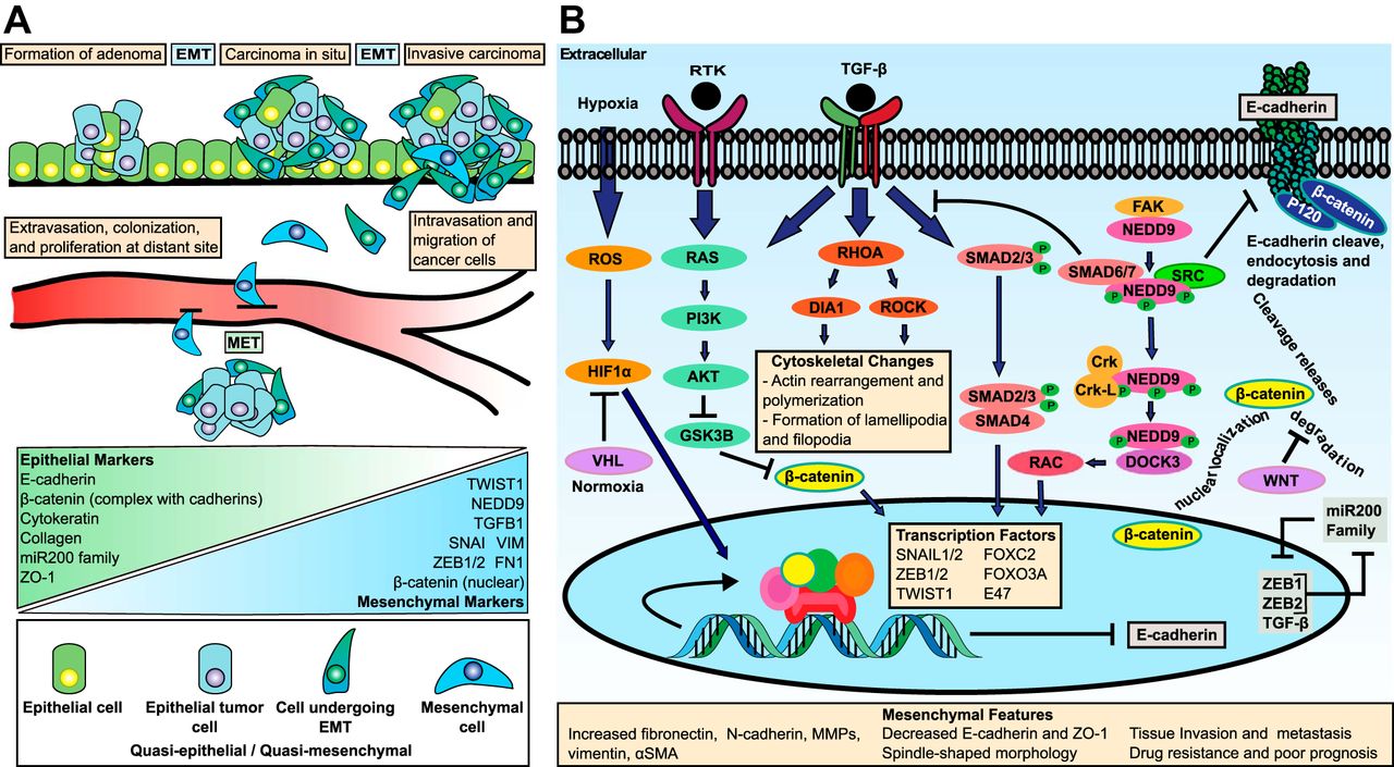
Here’s another diagram:
another EMT diagram

According to Actin stress fibres and cell-cell adhesion molecules in tendons: organisation in vivo and response to mechanical loading of tendon cells in vitro.

“Tendons consist of parallel longitudinal rows of cells separated by collagen fibres. The cells are in intimate contact longitudinally within rows, and laterally via sheet-like lateral cell processes between rows. At points of contact, they are linked by gap junctions. Since tendons stretch under load, such cell contacts require protection. Here we describe the organisation of the actin cytoskeleton and actin-based cell-cell interactions in vivo and examine the effect of cyclic tensile loading on tendon cells in vitro. Cells within longitudinal rows contained short longitudinally running actin stress fibres. Each fibre was aligned with similar fibres in the cells longitudinally on either side, and fibres appeared to be linked via adherens junctions. Overall, these formed long oriented rows of stress fibres running along the rows of tendon cells. In culture, junctional components n-cadherin{this increase is actually not good news for creation of epithelial cells as this is a mesenchymal marker} and vinculin and the stress fibre component tropomyosin increased in strained cultures, whereas actin levels remained constant.  (1) cells are linked via actin-associated adherens junctions along the line of principal strain; and (2) under load, cells appear to attach themselves more strongly together, and assemble more of their cytoplasmic actin into stress fibres with tropomyosin. Cell-cell contacts are protected during stretch, and also that the stress fibres, which are contractile, may provide an active mechanism for recovery from stretch. In addition, stress fibres are ideally oriented to monitor tensile load and thus may be important in mechanotransduction and the generation of signals passed via the gap junction network.”

So according to this in response to load cells may establish more cell-cell contact characteristic of epithelial cells.

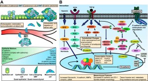
Mechanism of the Mesenchymal–Epithelial Transition and Its Relationship with Metastatic Tumor Formation

epithelial characteristics were dramatically associated with increased bone and soft-tissue colonization after intracardiac or intratibial injection.”

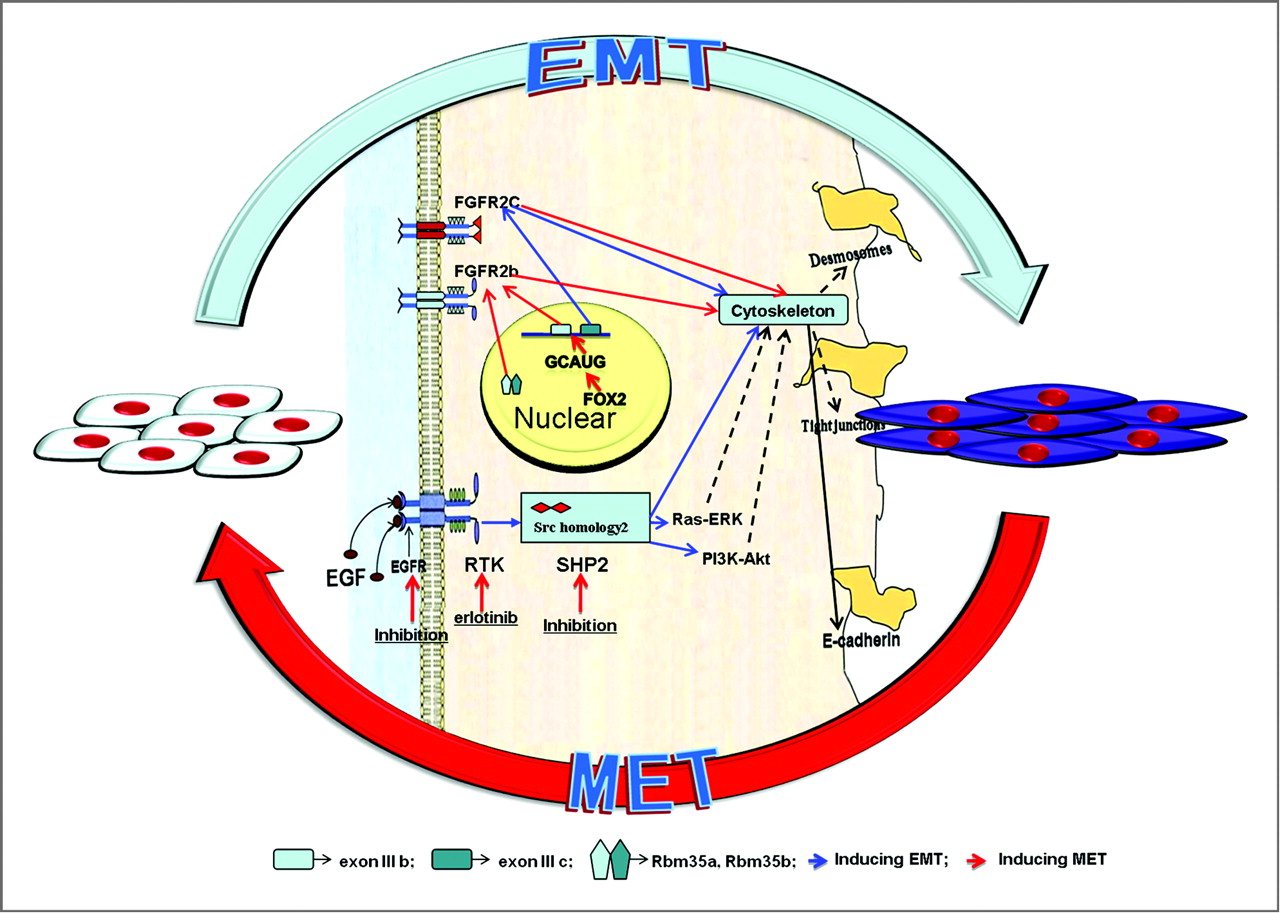
“Multiple complex signaling systems are required for the induction of EMT and are also closely related with MET. The FGFR2 gene, which is located at human chromosome 10q26, encodes for FGFR2b and FGFR2c isoforms due to alternative splicing and mutually exclusive use of exon IIIb or exon IIIc. FGFR2b primarily binds FGF10 and FGF7 and is the isoform of choice in epithelial cells, whereas FGFR2c binds FGF2 and is mainly expressed in cells of mesenchymal origin. FGF/FGFR2 signaling governs the EMT that is required for organogenesis in mouse embryos.”

“expression of FGFR2b induced MET [induced cancer in one instance]”

“As for the regulation of FGFR2 isoforms’ alternative splicing, a highly conserved GCAUG element was shown to be required for efficient exon IIIb activation. Afterward, Fox protein family members, especially Fox-2, were shown to regulate the FGFR2 exon choice, and this regulation was absolutely dependent on the GCAUG elements present in the FGFR2 pre-mRNA. Fox-2 induced the FGFR2c to FGFR2b switch, accompanied by molecular and morphological changes consistent with MET”

“2 paralogous epithelial cell type–specific RNA binding proteins, Rbm35a and Rbm35b, which are essential regulators of FGFR2 splicing. Ectopic expression of either protein in cells that express FGFR2c caused a switch in endogenous FGFR2 splicing to the epithelial isoform”<-Note it’s FGFR3 that’s typically associated with dwarfism.

MET

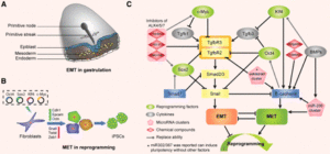
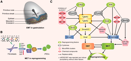
EMT and MET as paradigms for cell fate switching

“Cell fate determination is a major unsolved problem in cell and developmental biology. The discovery of reprogramming by pluripotent factors offers a rational system to investigate the molecular mechanisms associated with cell fate decisions. The idea that reprogramming of fibroblasts starts with a mesenchymal-epithelial transition (MET) suggests that the process is perhaps a reversal of epithelial to mesenchymal transition (EMT) found frequently during early embryogenesis. As such, we believe that investigations into MET-EMT may yield detailed molecular insights into cell fate decisions, not only for the switching between epithelial and mesenchymal cells, but also other cell types.”

“In any given animal tissue, one may find two very common cell types: the epithelial cells that line the surface of a tissue or organ and mesenchymal cells that are embedded in the three-dimensional matrix. “<-Growth plate cells line the bone matrix.

“the epithelial cells are attached to the basement membrane, establish an apical–basal axis of polarity, and communicate with each other through the gap junction. Across and underneath the basement membrane, there is the stroma made of the three-dimensional extracellular matrix synthesized by the resident mesenchymal or stromal cells.”

“Inside the nuclei, Snail genes are considered as the most important downstream targets of the nodal-SMAD2/3 pathway during gastrulation. The Snails may in fact be the guardians of the mesenchymal phenotype by activating mesenchymal genes and suppressing epithelial genes. Indeed, Snails have been shown to down-regulate E-cadherin effectively, which is one of hallmarks for epithelial cells. Snail-deficient embryos could not proceed through gastrulation and form mesodermal cells as they could not down-regulate the expression of E-cadherin in the primitive streak. It is generally recognized that the embryonic EMT process is orchestrated and maintained through the collaboration of extracellular signals and intracellular transcription factors.”

F1.medium

Fluid Flow enhances Osteoclast Production

Cortical bone is detrimental to longitudinal bone growth as it is not capable of interstitial growth.  Osteoclasts degrade cortical bone.  Degradation of cortical bone in combination with generation of tissue capable of interstitial growth is key to height growth.  Thus if fluid flow increase osteoclastgenesis this should be beneficial.

SUPRAPHYSIOLOGICAL LOADING INDUCES OSTEOCYTE-MEDIATED  OSTEOCLASTOGENESIS IN A NOVEL IN VITRO MODEL FOR BONE IMPLANT LOOSENING

“We aimed to develop an in vitro model for bone implant loosening, allowing analysis of
biophysical and biological parameters contributing to mechanical instability-induced
osteoclast differentiation and peri-implant bone loss. MLO-Y4-osteocytes were mechanically
stimulated for 1h by fluid shear stress using regimes simulating 1) supraphysiological loading
in the peri-prosthetic interface (2.9+/-2.9Pa, 1Hz, square wave),{This is below the minimum chondregenic range which is about 0.1MPa or 100Pa} 2) physiologic loading in the
cortical bone (0.7+/-0.7Pa, 5Hz, sinusoidal wave), and 3) stress shielding. Cellular
morphological parameters, membrane-bound RANKL expression, gene expression
influencing osteoclast differentiation, nitric oxide release and caspase 3/7-activity were
determined. Either Mouse bone marrow cells were cultured on top of loaded osteocytes or
osteocyte-conditioned medium was added to bone marrow cells. Osteoclast differentiation
was assessed after 6 days. We found that osteocytes subjected to supraphysiological loading
showed similar morphology and caspase 3/7-activity compared to simulated physiological
loading or stress shielding. Supraphysiological stimulation of osteocytes enhanced osteoclast differentiation by 1.9-fold compared to physiological loading when cell-to-cell contact was permitted{this is not a bad thing as degradation of bone could potentially be beneficial for height growh}. In addition, it enhanced the number of osteoclasts using conditioned medium by 1.7-fold, membrane-bound RANKL by 3.3-fold, and nitric oxide production by 3.2-fold. The stimulatory effect of supraphysiological loading on membrane-bound RANKL and nitric oxide production was higher than that achieved by stress shielding.  In conclusion, the in vitro model developed recapitulated the catabolic biological situation in the peri-prosthetic interface during instability that is associated with osteoclast differentiation and enhanced RANKL expression. The model thus provides a platform for pre-clinical testing of pharmacological interventions with potential to stop instability-induced bone implant
loosening.”

“[There’s] a specific role for osteocyte communication with osteoclasts “<-Osteoclasts, the cells that break down and reabsorb bone, stem from monocytes and macrophages rather than osteogenic cells.

“The decoy receptor for RANKL, osteoprotegrin (OPG), is released by osteocytes upon
physiological mechanical loading.

“Supraphysiological loading had a flow velocity that was 5.6-fold higher, and the wall shear stress rate that was 4.0-fold higher compared to physiological loading”

“Sixty minutes of supraphysiological loading on MLO-Y4 osteocytes did not change the
cell area, cell perimeter or cell feret’s diameter compared to physiological loading or stress
shielding”<-it would be beneficial if it did because it would indicate that the stem cells could possibly change in diameter.

Osteoblasts, Osteoclasts, and Osteocytes: Unveiling Their Intimate-Associated Responses to Applied Orthodontic Forces

“Histologically, the skeleton comprises 2 types of bone tissue: cortical (compact) and trabecular (cancellous) bone. The cortical bone makes up 80% of the volume in the adult skeleton and the trabecular bone makes up the remaining 20%. The cortical bone forms a peripheral shell in all kinds of bone.”
“The vertebrae are most rich in trabecular bone”
“Modeling of the bone is a process that changes the size and shape of bone either by bone resorption without subsequent bone formation or bone formation without previous bone resorption.”
“microcracks in bone and subsequent apoptosis in osteocytes will activate lining cells in
the remodeling process to stimulate osteoclast formation and bone resorption of the microdamaged area. During the resorption process, growth factors in bone are released, which attract and activate osteoblasts to form new bone under a canopy of bone lining cells.”
“. Osteoblasts form a 1 cell layer covering all bone surfaces. One function is to synthesize an extracellular matrix, consisting of type I collagen fibers and several other proteins, and to subsequently mineralize this matrix into bone”
“Osteoclasts are the only cells in nature that can degrade mineralized bone tissue and are important for physiological remodeling and modeling processes”

Teriparatide potential height increase tool

Teriparatide Improves Trabecular Osteoporosis but Simultaneously Promotes Ankylosis of the Spine in the Twy Mouse Model for Diffuse Idiopathic Skeletal Hyperostosis.

Full study->teriparitadeboneformation

“Diffuse idiopathic skeletal hyperostosis (DISH) is a common skeletal disorder in the elderly, which can develop into periosteal hyperostosis and paradoxically into immobilization-associated trabecular osteoporosis. The bone anabolic agent, teriparatide (TPD), seems to be a rational treatment for the immobilization-associated osteoporosis. However, it can lead to development of hyperostosis lesions in DISH patients. Here, we demonstrate TPD effectively treats trabecular osteoporosis while simultaneously promoting ankylosis of the spine in DISH model tiptoe-walking Yoshimura (twy) mice, compared with the ICR mice. Eighteen male twy mice were divided into three groups, and ICR mice were used as a normal control. Subcutaneous injections of TPD or phosphate-buffered saline (PBS) were performed according to three dosing regimens; 40 µg/kg once daily (TPD × 1 group), 40 µg/kg three times daily (TPD × 3 group), and PBS (control; Ctl group). Treatment was commenced at the age of 7 weeks and continued for 5 weeks. Micro-computed tomography (µCT) and histological analysis were performed. Longitudinal µCT study revealed that trabecular bone volume in both the vertebral body and distal femur decreased with time in the Ctl group, but increased dramatically in the TPD × 3 group. The twy mice developed ankylosis of the spine, the progression of which was accelerated with TPD therapy. We also confirmed that TPD therapy promoted ossification of spinal ligaments. Histomorphometrical study revealed that TPD treatment increased bone formation at the vertebrae enthesis region{This is the key to where this has promise} and in the trabecular bone. TPD therapy effectively treats trabecular osteoporosis, but potentially promotes ankylosis of the spine in patients with DISH.”

“The twy mice are mutant mice showing multiple osteochondral lesions, and have been used as a model for DISH and ossification of the posterior longitudinal ligament”

Teriparatide increased ectopic calcification(Fig3C).

“both TPD × 1 and TPD × 3 treatments promoted ectopic calcification. Histologically, there were no cells inside the ectopic calcification but there were fibroblast- or osteoblast-like cells around the ectopic calcification.”<-Fibroblast cells could be precursors to chondrogenic cells.

Here you can see the effects of teraparatide, it can increase height of the spine but also can cause scoliosis:


teriparatide effects