Author Archives: Tyler

Sesamoid Bone

A sesamoid is a bone formed within a tendon or muscle while we’re looking for is more interosseous chondroification(the formation of new cartilage tissue within the bone) or probably maybe even first interosseous epithelialification as you might need to transition mesenchymal to epithelial tissues first.

Coincident development of sesamoid bones and clues to their evolution.

“Sesamoid bones form within tendons in regions that wrap around bony prominences. They are common in humans but variable in number. Sesamoid development is mediated epigenetically by local mechanical forces associated with skeletal geometry, posture, and muscular activity. In this article we review the literature on sesamoids and explore the question of genetic control of sesamoid development. Examination of radiographs of 112 people demonstrated that the relatively infrequent appearances of the fabella (in the lateral gastrocnemius tendon of the knee) and os peroneum (in the peroneus longus tendon of the foot) are related within individual. This finding suggests that the tendency to form sesamoids may be linked to intrinsic genetic factors. Evolutionary character analyses suggest that the formation of these sesamoids in humans may be a consequence of phylogeny. These observations indicate that variations of intrinsic factors may interact with extrinsic mechanobiological factors to influence sesamoid development and evolution.”

“As many as 42 sesamoid bones can be found in some individuals. Mechanically, sesamoid bones serve to protect the tendon from damage and, in some cases, increase the efficiency or mechanical advantage of their associated muscle.”

“Most sesamoid bones in humans are 5 to 10 mm in diameter or smaller and are present in 1 to 100% of individuals.”

“Tendon chondrometaplasia[tendon turns into cartilage] and sesamoid bones tend to develop within tendons in areas that experience both tensile strain and hydrostatic compressive mechanical stresses.”<-So tendonous tissue may help form neo growth plates.  Tendon enthesis attach into the bone.  Tendon is capable of undergoing  chondrometaplasia.  So potentially you could form neo growth plates at these tendon entheses.  Originally I thought that ligament enthesis had more potential for neo-growth plate formation but tendon enthesis may have potential too.

” fibrous tendon tissue can form regions of fibrocartilage in areas that wrap around bony prominences (fibrocartilage is a tissue whose phenotype is intermediate between fibrous and cartilaginous tissue, consisting of chondrocytes embedded in aligned bundles of type I collagen)”

“compression loading and treatment with transforming growth factor beta (TGF-β) each resulted in upregulation of aggrecan and biglycan synthesis in fetal bovine tendon, suggesting that one aspect of the response of cells to compressive load is increased TGF-β synthesis which, in turn, stimulates synthesis of extracellular matrix proteoglycans and leads toward fibrocartilage formation. The process of fibrocartilaginous metaplasia in tendons, which is a direct response to an altered mechanical loading environment, appears to represent an intermediate step in the formation of a sesamoid cartilage.”

Sesamoid bones:

sesamoid-bone

Their position won’t help you grow taller but they could if they were in the articular cartilage.  Maybe if tendonous tissue was inserted in the articular cartilage.

“Hox A11, had profound effects on the developing mouse skeleton, including abnormal sesamoid bone development in both the forelimbs and hindlimbs.”

How might we turn tendons(and especially tendenous enthesis) into cartilage tissues.  What if we form a sesamoid bone within the enthesis?

Expression of Bone Morphogenetic Protein-2 in the Chondrogenic and Ossifying Sites of Calcific Tendinopathy and Traumatic Tendon Injury Rat Models

“Ectopic chondrogenesis and ossification were observed in a degenerative collagenase-induced calcific tendinopathy model and to a lesser extent, in a patellar tendon traumatic injury model. We hypothesized that expression of bone morphogenetic protein-2 (BMP-2) contributed to ectopic chondrogenesis and ossification. This study aimed to study the spatial and temporal expression of BMP-2 in our animal models.
Seventy-two rats were used, with 36 rats each subjected to central one-third patellar tendon window injury (C1/3 group) and collagenase-induced tendon injury (CI group), respectively. The contralateral limb served as controls. At week 2, 4 and 12, 12 rats in each group were sacrificed for immunohistochemistry and RT-PCR of BMP-2.
For CI group, weak signal was observed at the tendon matrix at week 2. At week 4, matrix around chondrocyte-like cells was also stained in some samples. In one sample, calcification was observed and the BMP-2 signal was observed both in the calcific matrix and the embedded chondrocyte-like cells. At week 12, the staining was observed mainly in the calcific matrix. Similar result was observed in C1/3 group though the immunopositive staining of BMP-2 was generally weaker. There was significant increase in BMP-2 mRNA compared to that in the contralateral side at week 2 and the level became insignificantly different at week 12 in CI group. No significant increase in BMP-2 mRNA was observed in C1/3 group at all time points.
Ectopic expression of BMP-2 might induce tissue transformation into ectopic bone/cartilage and promoted structural degeneration in calcific tendinopathy.”

“the presence of chondrocyte phenotype and ectopic ossification in a collagenase-induced patellar tendon injury model”

Here’s potential chondrogenic lesions:


Unfortunately none of those will add to height.

Hiroki Yokota Grant progress on LSJL

What can we discern about the plans from LSJL from the grants?  Yokota is one of the primary scientists behind the study Lengthening of Mouse Hindlimbs with Joint Loading.  There does not seem to be very much on the LSJL length effects since the expiring of Ping Zhang’s 2010 grant.  We either have to study the lengthening effects on our own or help Ping Zhang get more funding.

Yokota doesn’t mainly study the lengthening effects which are primarily studied by Ping Zhang as shown by this grant.

Here’s Hiroki Yokota’s 2015 grant:

“The long-term objective of the proposed studies is to elucidate the mechanism of mechanotransduction in bone. Our present bioengineering-oriented project developed a high-resolution piezoelectric mechanical loader and evaluated the role of mechanical stimulation in bone using cultured osteoblasts. The results reveal that (a) deformation of 3D collagen matrix can induce strain-induced fluid flow;(b) strain-induced fluid flow, and not strain itself, predominantly activates the stress-responsive genes in osteoblasts;and (c) architecture of 3D collagen matrix establishes a pattern of strain-induced fluid flow and molecular transport{We are not interested so much on the effects on osteoblasts but more on the effects of fluid flow on bone degradation and fluid flow on mesenchymal stem cells to create neo growth plates}. Many lines of evidence in animal studies support enhancement of bone remodeling with strain of 1000 – 2000 microstrains. An unclear linkage between our in vitro studies and these animal studies is the role of strain and fluid flow in bone remodeling. In vitro osteoblast cultures including our current studies use 2D substrates or 3D matrices that hardly mimic the strain-induced fluid flow in vivo. This difference between in vitro and in vivo data makes it difficult to evaluate the role of strain and fluid flow in bone remodeling and anti-inflammation. First, microscopic strain in bone might be higher than the macroscopic strain measured with strain gauges. A local microscopic strain higher than 1000 – 2000 microstrains may therefore drive fluid flow in bone. Second, the lacunocanalicular network in bone could amplify strain-induced fluid flow in a loading-frequency dependent fashion{This we should try to modify the frequency of LSJL to amplify strain}. Lastly, interstitial fluid flow in bone might be induced by in situ strain as well as strain in a distant location, such that deformation of relatively soft epiphyses induces fluid flow in cortical bone in diaphyses{We are more interested in deformation of the epiphysis as that’s where growth plates typically occur but deformation of the epiphysis in one end may induce fluid flow in the epiphysis in the other}. This renewal proposal will use mouse ulnae ex vivo as well as mouse in vivo loading to examine the above possible explanations for the data divergence.
Specific aims i nclude: (1) fabricating a piezoelectric mechanical loader for ex vivo and in vivo use;(2) quantifying ex vivo macroscopic and microscopic strains using electronic speckle pattern interferometry as well as molecular transport using fluorescence recovery after photobleaching;(3) conducting bone histomorphometry to evaluate ex vivo data;and (4) examining load-driven adverse effects with gene expression and enzyme activities (e.g., matrix metalloproteinases). Mechanical loads will be given in the ulna-loading (axial loading) and elbow-loading (lateral loading) modes{he’s planning on doing another LSJL loading study!}. These two modes have been shown to enhance bone remodeling in the diaphysis with different patterns of strain distribution. Successful completion of the proposed renewal proposal will provide basic knowledge about induction of fluid flow in bone and establish a research platform for devising therapeutic strategies for strengthening bone and preventing bone loss.”

Here’s the Hiroki Yokota 2014 grant:

Mechanical Loading and Bone

“The long-term objective of this study is to elucidate the mechanisms underlying loading-induced bone remodeling and develop unique loading-based therapies for preventing bone loss. The specific goal of this study, based on our most recent observations, is to determine how mechanical loading to the knee (knee loading – application of mild lateral loads to the knee) may exert global suppression of osteoclast development not only in the loaded (on-site) bone but also in the non-loaded (remote) bone{This is unfortunately not very promising for height growth as osteoclast driven remodeling is pretty significant for growth plate formation}. As a potential regulatory mechanism, we will focus on secretory factors (e.g., Wnt3a, NGF?, TNF?, etc.) and low-density lipoprotein receptor-related protein 5 (Lrp5) mediated signaling. In the parent project, we have shown that knee loading enhances bone formation in the tibia and the femur through the oscillatory modulation of intramedullary pressure. However, its effects on bone resorption have not been well understood. Preliminary studies using a mouse ovariectomized model, which mimics post-menopausal osteoporosis, indicate that knee loading can suppress development of multi-nucleated osteoclasts from bone marrow cells, and the loading effects are observed not only in the loaded femur but also in the non-loaded contralateral femur. In this competitive renewal project, we will test the hypothesis that joint loading (knee/elbow loading) can suppress an OVX-induced osteoclastogenesis in a systemic manner through Lrp5-mediated Wnt signaling with Wnt3a as a secretory factor, as well as interactions with other secretory factors. To examine this hypothesis, we propose two specific aims using a mouse loading model (knee loading, elbow loading, ulna bending, and tibia loading), and assays for bone remodeling and primary bone marrow cells.
Aim 1 : Determine the local and global effects of joint loading on osteoclastogenesis Aim 2: Evaluate the role of load-modulated secretory factors in osteoclastogenesis In response to mechanical loading, we will conduct X-ray imaging and colony forming unit assays{The x-rays will be highly useful in determining whether LSJL can induce neo-growth plate formation although the effects would have to be large to show up on the xray}. We will also examine expression of critical secretory factors such as Wnt3a, NGF?, TNF?, OPG, RANKL, etc. in the serum. Primary bone marrow cells will be cultured, and the mechanisms underlying loading-driven regulation of osteoclastogenesis will be investigated. We will examine expression of regulatory factors, including NFATc1 (master transcription factor for osteoclastogenesis) and osteoclast markers such as OSCAR, cathepsin K, etc. We will employ Lrp5 KO mice (global, and conditionally selective to osteocytes), as well as neutralizing antibodies and RNA interference (loss of a function), and plasmids (gain of a function). We expect that this project will contribute to our basic understanding of load-driven regulation of bone resorption and development of loading regimens useful for global prevention of bone loss. ”

Let’s look at Hiroki Yokota’s 2013 Grant:

“The long-term objective of the proposed studies is to elucidate the mechanism of mechanotransduction in bone. Our present bioengineering-oriented project developed a high-resolution piezoelectric mechanical loader and evaluated the role of mechanical stimulation in bone using cultured osteoblasts. The results reveal that (a) deformation of 3D collagen matrix can induce strain-induced fluid flow{If it is fluid flow that can induce neo-growth plate formation via stem cell simulation then we need to make sure that LSJL deforms the 3D collagen matrix};(b) strain-induced fluid flow, and not strain itself, predominantly activates the stress-responsive genes in osteoblasts;and (c) architecture of 3D collagen matrix establishes a pattern of strain-induced fluid flow and molecular transport. Many lines of evidence in animal studies support enhancement of bone remodeling with strain of 1000 – 2000 microstrains{2000 microstrain is about a 0.2% change in bone length.  LSJL laterally compresses the bone so the compression has to be by at least .1 or .2% to work}. An unclear linkage between our in vitro studies and these animal studies is the role of strain and fluid flow in bone remodeling. In vitro osteoblast cultures including our current studies use 2D substrates or 3D matrices that hardly mimic the strain-induced fluid flow in vivo. This difference between in vitro and in vivo data makes it difficult to evaluate the role of strain and fluid flow in bone remodeling and anti-inflammation. First, microscopic strain in bone might be higher than the macroscopic strain measured with strain gauges. A local microscopic strain higher than 1000 – 2000 microstrains may therefore drive fluid flow in bone. Second, the lacunocanalicular network in bone could amplify strain-induced fluid flow in a loading-frequency dependent fashion. Lastly, interstitial fluid flow in bone might be induced by in situ strain as well as strain in a distant location, such that deformation of relatively soft epiphyses induces fluid flow in cortical bone in diaphyses{of course our goal is to create new growth plates in the epiphysis but the fluid flow from compressing the ends of the epiphysis may flow deeper helping to induce mesenchymal condensation to induce neo growth plates closer to where the epiphysis meets the diaphysis}. This renewal proposal will use mouse ulnae ex vivo as well as mouse in vivo loading to examine the above possible explanations for the data divergence.
Specific aims include: (1) fabricating a piezoelectric mechanical loader for ex vivo and in vivo use;(2) quantifying ex vivo macroscopic and microscopic strains using electronic speckle pattern interferometry as well as molecular transport using fluorescence recovery after photobleaching;(3) conducting bone histomorphometry to evaluate ex vivo data;and (4) examining load-driven adverse effects with gene expression and enzyme activities (e.g., matrix metalloproteinases). Mechanical loads will be given in the ulna-loading (axial loading) and elbow-loading (lateral loading) modes. These two modes have been shown to enhance bone remodeling in the diaphysis with different patterns of strain distribution. Successful completion of the proposed renewal proposal will provide basic knowledge about induction of fluid flow in bone and establish a research platform for devising therapeutic strategies for strengthening bone and preventing bone loss. ”

The grants from 2013-2006 are virtually the same.  It’s only 2014 which is different however it’s unfortunate that it’s not focusing on the lengthening effects.

Unfortunately Ping Zhang’s grant Load-Driven Bone Lengthening only ran from 2008-2010.

Muscle Pump and Hydrostatic Pressure

Getting a pump in your muscles may be a way to induce hydrostatic pressure but it is unlikely as lots of bodybuilders work towards achieving the pump so it’s something that occurs very frequently physiologically.  So if the muscular pump did affect height it would likely be a phenomenon that would be noticed by now.  But the goal of the pump is get the blood to the muscle under target not the bone.  The pump is very localized to the muscle under tension so this may be why the muscular pump does not increase height.  The target area for a hydrostatic pressure increase is the bone and the pump targets the muscle.  This does not mean that an understanding of a muscular pump could help us understand how to increase hydrostatic pressure in the bone.

A bone fluid flow hypothesis for muscle pump-driven capillary filtration: II. Proposed role for exercise in erodible scaffold implant incorporation.

“A model is presented for enhancement of fluid flow through bone matrix and any porous tissue engineering scaffold implanted within it. The mechanism of enhancement is the skeletal muscle pump in compartments adjacent to the bone. Pressure waves from muscle pump contractions aided by increased blood pressure during exercise coupled with temporary occlusion of arteries leading to and veins from the bone, increase hydraulic pressure in cortical bone capillaries so as to amplify capillary filtration. It is proposed that capillary filtration increase is sufficiently convective to contribute to bone fluid flow and associated percolation through tissue engineered scaffold matrix implants. Importance of this contribution is its relative role in maintaining seeded cells in bioreactor scaffolds. Validation of the hypothesis starts at a minimum level of demonstrating that capillary filtration is convective. At a maximum level confirmation of the hypothesis requires demonstration that capillary filtration-based interstitial flow is sufficient to stimulate not only host bone cells (as proposed in part I of the hypothesis) but bioreactor-seeded cells as well. Preliminary data is presented supporting the prediction that skeletal muscle contraction generates convective capillary filtration.”

Although we don’t really want increased hydrostatic pressure in the capillaries that’s more likely to just deliver more nutrients to the bone.  Increased hydrostatic pressure in capillaries increases capillary filtration getting more nutrients to the interstitial fluid.  What we want is increased hydrostatic pressure in the interstitial fluid itself to encourage chondrogenic differenetiation.

“Nutrient exchange is not the sole function of transport in bone. There is increasing evidence that interstitial fluid flow is sensed by and modulates the behavior of bone cells. Percolation through bone matrix and associated implants is referred to as bone interstitial fluid flow
(BIFF). Two mechanisms for bone cell sensing of BIFF have been proposed; one mechanical and the other electrokinetic. The electrokinetic model focuses on streaming potentials that are putatively sensed by electrokinetic receptors in bone cell membranes. The mechanical model focuses on shear stress at the membrane-fluid interface, which is transmitted to second messenger by mechano-receptors”

“osteocytes and their processes are surrounded by relatively thin fluid (not necessarily Newtonian) annuli in the lacunar and canalicular compartments, rather than relatively large volumes of flowing blood.”

“muscle pump and exercise effects combine to increase capillary filtration sufficiently to add a significant component to BIFF.  We reason that skeletal muscle, acting through a muscle pump mechanism, increases the rate of capillary filtration by increasing capillary hydraulic pressure via contraction of skeletal muscle in compartments adjacent to bone. Exercise magnifies the affect by increasing baseline blood pressure through increased heartrate and muscle pump activity. Two anatomical circumstances suggest how the mechanism operates: (1) bone influx and efflux vessels outside bone are contained within fascia bounded compartments, which include skeletal muscle, and (2) efflux vessels (veins) are valved.”

“During the same exercise vascular resistance in bone increases two to fourfold while vasodilation in adjacent muscle increases”<-So blood flow in muscles increase while blood flow in bone decreases.  This could in fact increase hydrostatic pressure in bone as hydrostatic pressure is the force exerted by a fluid at rest and vascular resistance implies that there’s more fluid at rest.

“Solitons in arteries propagate to capillary beds where they increase intravascular hydraulic pressure in fluid unable to escape through veins.  In any given osteon or Haversian canal capillary filtration is increased driving extravascular fluid over perivascular
tissue and through nearest canaliculi. Pressures generated during exercise above heartbeat baselines can be considerable; interstitial values as high as 570 mmHg”

” IMP is a poor indicator of blood flow in bone the blood pressure changes associated with its increase are significant. (2) Blood flow to limb bones increases during exercise. (3) Vascular resistance in limb bones increases during exercise”

“contraction of the quadriceps muscle causes a 30 mmHg or more rise in femur IMP”

The Key Role of the Blood Supply to Bone.

“Blood supplies oxygen, nutrients and regulatory factors to tissues, as well as removing metabolic waste products such as carbon dioxide and acid. Bone receives up to about 10% of cardiac output, and this blood supply permits a much higher degree of cellularity, remodelling and repair than is possible in cartilage, which is avascular. The blood supply to bone is delivered to the endosteal cavity by nutrient arteries, then flows through marrow sinusoids before exiting via numerous small vessels that ramify through the cortex. The marrow cavity affords a range of vascular niches that are thought to regulate the growth and differentiation of hematopoietic and stromal cells, in part via gradients of oxygen tension. The quality of vascular supply to bone tends to decline with age and may be compromised in common pathological settings, including diabetes, anaemias, chronic airway diseases and immobility, as well as by tumours. Reductions in vascular supply are associated with bone loss. This may be due in part to the direct effects of hypoxia, which blocks osteoblast function and bone formation but causes reciprocal increases in osteoclastogenesis and bone resorption. Common regulatory factors such as parathyroid hormone or nitrates, both of which are potent vasodilators, might exert their osteogenic effects on bone via the vasculature. These observations suggest that the bone vasculature will be a fruitful area for future research.”

” impairment of the blood supply is well-known to reduce growth and repair, cause bone loss and, ultimately, necrosis”<-Maybe this could actually be a good thing as hypoxia is often associated with chondrogenesis.

hypoxia

With severe bone loss comes the space for neo-growth plate formation.

“drugs used to treat hypertension[high blood pressure] can increase systemic blood flow”

Mechanotransduction in musculoskeletal tissue regeneration: effects of fluid flow, loading, and cellular-molecular pathways

“Dynamic fluid flow induced by mechanical loading has been shown to have the potential to regulate bone adaptation and mitigate bone loss.”

“High physical activity level has been associated with high bone mass”

“Fluid flow-induced shear stress within bone has been considered as the source of how bone cells sense mechanical stimulation. Bone interstitial fluid is filling a variety of voids and channels within the bone matrix, including lacunae-canaliculi, bone tubules, Haversian canal and Volkmann canal, and osteon. Mechanical loading-induced interstitial bone fluid flow may play a role in mechanical sensing, bone cells response, signal transmission, transfer of nutrients, and so forth.”

“increased venous pressure can promote new bone formation in the periosteum. The data indicated that increased venous pressure will increase blood supply from the capillaries to the bone tissue”

“muscle force alone, if applied at a low rate, such as resistant weight lifting with high intensity, would not be able to generate sufficient strain and fluid pressure in bone. MS with a relatively high rate and a small magnitude, however, can trigger significant fluid pressure in the skeleton.”

“mechanical loading has been shown to reduce sclerostin levels in bone”

How bone knows how to grow

How does bone know how to be the proper shape and size for development?  Can we manipulate this to grow taller?

I received this email from the author regarding how distraction osteogenesis would affect how bone manipulates growth in regards to maintaining placement of superstructures:

“It is indeed an interesting question as it challenges the system with an unnatural manipulation – i.e. interstitial growth.

The simple answer is: we haven’t tried, so I can’t say for sure.

If the relative locations of ligament and tendon insertions are what you are interested in, then previous works show that the periosteum is involved in regulation of their positions (see list below). Moreover, if the balance between proximal and distal growth rates is what you are interested in, then other works show that cross-sectional cutting and stripping of the periosteum can cause temporal acceleration in overall growth rate of the bone (also in humans, if I remember correctly), followed by a potential change in proximal to distal growth balance (I don’t think that these works test how these influence the positioning of superstructures in the bone; see list below).
Therefore, if the operation you are applying includes anchoring of the periosteum to the bone or its cutting and stripping, this is something that may influence the scaling of the bones.”

Isometric Scaling in Developing Long Bones Is Achieved by an Optimal Epiphyseal Growth Balance.

“One of the major challenges that developing organs face is scaling, that is, the adjustment of physical proportions during the massive increase in size. Although organ scaling is fundamental for development and function, little is known about the mechanisms that regulate it. Bone superstructures are projections that typically serve for tendon and ligament insertion or articulation and, therefore, their position along the bone is crucial for musculoskeletal functionality. As bones are rigid structures that elongate only from their ends, it is unclear how superstructure positions are regulated during growth to end up in the right locations. Here, we document the process of longitudinal scaling in developing mouse long bones and uncover the mechanism that regulates it. To that end, we performed a computational analysis of hundreds of three-dimensional micro-CT images, using a newly developed method for recovering the morphogenetic sequence of developing bones. the relative position of all superstructures along the bone is highly preserved during more than a 5-fold increase in length, indicating isometric scaling. It has been suggested that during development, bone superstructures are continuously reconstructed and relocated along the shaft, a process known as drift.  most superstructures did not drift at all. Instead, we identified a novel mechanism for bone scaling, whereby each bone exhibits a specific and unique balance between proximal and distal growth rates, which accurately maintains the relative position of its superstructures. Moreover, we show mathematically that this mechanism minimizes the cumulative drift of all superstructures, thereby optimizing the scaling process. [There’s] a general mechanism for the scaling of developing bones. More broadly, these findings suggest an evolutionary mechanism that facilitates variability in bone morphology by controlling the activity of individual epiphyseal plates.”

If we can trick the bone into thinking it’s drifting maybe we can convince it to grow to maintain the position of the superstructure.  For example, dislocating the bone or similar means.

Although the molecular mechanisms regulating each growth plate for different bones are similar the bones still have different elongation rates.

“superstructures, known as bone ridges, tuberosities, condyles, etc., are necessary for the attachment of tendons and ligament as well as for articulation. To perform these functions they are located at specific positions along the bone. Bone superstructures emerge during early skeletogenesis . During growth, bones elongate extensively by advancement of the two growth plates away from the superstructures. It is therefore expected that during elongation, superstructures would remain at their original position near the center of the bone. Nevertheless, the end result is proper spreading of superstructures along the mature bone, which clearly implies the existence of a morphogenetic mechanism that corrects their locations.”

It’d be interesting to see what happens to bone superstructures during distraction osteogenesis.

“An ossified bone is a rigid object and so are the superstructures protruding from it, implying that they cannot be relocated by means of cell migration or proliferation. Therefore, any scaling mechanism must be adapted to overcome these physical restrictions.”<-So we have to make the bone less rigid.

“forelimb bones tend to grow away from the elbow joint, whereas bones in hind limbs tend to grow toward the knee joint.”

” Because the periosteal sheath is stretched over the entire external surface of the bone, including both the superstructures and the growth plates, it can pass to the growth plates signals concerning the relative position of superstructures.”<-Then perhaps we can manipulate longitudinal bone growth by manipulating the periosteal sheath.

“periosteal tension down-regulates growth plate activity, as the higher the tension level, the more inhibited growth plate activity is.  Damaged periosteum forms a scar tissue at the site of destruction. This scar tissue, which anchors the periosteum into the bone, creates an independent tension level near each growth plate. As a result, a new growth balance is formed, which equals the ratio between the distances from the site of the scar to the two ends of the bone, therefore maintaining the relative position of the scar site.. Superstructures can be considered as natural anchoring points for the periosteum into the ossified bone, either due to the insertion of tendons through them into the bone cortex, or by means of steric interference, such as in the tibiofibular junction. This results in a regulatory loop whereby the superstructures determine the tension levels of the two periosteal segments, which control the ratio of growth rates by inhibiting growth plate activity, which in turn maintains the relative position of the superstructure.”

Mechanical regulation of musculoskeletal system development 

“During embryogenesis, the musculoskeletal system develops while containing within itself a force generator in the form of the musculature. This generator becomes functional relatively early in development, exerting an increasing mechanical load on neighboring tissues as development proceeds. A growing body of evidence indicates that such mechanical forces can be translated into signals that combine with the genetic program of organogenesis. This unique situation presents both a major challenge and an opportunity to the other tissues of the musculoskeletal system, namely bones, joints, tendons, ligaments and the tissues connecting them. Here, we summarize the involvement of muscle-induced mechanical forces in the development of various vertebrate musculoskeletal components and their integration into one functional unit.”

“These forces [on the cell cytoskeleton], which can be translated into biochemical signals by molecules possessing mechanotransduction capabilities, are transmitted across transmembrane receptors into the extracellular matrix (ECM) and can also reach neighboring cells.”

“In the case of musculoskeletal development, exogenous forces acting on tendons and the skeleton are generated by muscle contraction.”

“Bone morphology is regulated by mechanical forces at different levels, as demonstrated by the various developmental and functional aberrations that arise in the absence of muscle contraction. (1) Bone elongation is impaired due to reduced chondrocyte proliferation in the growth plate. (2) Additionally, the organization of resting chondrocytes into columns is impaired, which can also affect skeletal elongation. (3) Bone eminence growth is arrested, resulting in smaller or absent eminences. (4) Differential appositional growth is lost, resulting in a circular circumferential shape. (5) Joint formation is impaired during embryonic development, leading to joint fusion.”

“These effects on chondrocyte proliferation could be mediated by yes-associated protein 1 (YAP1), a mechanosensor that is part of the Hippo signaling pathway. Indeed, changes in YAP cellular localization in chondrocytes were identified in vitro in response to matrix stiffness, and YAP was shown to regulate bone size, promote chondrocyte proliferation and inhibit chondrocyte differentiation in vitro and in vivo by suppressing collagen type X”

“mechanical forces can regulate both the content and the dynamics of proteoglycan and collagen production by these cells”

“Bones grow in width by preferential periosteal growth, which involves repetitive steps of strut-and-ring construction by mineral deposition.”

“non-selective mechanosensitive cationic channels, PIEZO1 and PIEZO2, which are expressed in a variety of tissue types, including chondrocytes”

“the attachment between the very elastic tendon and the very rigid bone creates a point of high stress concentration during force transfer, which could lead to detachment. Dissipation of this stress is achieved either by the formation of fibrous attachments, in which tendon fibers are inserted into the cortical bone in a structure that resembles a root system, or by the formation of a fibrocartilaginous attachment composed of different layers that gradually change in stiffness. Although enthesis development begins in the embryo, the formation of the unique transitional tissue and its subsequent mineralization occur postnatally.”

Growth and mechanobiology of the tendon-bone enthesis

“In the mature skeleton, the tendon-bone enthesis is an interfacial zone of transitional tissue located between compliant, fibrous tendon to rigid, dense mineralized bone. This transitional tissue provides a mechanism of stress and strain reduction at the interface between two mechanically dissimilar tissues”

“Fibrous entheses are generally found at insertion sites of stabilizing tendons, whereas fibrocartilaginous entheses are typically found at insertions of tendons that contribute to joint movement. Fibrous enthesis attach directly to bone and typically form Sharpey’s fibers, which are perforating fibers that embed into bone’s periosteal surface”

“Fibrocartilaginous entheses consist of four distinct histological zones, including aligned tendon, unmineralized fibrocartilage, mineralized fibrocartilage, and subchondral bone. A smooth and uniform basophilic tidemark distinguishes the transition between the two fibrocartilaginous zones, and this tidemark is disrupted and irregular in enthesopathy. The fibrocartilage enthesis matures during postnatal growth in response to mechanical loads from skeletal muscle and consists of cells that express both tenogenic and chondrogenic factors”

“Morphologically, the development of the enthesis has been likened to a “miniature” or arrested growth plate. However, unlike growth plates in long bones which eventually fuse at skeletal maturity, the fibrocartilage of the enthesis retains the morphological features of fibrocartilage and maintain Gli1+ cells at the interface throughout postnatal growth”

Lasting organ-level bone mechanoadaptation is unrelated to local strain

“Bones adapt to mechanical forces according to strict principles predicting straight shape. Most bones are, however, paradoxically curved. To solve this paradox, we used computed tomography–based, four-dimensional imaging methods and computational analysis to monitor acute and chronic whole-bone shape adaptation and remodeling in vivo. We first confirmed that some acute load-induced structural changes are reversible, adhere to the linear strain magnitude regulation of remodeling activities, and are restricted to bone regions in which marked antiresorptive actions are evident.{this is actually a good finding because it emphasizes that bone is an adaptive tissue} We make the novel observation that loading exerts significant lasting modifications in tibial shape and mass across extensive bone regions, underpinned by (re)modeling independent of local strain magnitude, occurring at sites where the initial response to load is principally osteogenic. This is the first report to demonstrate that bone loading stimulates nonlinear remodeling responses to strain that culminate in greater curvature adjusted for load predictability without sacrificing strength.”

“Wolff’s law is at odds with the curved overall shape of most bones, however, because adherence to these principles would predict straighter bones.”

“bones is instead optimized for load predictability. This would lead to the prediction that as bone curvature increases and predictability of the bending direction is augmented, its strength decreases”

“bone acts as an organ to acquire lasting modifications in shape and strength with greater bending predictability, which involves coordination of spatial remodeling that is unrelated to local strain magnitude.”<-it could be unrelated to local strain magnitude because of fluid forces.

“acute bone gains are lost within 6 weeks after load and extend this by showing that this is achieved through a simple reversal, involving greater resorption and lower formation in the proximal tibia in the chronic phase”

“net load-induced acute gains in the mid-to-distal tibia are, in contrast, lasting. Our 4D analyses reveal that this gain in bone mass is achieved predominantly by an enhanced formation in the acute phase, with only minor subsequent modifications in remodeling in the chronic postload phase. Comparing the remodeling activities in these two diverse, proximal and distal, locations by our novel application of whole-bone 4D analyses leads us to propose that the rapidity of load-induced acute changes in bone remodeling and the rate of their chronic postload reversal are linked. Reversibility in the proximal tibia is aligned to a marked load-related antiresorptive effect, while, contrarily, the lasting adaptation in the mid-to-distal region is principally linked with load-induced osteogenesis. It is intriguing that only the latter leads to permanent modification in bone shape.”

“Long-term curvature changes in nonbiological materials can involve creep deformation.”

“We show that some acute load-induced structural changes are reversible and adhere to linear strain magnitude feedback-loop regulation of remodeling activities. On the other hand, a vast proportion of the bone retains lasting structural memory of loading to generate nonlinear, strain magnitude–independent remodeling to achieve greater curvature optimized for load predictability without sacrificing strength.”<-some bone changes may be due to creep deformation thus being permanent.

Heterotopic Ossification

Heterotopic ossification is endochondral ossification that occurs outside the bone.  Understanding why it occurs can help us find ways to induce endochondral ossification within the bone.  The biggest issue with inducing a new growth plate in bone is the permissive local environment criteria.  The bone likely has to be degraded in some way to induce a neo-growth plate as the existing bone environment likely puts a constraining factor on growth.

Identifying the Cellular Mechanisms Leading to Heterotopic Ossification.

“Heterotopic ossification (HO) is a debilitating condition defined by the de novo development of bone within non-osseous soft tissues, and can be either hereditary or acquired. The hereditary condition, fibrodysplasia ossificans progressiva is rare but life threatening. Acquired HO is more common and results from a severe trauma that produces an environment conducive for the formation of ectopic endochondral bone. Despite continued efforts to identify the cellular and molecular events that lead to HO, the mechanisms of pathogenesis remain elusive. It has been proposed that the formation of ectopic bone requires an osteochondrogenic cell type, the presence of inductive agent(s) and a permissive local environment. To date several lineage-tracing studies have identified potential contributory populations. However, difficulties identifying cells in vivo based on the limitations of phenotypic markers, along with the absence of established in vitro HO models have made the results difficult to interpret. The purpose of this review is to critically evaluate current literature within the field in an attempt identify the cellular mechanisms required for ectopic bone formation. The major aim is to collate all current data on cell populations that have been shown to possess an osteochondrogenic potential and identify environmental conditions that may contribute to a permissive local environment. This review outlines the pathology of endochondral ossification, which is important for the development of potential HO therapies and to further our understanding of the mechanisms governing bone formation.”

“of the 80 % of war victims who suffer major extremity trauma during combat injury, approximately 64 % of these patients go on to develop some degree of HO”

“Current evidence suggests that the formation of ectopic bone in vivo requires three primary conditions: (1) a cell type capable of osteogenic differentiation, (2) the presence of inductive agents and (3) a permissive local environment”

“o date many contributory biological factors have been implicated in the aetiology, including the bone morphogenetic proteins (BMPs), inflammation, prostaglandin E2, hypercalcemia, hypoxia, abnormal nerve activity, immobilisation and dysregulation of hormones”
heterotropic endochondral ossification factors

“Tissue damage leads to the infiltration of immunological cells (monocytes, neutrophils and leukocytes) through the local vasculature. Resulting fibro-proliferation of an as yet unknown cell population is accompanied by hypoxia and the generation of brown adipose tissue at the site of damage. The presence of adipose tissue is hypothesised to lower the local oxygen tension leading to the establishment of a chondrogenic environment. Neovascularisation accompanies chondrogenesis and provides an avenue through which systemic cell types (endothelial cells, pericytes etc.,) may enter the injury site, and potentially contributed to osteochondrogenic differentiation. A subsequent increase in local oxygen tension promotes chondrocyte maturation and hypertrophy. The collagenous matrix deposited by these cells is then remodelled and ossified to form endochondral bone”<-If we induce such factors in the bone we can create new growth plates in there too.

“MSCs have frequently been shown to form endochondral bone when cultured under appropriate conditions (e.g. under hypoxia and/or in the presence of TGF-β)”

“MSCs may also contribute to chondrocyte hypertrophy and the progression of HO via their immunomodulatory effects, primarily through the production of anti-inflammatory cytokines and nitric oxide (NO)”

Several cell types are listed that are capable of heterotopic ossification are likely present in bone.

“Bone marrow HSC side population    Lin−/Sca-1+/cKit+/CD45+”

“Mesenchymal precursor cell (MPC)    CD44+/CD49e+/CD73+/CD90+/CD105+”

“MSC    CD73+/CD90+/CD105+”

“cells presenting the glutamate transporter GLAST were found to contribute to the formation of ectopic bone, and that these GLAST+ cells appeared to be distinct from the Tie2+ population”

” a significant upregulation in transcriptional activity in key osteogenesis-related genes (ALPL, BMP-2, BMP-3, COL2A1, COLL10A1, COL11A1, COMP, CSF2, CSF3, MMP8, MMP9, SMAD1 and VEGFA) in patients that developed HO compared to those who did not.”

Influence of transcutaneous electrical stimulation on heterotopic ossification: an experimental study in Wistar rats.

“Heterotopic ossification (HO) is a metaplastic biological process in which there is newly formed bone in soft tissues, resulting in joint mobility deficit and pain. Different treatment modalities have been tried to prevent HO development, but there is no consensus on a therapeutic approach. Since electrical stimulation is a widely used resource in physiotherapy practice to stimulate joint mobility, with analgesic and anti-inflammatory effects, its usefulness for HO treatment was investigated. We aimed to identify the influence of electrical stimulation on induced HO in Wistar rats. Thirty-six male rats (350-390 g) were used, and all animals were anesthetized for blood sampling before HO induction, to quantify the serum alkaline phosphatase. HO induction was performed by bone marrow implantation in both quadriceps of the animals, which were then divided into 3 groups: control (CG), transcutaneous electrical nerve stimulation (TENS) group (TG), and functional electrical stimulation (FES) group (FG) with 12 rats each. All animals were anesthetized and electrically stimulated twice per week, for 35 days from induction day. After this period, another blood sample was collected and quadriceps muscles were bilaterally removed for histological and calcium analysis and the rats were killed. Calcium levels in muscles showed significantly lower results when comparing TG and FG (P<0.001) and between TG and CG (P<0.001). Qualitative histological analyses confirmed 100% HO in FG and CG, while in TG the HO was detected in 54.5% of the animals. The effects of the muscle contractions caused by FES increased HO, while anti-inflammatory effects of TENS reduced HO.”

“The formation of heterotopic bone may be due to muscle trauma (myositis ossificans). It is common in people who have undergone total hip arthroplasty , those with spinal cord injuries, and victims of head trauma, all of which often lead to long periods of immobilization of the affected limbs.”

“skeletal muscle serves as a physical safeguard for the other organs and is anatomically located immediately beneath the skin, so it represents the most damaged organ in the body. Although skeletal muscle is characterized by the presence of fatty and connective tissues that originated from nonmyogenic mesenchymal progenitors, those progenitors were initially identified in BM”

“Muscle contraction occurs by the deposition of calcium in muscle tissue, and this stimulates the sliding of actin and myosin myofibrils, which characterizes the contractile process”

“electrical stimulation helps the deposition of calcium, causes changes in oxygen content and pH, stimulates expression of growth factors, and recruits help in osteoblast migration and secretion of extracellular matrix (ECM), leading to bone formation.”

“Mechanotransduction refers to the process by which the body converts a mechanical stimulus into a cellular response”

Cholesterol accumulation caused by low density lipoprotein receptor deficiency or a cholesterol-rich diet results in ectopic bone formation during experimental osteoarthritis.

“Osteoarthritis (OA) is associated with the metabolic syndrome, however the underlying mechanisms remain unclear. We investigated whether low density lipoprotein (LDL) accumulation leads to increased LDL uptake by synovial macrophages and affects synovial activation, cartilage destruction and enthesophyte/osteophyte formation during experimental OA in mice.

LDL receptor deficient (LDLr−/−) mice and wild type (WT) controls received a cholesterol-rich or control diet for 120 days. Experimental OA was induced by intra-articular injection of collagenase twelve weeks after start of the diet. OA knee joints and synovial wash-outs were analyzed for OA-related changes. Murine bone marrow derived macrophages were stimulated with oxidized LDL (oxLDL), whereupon growth factor presence and gene expression were analyzed.

A cholesterol-rich diet increased apolipoprotein B (ApoB) accumulation in synovial macrophages. Although increased LDL levels did not enhance thickening of the synovial lining, S100A8 expression within macrophages was increased in WT mice after receiving a cholesterol-rich diet, reflecting an elevated activation status. Both a cholesterol-rich diet and LDLr deficiency had no effect on cartilage damage; in contrast, ectopic bone formation was increased within joint ligaments (fold increase 6.7 and 6.1, respectively). Moreover, increased osteophyte size was found at the margins of the tibial plateau (4.4 fold increase after a cholesterol-rich diet and 5.3 fold increase in LDLr−/− mice). Synovial wash-outs of LDLr−/− mice and supernatants of macrophages stimulated with oxLDL led to increased transforming growth factor-beta (TGF-β) signaling compared to controls.

LDL accumulation within synovial lining cells leads to increased activation of synovium and osteophyte formation in experimental OA. OxLDL uptake by macrophages activates growth factors of the TGF-superfamily.”

“multiple injections of members of the TGF-super family, such as TGF-β or BMP-2, directly into the knee joint of the mouse caused abundant enthesophyte/osteophyte formation”

New Yokota/Zhang patent and New potential Height Increase device

The first Yokota/Zhang patent doesn’t provide that much insight but the second study provides a potential device that gives an alternative method of bone lengthening.  The study is confusing so I’d appreciate any second opinions.

Mechanical bone loading to reduce arthritic pain

“mechanical loading of the knee to downregulate nerve growth factor beta (NGFb), which is believed to be a major cause of pain in arthritic joints.”

“the joint loading may be performed at between 0.5 N and IO N, preferably at 1 N, and the fluid flow may be performed at, for example, 5 dyn/cm2. In one aspect, the results described herein suggest that gentle knee loading analogous to massage therapy is beneficial not only to enhancing bone formation and accelerating wound healing but also to preventing NGFP-induced nerve growth and pain perception in cartilage.”

” it has been recently suggested that a consequence of compressive loading is production of hydrostatic pressure as well as fluid flow to cartilage.”<-Hydrostatic pressure in bone could be a key to induce neo-growth plates.

“In osteoarthritis, chondrocytes are known to be exposed to flow shear presumably due primarily to synovial fluid and high amplitude of fluid flow reproduces the hallmarks of osteoarthritis in vitro. The frequency of 5 Hz might not be representative of massage to humans by hands but more pertinent to those by vibrator for foot massage. In another embodiment as described herein, the levels of loading in vivo have been optimized herein to produce anabolic response in the bone and cartilage.”

“Cyclic compression was applied to the mouse right knee using a custom-made piezoelectric loading device following reported methods. The mouse was mask-anesthetized using 2% isoflurane, and lateral loads to the knee were applied for 5 min at 5 Hz with a peak-to-peak force of 1 and 3 N.”

“Knee loading at 1 N but not at 3 N decreased the phosphorylation level of p38 (p- p38) in the cartilage”

“it was discovered herein that joint loading, illustratively, of a knee at 1 N, reduced mRNA levels of NGF and its low affinity receptor, p75 in cartilage and subchondral bone. Additionally, it was discovered that, in cartilage, joint loading, illustratively, of a knee at 1 N, reduced the phosphorylation level of p38 MAPK (p38-p) and activity of Racl GTPase. Additionally, it was discovered that, fluid flow at, for example, 5 and 10 dyn/cm2, reduced mRNA levels of NGFP and p75{neuron related gene} in C28/I2 human chondrocytes.”

“Nerves are known to exist in trabecular bone of the epiphysis, and are believed to grow in response to NGF{it’s possible that the growth of these nerves could affect height growth} . Although healthy cartilage is not believed to consist of vascular or neural tissues, arthritic cartilage is believed to lose its ability to remain aneural and avascular. It has been reported that dynamic loading to cartilage evokes stimulation of matrix synthesis{Could enough matrix synthesis increase height}, as well as regulation of enzymatic activities of matrix metalloproteinases. In addition to the reported regulatory role in matrix homeostasis, in one embodiment of the invention herein the results herein point out that mechanical stimuli at moderate amplitudes regulate transcription of NGF and its receptor in cartilage and chondrocytes.”

“both gentle mechanical loading and salubrinal share the Racl -mediated signaling pathway for – mRNA expression of NGF. In myocardial remodeling, it is reported that deficiency of Racl reduces stress to the endoplasmic reticulum. Since the elevated phosphorylation level of eIF2ot by salubrinal also suppresses stress to the endoplasmic reticulum, the observed linkage of salubrinal to Racl appears to be consistent with downregulation of NGF .”  Note that an increase in Rac1 expression was linked to an increase in chondrogenic marker genes.  This suggests that gentle mechanical loading may not be best for inducing exogenic bone mesenchymal chondrogenesis(neo-growth plate) and more extreme load may be needed.

This next paper is listed at the end as a related method:

System and Method for Joint Restoration by Extracapsular Means

“A system and method for joint restoration by extracapsular means includes an actuator operable to apply a force to a portion of a bone to effect a change in the joint space geometry. One embodiment of the system includes an actuator operable to apply a cyclic loading to subchondral bone of a femur, wherein loads of a predetermined magnitude are alternately applied and released. Between periods of cyclic loading, rest periods are provided where no load is applied. Over time, the femoral joint surface is remodeled in accordance with the location, direction, magnitude, and frequency of the loading.”

“Osteocytes sense the increased strain environment, and respond accordingly. When bone tissue is damaged as in the micro-cracking that occurs in the presence of excessive stress or strain, osteoclasts remove the necrotic osteocytes. This activates growth factors held in the osteocytes, such as bone morphogenic protein (BMP) or transforming growth factor (TGF) beta 1.”

At sufficiently high stress levels, deformation will occur with time, leading to “creep-failure”, or deformation that does not recover once the load is removed. The creep response of bone is significantly larger in younger bones as compared to older bones.”

“Similarly, when bone is measured on a large scale, it exhibits very classical (single elastic constant) behavior, but when the scale is reduced down to the trabecular level or below, the behavior becomes much more viscoelastic in nature, and tends to follow a Cosserat (multiple elastic constants) curve. This allows for much higher than predicted (by the classical approach) strain limits before failure occurs. In order for bone formation to be initiated, the magnitude of mechanical strain of the bone must surpass some threshold. Therefore, for restorative remodeling to occur, this threshold must be exceeded, while not causing failure”

” Trabecular bone can be found inside the condylar region of a femur, and alongside the cortical bone. The trabecular bone transfers the loads from the subchondral bone to the cortical bone, and the subchondral bone is that bone which supports the articular regions of the joint surfaces. Each different type of bone may undergo different deformation mechanisms. For example, cortical bone in particular exhibits “cement line slippage” between the osteons, which accounts for an ISF type (almost viscoelastic) behavior when applied to localized regions. This is typically considered the reason bone is a “tough, non-brittle” material. It is also a response that is dependent on the direction of the applied load-a result of the oriented structure of bone”

” a more rapid load onset results in a more rapid bone change. Conversely, a slower application of a load results in a smaller change, but thickening of the bone to handle the higher stress. Thus, a static load may build more dense bone, but a dynamic load may cause greater overall deformation of the bone.”<-Thus we should probably try to make sure that the clamping is the least static possible.  Constantly increasing clamping force is one way.

” the system components described herein can take advantage of the properties of bone that allow the bone to deform under constant stress via a “creep” or plastic deformation mechanism. The system components can push on the underside—e.g., the trabecular side—of the deformed subchondral bone, forcing a change of surface dimension on the joint surface (opposing) side of the subchondral bone. The subchondral bone may be softened to facilitate the reshaping process by drilling, cracking, laser etching, ultrasonically, biologically or by chemically treating the subchondral or the underlying cancellous bone, or by any other means in conjunction with the use of the system of the present invention, either to facilitate the initial movement, or during subsequent treatments. The devices according to the present invention may be permanently implanted in the bone, or can be removed after the desired results are obtained.”<-Can we induce a similar plastic deformation mechanism but in order to increase height.

“the term “static load” as used herein does not imply that a load that can or will never change; rather, the term refers to a load that is either constant for some period of time, or a load that is applied so slowly as to approximate a constant load. This is distinguished from a dynamic load, which may be a single load applied very quickly, or may be a cyclic load of constant amplitudes and/or frequency, or one of varying amplitudes and/or frequency.”<-So we may want to rapidly clamp then unclamp in order to get a single load applied rapidly.

the present invention has applications where shortening or lengthening of bone is desired to restore a normal joint geometry, and little or no joint surface remodeling is required. For example, a system including piezoelectric actuators can be applied to one or both sides of a joint to correct an angular displacement. ”

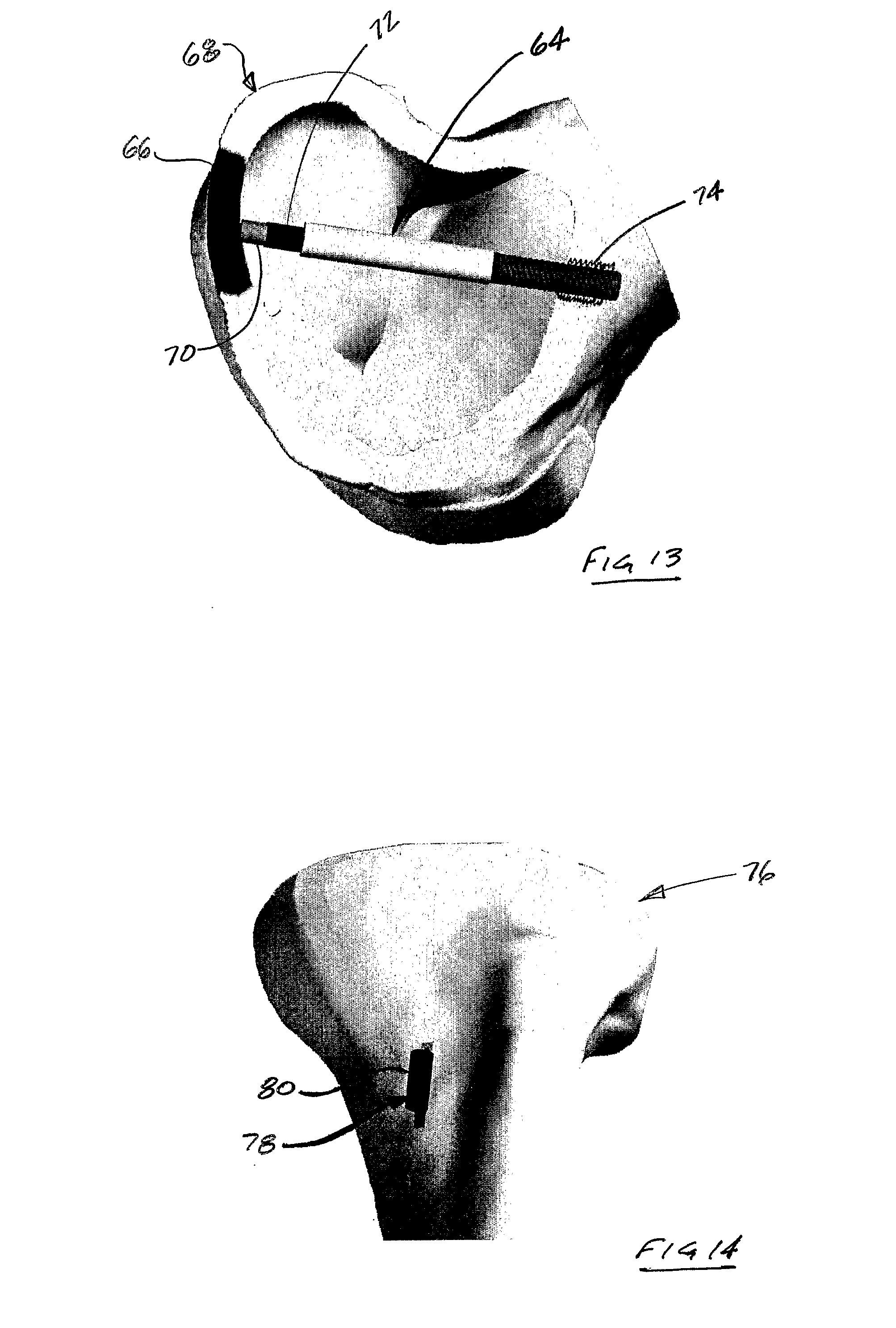
” For example FIG. 14 shows a tibia 76 having a system 78 in accordance with the present invention attached to it. The system 78 includes a linear actuator 80, which can be used to apply a static load, a cyclic load, or some combination thereof to the tibia 76. When a system, such as the system 78, includes two or more such actuators, one can be inserted in the cortical region and over time “grow” one side—e.g., the lateral side—and another can be inserted on the medial side to contract the bone. This effects an angular change at the joint line, and restores a more appropriate mechanical joint alignment.”<-For our purposes, we’d just set the two actuators to length bone.

ActuatorBone


FIG. 14 shows a system in accordance with another embodiment of the present invention, the system including a linear piezoelectric actuator to increase the length of a tibia;

actuators to increase tibia length

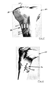
“FIG. 15 shows a system in accordance with another embodiment of the present invention, the system including a plurality of linear piezoelectric actuators to increase the length of a tibia as an alternative to an osteotomy”

Nowhere does it state that this would be limited to individuals with opten growth plates and in fact there are no visible growth plates on this bone.

This is an example of a linear actuator:

Note that I have no idea whether this actuator is sufficient in any way to provide a lengthening force on the bone.  It is just an example.

Note that the linear actuator is used in addition to the invention.  It seems that MIchael has considered using a linear actuator for a height increase device before.

” a swelling memory polymer can be used to provide expansion in a predetermined direction to a predetermined volume, thereby exerting pressure against the containing tissues. Shape memory alloys, such as Nitinol (Ni-TI) can also be used. Such alloys, commonly used in bone staples, can be formed as “muscle wires” and inserted into the cortical bone, where they lengthen in response to outside stimuli.”

“A shape memory alloy could also be formed as a spring, and configured to lengthen (or contract) upon application of an electrical current, for example, an 80 mA current at 20C”

This is how he describes inserting the device into the bone:

“creating an aperture[opening] in the bone proximate the articular surface, thereby making accessible an internal portion of the bone generally opposite the articular surface;
accessing the internal portion of the bone through the aperture in the bone; and
applying the at least one loading condition to the internal portion of the bone, thereby facilitating structural changes in the bone supporting the articular surface”

Then he describes inserting the device into the bone:

“wherein the at least one loading condition is applied to the bone with a joint restoration system including a housing having an aperture therethrough, and an elongate member configured for insertion into the aperture in the housing, the method further comprising attaching the housing to the bone such that the aperture in the housing is generally aligned with the aperture in the bone, and
wherein applying the at least one loading condition to the internal portion of the bone includes inserting the elongate member through the apertures such that the elongate member contacts the internal portion of the bone and applies a force thereto.”

“the joint restoration system further including a compression member configured to cooperate with the housing to apply a force to the elongate member, the method further comprising inserting the compression member into the housing such that it contacts the elongate member and imparts a force thereto, thereby facilitating the application of the force to the internal portion of the bone by the elongate member.”

Here he describes the bone lengthening method:

“The method of claim 1, wherein the at least one loading condition is applied to an external portion of the bone such that the certain structural change includes at least one of an increase in a length of the bone or a decrease in a length of the bone.”

” The method of claim 6, wherein the at least one loading condition is applied to the bone with a joint restoration system including an electromechanical actuator, the method further comprising:
attaching the actuator to the external portion of the bone; and operating the actuator to apply the at least one loading condition to the external portion of the bone.”

” piezoelectric devices will often have displacements in the 100’s of micrometers, which will not provide enough travel to effect desired bone growth in many patients. To overcome this limitation, the actuator is provided with a secondary movement mechanism. The secondary mechanism is configured to provide a ratcheting, positive lock that outwardly extends the extendable component by some discrete amount. This allows the application of a stepwise series of 100 micrometer piezoelectric adjustments, until a total bone displacement of 1-5 mm displacement is achieved.”

The paper A LINEAR ACTUATED TORSIONAL DEVICE TO REPLICATE CLINICALLY RELEVANT SPIRAL FRACTURES IN LONG BONES, describes one potential way a linear actuator can be applied to bone.  Although the device therein does not seem to be applied along the longitudinal axis as suggested by the patent but rather on the top and bottom of the bone.

This paper describes the use of a linear actuator to move a nail in distraction osteogenesis.  Note that in the patent above it specifically states there there is no osteomy required for this device to lengthen bone.