Author Archives: Tyler

A paper from the LSJL scientists with a potential design of a knee loading device

I found another paper that studied the LSJL knee device here.

I happened upon this paper however which was published at a conference and has potential new insights.

FEA analysis of a portable knee rehabilitation device

“The knee loading device examined in this paper only remains effective for small levels of deformation. The intended displacement of the working device is very small, a maximum of only 6.35mm, and therefore the device cannot accommodate large deformations as such deformations will greatly decrease the effective range of motion of the device.”<-So the device mentioned in this paper can only deform the bone by a maximum 6.35mm and likely less than that as just because the device can be displaced by 6.35mm doesn’t mean it will displace the bone by 6.35mm.

According to mechanostat theory, you have to deform the bone by at least 1500microstrain or 0.15% of a bones original length to get into the plastic deformation range to actually stretch the bone out permanently.

The average adult femur is 2.34 cm in diamater and the maximum displacement is the same regardless of femur shape and size.  The diameter of the femur is what matters as the device loads laterally.  The maximum displacement force is well over what is needed to plastically deform the bone.  But remember that the maximum displacement force is not actually how much the bone is deformed and the bone is being loaded laterally and not being stretched.  By loading laterally a moderate stretching force should be applied but not by the entirety of the deformation.

So it is not that the device can induce an increase in deforming the bone but it is possible by changing the microenvironment via degradation of cortical bone, initiation of mesenchymal condensation, and an increase in chondrogenic signaling.

If you click on the link above to see the study and look at figures 2 and 3, you can see that the device looks remarkably like a clamp.

“The described joint loading modality applies lateral loads to synovial joints.”

“To apply such [the needed] load, a device would need to have a means of producing a transverse force directly to the end of a long bone, such as at the knee{My current working theory is the load needs to be applied at the the epiphysis with the intent to maximize bone on bone contact and not on the synovial joint} . A cyclic force applied in such an area would force a slight shift of the fluid within the bone towards the opposite end of the bone in a controlled fashion.  While no such device currently exists for use on humans{I think a clamp device can serve such a role}, a new joint study seeks to develop a portable device designed for human use to be used in future testing.”<-For our purposes we would likely want more fluid flow as to induce cortical bone degradation and mesenchymal condensation.

“a pressure of approximately 6.90KPawas used as the load for each vertical pad [in a potential joint loading device]. The pressure equates to approximately 40 N over the entire surface area which is the desired maximum load for the device.”

An Irwin Quick Grip 12-inch can generate 300lbs of force.

300lbs is 1334N which is well over 40N but it is likely that you are not going to be able to generate that force however it is also likely that you will generate over 40N of force.

“stress is equal to force divided by cross-sectional area and strain is equal to change in length divided by original length. Stress and strain are related by Hooke’s Law, which states that stress is directly related to strain by a factor known as the Modulus of Elasticity, which is unique to every material.”

To effectively measure strain we’d have to be able to measure microchanges in bone length which I do not see as being possible at this juncture.

“Given the duty cycle of 5 minutes of daily operation per patient with a 1
Hz frequency loading function during operation; or 300 cycles per operation, this device is designed to last for 2683 uses. The choice of 1 Hz as one example was linked to daily
human physical activities such as walking. The device is able to induce loads up to 20 Hz, and it is a future task to evaluate appropriate loading frequencies.”<-Given that walking does not traditionally increase bone length we would likely use a different frequency.  But using a clamp it’s very hard to get such frequencies.  20Hz is equal to 20times per second and it would be virtually impossible to rapidly unclamp and reclamp in that amount of time.

What my current LSJL method instead tries to progressively clamp harder and harder(while still being mindful not to clamp to the point of too much pain) to increase the number of “cycles”.

It should be noted that nothing in this study mentions using this device for longitudinal bone growth but the other studies that Yokota et al. have done on the joint loading modality suggests that it could.  If you look at figure 1B(in the study link above), you can see that joint loading puts pressure on the cortical bone from the medullary cavity.  At a sufficient enough pressure, there could be degradation of the cortical bone and there was evidence of this in a diagram in one LSJL study.  The degradation of cortical bone is highly significant as one of the key events of fusion is the joining of the cortical bone of the epiphysis to the diaphysis.  By degrading cortical bone, we can reverse some of the constraining effects of cortical bone on future longitudinal bone growth.  After all, a key event in distraction osteogenesis is the inducement of a cortical bone fracture.

New height increase supplement

Here’s a paper from Korea about a new longitudinal bone growth supplement that may be promising:

Effect of KH-BaRoKer-SeongJangTang based on traditional medicine theory on longitudinal bone growth

“KH-BaRoKer-SeongJangTang (KBS) is a recently developed formulation by using traditional drugs considering traditional medical theory of Oriental books such as ShinNongBonChoGyeong and JuRye, which has been used to improve the growth of child in Korea. Although KBS is usually prescribed to many children who are in retard for their age, its pharmacological effects have not been fully understood in experimental models. The aim of this study was to evaluate the effects of KBS on bone growth. Growth plate thickness and bone parameters such as bone volume/tissue volume (BV/TV), trabecular thickness (Tb.Th), trabecular number (Tb.N), connection density (Conn.D), and total porosity were analyzed by means of microcomputed tomography. Serum insulin-like growth factor-I (IGF-I) levels were measured by enzyme-linked immunosorbent assay. Hepatic IGF-I mRNA expression was analyzed by real-time polymerase chain reaction. Phosphorylation of signal transducer and activator of transcription5 (STAT5) was investigated using Western blot analysis and immunohistochemistry. The thickness of growth plate was increased by KBS. BV/TV, Tb.Th, TbN, Conn.D, and total porosity were improved by KBS. Hepatic IGF-I mRNA and serum IGF-I levels were elevated by KBS. Phosphorylation of STAT5 was increased with administration of KBS. These results suggest that KBS would be helpful to children who are in retard for their age through the elevation of IGF-I.”

Increase in Stat5 phosphorylation, increase in IGF-1, and increase in growth plate thickness could be indicative that the supplement could increase growth.

“Male ICR mice (4 weeks old) and diets were purchased from Dae-Han Experimental Animal Center (Eumsung, Republic of Korea), acclimated for 7 days, and then randomly assigned for 2 weeks to adequate protein (CON, 20% protein) or low protein diet (PEM, 4% protein) . The protein source used was casein. Except for the protein content, the two diets were identical and isocaloric. After 2 weeks, mice were divided into five groups, CON (adequate protein diet + distilled water (DW)-administered group); PEM (low protein diet + DW-administered group); KBS (low protein diet + KBSadministered group); Arg (low protein diet + Arg-administered group); Glu (low protein diet + Glu-administered group). The mice were fed indicated diet, administered each material three times a week for 12 weeks, housed four to six per cage in a laminar air-flow room, and maintained at a temperature of 22 ± 1℃, a relative humidity of 55 ± 1% throughout the study.”

So it seems that this supplement is an IGF-1 mimetic.  Whether this can increase height in children with normal IGF-1 levels depends on the bodies negative feedback mechanisms.

“Binding of GH to GHR activates receptor-associated intracellular tyrosine protein kinase Janus kinase 2 (JAK2), which phosphorylates signal transducer and activator of transcription 5 (STAT5). The phosphorylated STAT proteins translocate to the nucleus, where they bind to specific DNA sequences and regulate gene transcription. Among the signal cascades from the GHR, the JAK2-STAT5 pathway is regarded as a major pathway that mediates the action of GH on gene transcription in the liver. This pathway was shown to be responsible for the transcriptional action of GH on IGF-I. IGF-I is a mitogenic factor for various cells and plays an important role in cell growth and survival, and the majority of plasma IGF-I is biosynthesized in the liver”

Two of the ingredients of the supplement are listed to be arginine and glutamine which aren’t exactly game changing.  A detailed list of the ingredients are given in Table 2 of the paper(link provided).

KBS on bone thickness


The kbs growth plate and the general bone architecture does look a little bit better than the Arginine group.

“Effect of KBS on tibial growth plate thickness. (A) Representative 3D CT images of knee joint showing growth plate. (B) The thickness of excised bone growth plate was determined on five points. CON, adequate protein diet + DW-administered group; PEM, low protein diet + DW-administered group; KBS, low protein diet + KBS-administered group; Arg, low protein diet + Arg-administered group; Glu, low protein diet + Glu-administered group”

So the supplement didn’t have much more benefit over arginine.

“The lengths of proximal tibia growth plate in the CON and PEM groups were 112.82+/- 4.18 and 86.43+/-1.47, respectively. The growth plate lengths in the KBS, Arg, and Glu groups were 119.05+/- 6.48, 118.75+/- 4.81, and 87.82+/- 6.38, respectively. KBS and Arg significantly enhanced the longitudinal bone growth, whereas Glu did not”

Interestingly, the serum IGF-1 levels were lower than the contAnother major ingredient was a variation on the Hominis Placenta Extract.  Given that the source is the human placenta which is very powerful, it is possible that this extract has of yet undocumented effects.  And many species eat the placenta.rol group in the KBS group and lower than that in the Arginine group.

“Carthami Tinctorii Fructus increased the level of serum IGF-I and lengths of femur and tibia, however, its effect was very small and transient”

Something we can ascertain is that maybe excess IGF-1 levels don’t have that large of a benefit on height.  As the excess IGF-1 levels of KBS versus arginine groups did not result in increased height.

KBS and Arginine increased growth plate thickness by about the same amount so it’s hard tell whether KBS has additional effects beyond arginine.  The difference between KBS and Arginine growth plates are so small and the variance is so large that it’s hard to attribute the difference to anything other than normal variation.

It’s possible that KBS could increase longitudinal bone growth but whether that affect is greater than Arginine is unknown

Here’s the study on Carthami Tinctorii Fructus:

Determination of mineral content in methanolic safflower (Carthamus tinctorius L.) seed extract and its effect on osteoblast markers.

“Safflower (Carthamus tinctorius L.) seeds are used as a folk medicine to enhance bone formation or to prevent osteoporosis in Korea. Therefore, the methanolic extract of safflower seeds (MESS) containing high mineral content, such as calcium (Ca), potassium (K) and phosphorous (P), was evaluated for the role on osteoblast (Ob) markers of Sprague-Dawley rats. In serum of 3 to 11 weeks (wks) old rats, both osteocalcin (OC) content and bone-specific alkaline phosphatase (B-ALP) activity increased to their maximum levels in 4-7 wks. Hence, 3 wks old rats were selected for 8 wks oral treatment of MESS, resulted in the significant increase of Ob markers in serum such as OC content (4-8 wks), B-ALP activity (1-2 wks) and insulin-like growth factor I (IGF-I) level (1 wk), and the growth parameter such as the length of femur (2-8 wks) and tibia (4 wks). On the basis of Pearson’s correlation coefficient, there were a moderate correlation between OC and B-ALP at 8 wks, a low correlation between OC and IGF-I at 1, 4 and 8 wks, a moderate correlation between OC and femur length at 1, 2 and 8 wks, and a moderate correlations between OC and tibia length at 1 and 8 wks of MESS-treated groups. The result reveals that the changes of OC correlated at low to moderate level with the changes of B-ALP activity, IGF-I content and femur and tibia length in the MESS-treatment period. On the other hand, there were a strong correlation between IGF-I and femur length at 2 wks and moderate correlation between IGF-I and tibia length at 1, 2 and 8 wks of MESS-treated groups.

“A Korean herbal formulation, Gami-Honghwain, is comprised of crude ingredients from safflower seeds and hominis placenta.”

“There are reports about chemical components of the safflower. Its leaves contain eight flavonoids, some of which showed potent antioxidant activities. Its seeds also contain numerous polyphenolic compounds such as lignans, glucosides, flavonoids and serotonins”

The increase in tibia and femur length ranged from 3-5%.  Note though that a 3% increase of someone who’s 5’9″ brings them to 5’11”.  And at all data points the treated group had longer bones than the control group.

“. In the aqueous extract from safflower seeds, there are K (2.306 μg/g), P (1.043 μg/g), Mg (0.474 μg/g), Al (0.175 μg/g), Fe (0.100 μg/g), Ca (0.075 μg/g), Zn (0.070 μg/g), Na (0.066 μg/g), Cu (0.055 μg/g) and Sr (0.022 μg/g). In the MESS, there are Ca (3.752 μg/g), K (1.313 μg/g), P (1.161 μg/g), Na (0.177 μg/g), Fe (0.170 μg/g), Zn (0.042 μg/g), Mg (0.023 μg/g), Al (0.019 μg/g), Cu (0.015 μg/g) and Sr (0.002 μg/g).”<-None of these seem to be novel compounds except for Strontium which is found in small amounts in normal human food.  Since Strontium amounts are so small it would be possible to be deficient in it and extra dietary strontium may increase longitudinal bone growth.

The Questions that Must be Answered for a Height Increase Routine to Work

In evaluating potential height increase routines, there are questions that have to be answered true to potentially be effective.  Asking these questions before attempting a routine or supplement, will help to determine if that supplement or routine could possibly be effective although there are many supplements that can be synergestic.

1a.  Does the method increase longitudinal bone growth?

b. Or does the method stimulate an increase the thickness of the bone in a longitudinal direction(top of head or bottom of heel)?

c.  Or does the method target another tissue that is a determinant in height(for example: cartilage or skin at the top of the head)?

d. Does the method alter alignment(posture or loosening of ligaments via relaxin)?

The rest of these questions will involve assuming that 1a was the question that was answered yes to as b-d would have different follow up questions. Note that GH and IGF-1 are a maybe in terms of answering question 1a.  They tend to encourage tissue growth but do not necessarily encourage chondrogenic differentiation(although IGF-1 may).  Something like IGF-2 is something more likely to increase height as it is involved in an earlier development state.  And the growth plate is at an earlier development state than full bones.  GH and IGF-1 are present at all developmental states whereas something like IGF-2 is not.

2.  Does the method involve degradation of cortical bone?

b. Does the method stretch cortical bone(plastic deformation which requires extreme loads)?

This likely has to be an important criteria in the success of a height increase routine.  The end of growth involves the fusion of cortical bone between the diaphysis and the epiphysis.  Distraction osteogenesis involves the cracking of cortical bone.  Cortical bone is likely like a large constraint on longitudinal bone growth.  Note: LSJL likely degrades cortical bone via an increase in fluid flow.

bone loading without drillingD is the LSJL loaded bone and C is not.  There’s definitely degration of the trabecular bone in D and F.  In D the LSJL cortical bone looks thicker and stronger which would make it harder to grow taller.  However in F there are some signs of degradation.  First, note that in E, that the inner bone is a fairly solid circular object but in F there is a part that juts outward on the upper lateral region of F this could be a sign of cortical bone degradation.    And note that 50% is the dead center of the bone which is not a likely target for increasing bone length.  75% is much closer to the epiphysis and since most longitudinal bone growth occurs at the epiphysis this is very promising that the necessary cortical bone degradation can occur to enable bone elongation to occur.

The bone loading only occurred for 3 days.  If bone loading had occurred longer it is possible that the protrusion of the inner bone would extend far enough to reach the outer point of bone enabling a neo-growth plate to form.  The mice were 14 weeks old which is fairly fair along the skeletal maturity process.  Mice growth plates don’t fuse but they do become dysfunctional and this is the time when growth cessation occurs.  So a potential protrusion caused by cortical bone degradation at a late skeletal maturity state is promising for the possible effectiveness of LSJL on adults.

3.  Does the method stimulate chondrogenic differentiation?

Bones have not yet been shown to be capable of interstitial growth and only through an intermediary tissue(cartilage) are they able to grow longer.  It would be possible to grow taller through the articular cartilage(which is capable of endochondral ossification) at the longitudinal ends of the bones but there does not as of yet seem to be an effective way of doing that.

The creating of an intermediary tissue like cartilage within the bone seems to be necessary to make the bones grow longer.  LSJL upregulates chondrogenic genes.

In addition, the chondrogenic intermediary must progress through the various stages and undergo hypertrophy to push the bone apart.  But it appears that endochondral ossification tends to be the standard procedure for chondrogenic tissue.

Prx1

Prx1 seems to be a very promising target for height increase as it seems to have targets early in development.  Unfortunately, I couldn’t find any Prx1 stimulating substances but hopefully you can?  Srx1 is involved in the repair of Prx1 so that could be another target as well.

Regulatory divergence modifies limb length between mammals

“Natural selection acts on variation within populations, resulting in modified organ morphology, physiology, and ultimately the formation of new species. Although variation in orthologous proteins can contribute to these modifications, differences in DNA sequences regulating gene expression may be a primary source of variation. We replaced a limb-specific transcriptional enhancer of the mouse Prx1 locus with the orthologous sequence from a bat. Prx1 expression directed by the bat enhancer results in elevated transcript levels in developing forelimb bones and forelimbs that are significantly longer than controls because of endochondral bone formation alterations. Surprisingly, deletion of the mouse Prx1 limb enhancer results in normal forelimb length and Prx1 expression, revealing regulatory redundancy. These findings suggest that mutations accumulating in pre-existing noncoding regulatory sequences within a population are a source of variation for the evolution of morphological differences between species and that cis-regulatory redundancy may facilitate accumulation of such mutations.”

“One developmental control gene known to promote limb skeletal elongation is Prx1, also called MHox or Prrx1. Prx1 is a paired-related homeobox gene expressed in somites, craniofacial mesenchyme, and limb mesoderm during mouse development”

“the forelimbs of Prx1BatE/BatE mutants are on average ∼6% longer than their wild-type littermates at E18.5 ”

“The forelimbs of Prx1-null homozygotes are ∼12.5% shorter than controls at E18.5”

“average mutant long bones express ∼70% more Prx1 than wild-type siblings”

“Long bone chondrocyte proliferation is elevated by ∼6% at E15.5 in Prx1BatE/BatE mutants.”

“[The Prx1 induced] limb elongation arises at stages of mouse gestation when Prx1 expression is limited to the perichondrium of the developing limb skeletal elements. It is known that the perichondrium is an important regulator of endochondrial bone growth”<-Could this be related to the zone of Ranvier which should be connected to the perichondrium?  Since the zone of Ranvier is linked to an earlier developmental state of the growth plate it could mean that Prx1 could be helpful in inducing neo growth plates.

According to this grant, Prx1 inhibits bone formation so it favors an earlier developmental state overall.

Prx1-Expressing Progenitor Primary Cilia Mediate Bone Formation in response to Mechanical Loading in Mice

“Increases in mechanical loading can enhance the addition of new bone, altering geometry and density such that bones better withstand higher forces. Bone-forming osteoblasts have long been thought to originate from progenitors, but the exact source is yet to be identified. Previous studies indicate osteogenic precursors arise from Prx1-expressing progenitors during embryonic development and adult fracture repair. However, it is unknown whether this cell population is also a source for mechanically induced active osteoblasts. We first identified that Prx1 is expressed in skeletally mature mouse periosteum, a thin tissue covering the surface of the bone that is rich in osteoprogenitors. We then traced Prx1 progenitor lineage using a transgenic mouse model carrying both a Prx1-driven tamoxifen-inducible Cre and a ROSA-driven lacZ reporter gene. Cells that expressed Prx1 when compressive axial loading was applied were detected within the cortical bone days after stimulation, indicating osteocytes are of Prx1-expressing cell origin. In addition, we evaluated how these cells sense and respond to physical stimulation in vivo by disrupting their primary cilia, which are antenna-like sensory organelles known to enhance mechanical and chemical signaling kinetics. Although Prx1-driven primary cilium disruption did not affect osteoblast recruitment to the bone surface, the relative mineral apposition and bone formation rates were decreased by 53% and 34%, respectively. Thus, this cell population contributes to load-induced bone formation, and primary cilia are needed for a complete response. Interestingly, Prx1-expressing progenitors are easily extracted from periosteum and are perhaps an attractive alternative to marrow stem cells for bone tissue regeneration strategies.”

“periosteum, which surrounds bones and is rich in progenitor cells known to preferentially differentiate towards the osteogenic lineage”

“physical stimulation activates and encourages osteogenic differentiation of progenitors within the periosteum.”

“One potential mechanism by which progenitor cells may become mechanically activated is through the primary cilium. Primary cilia are antenna-like organelles that extend from the cell surface and serve as signaling microdomains.”

“osteogenic response to fluid shear is lost when periosteal progenitor primary cilia are disrupted in vitro”

“Prx1-expressing cells become embedded osteocytes in response to physical loading and this mechanism requires the primary cilium.”<-if Prx1 enhances longitudinal bone growth and Prx1 mainly effects on chondrogenic cells maybe there are non chondrogenic ways to enhance longitudinal bone growth.

Articular Cartilage does contribute to bone growth in juvenile mice

If we can figure out how to grow via articular cartilage that will be a lot easier than to try to create neo-growth plates.

Spatial Regulation of Gene Expression During Growth of Articular Cartilage in Juvenile Mice.

“In juvenile mammals, the epiphyses of long bones grow by chondrogenesis within the articular cartilage. A bWe used laser-capture microdissection to isolate chondrocytes from the superficial, middle, and deep zones of growing tibial articular cartilage in the 1-wk old mouse and then investigated expression patterns by microarray. To identify molecular markers for each zone of the growing articular cartilage, we found genes showing zone-specific expression and confirmed by real-time PCR and in situ hybridization.Bioinformatic analyses implicated ephrin receptor signaling, Wnt signaling, and BMP signaling in the spatial regulation of chondrocyte differentiation during growth. Molecular markers were identified for superficial (e.g. Cilp, Prg4), middle (Cxcl14, Tnn), and deep zone (Sfrp5, Frzb). Comparison between juvenile articular and growth plate cartilage revealed that the superficial-to-deep zone transition showed similarity with the hypertrophic-to-resting zone transition{This is interesting, the study speculates that both the resting zone and deep zone may come from the same pool of epiphyseal chondrocytes}.ConclusionsLaser capture microdissection combined with microarray analysis identified novel signaling pathways that are spatially regulated in growing mouse articular cartilage and revealed similarities between the molecular architecture of the growing articular cartilage and that of growth plate cartilage.”

“While cell proliferation occurs sparingly in adult articular cartilage, it is much more common in juvenile articular cartilage. Growth of the articular cartilage reportedly occurs appositionally from the articular surface, suggesting that the SZ may contain actively dividing chondrocytes, or a progenitor cell population that is capable of generating new chondrocytes”<-these properties could be exploited to induce new longitudinal bone growth

“DZ[Deep Zone Articular Cartilage] expressed a high level of Wnt5b, which promotes chondrogenesis and inhibits hypertrophy ”

The development of articular cartilage: evidence for an appositional growth mechanism

“It is well-established that cartilage grows by a combination of matrix secretion, cell hypertrophy and cell proliferation. The extent to which this growth is by appositional, as opposed to interstitial mechanisms, however, remains unclear. Using the knee joints of the marsupial Monodelphis domestica to study cartilage growth, we have combined an immunohistochemical study of the TGF-β family of cartilage growth and differentiation factors between 30 days postpartum to 8 months, together with a stereological analysis of cartilage morphology during growth. Furthermore, to gain an insight into the generation of the characteristic zones within cartilage, we have examined the effects of intra-articular administration of bromodeoxyuridine, an agent that is incorporated into DNA during cell division and blocks further cell cycling. During early growth, TGF-β2 and -β3 were widely expressed but TGF-β1 was less so. After the formation of the secondary centre of ossification, all isoforms became more restricted to the upper half of the tissue depth and their distribution was similar to that previously described for IGFs, and PCNA-positive cells. Stereological analysis of tissue sections from the femoral condylar cartilage at 3 and 6 months showed that there was a 17% increase in total cartilage volume but a 31% decrease in cell density on a unit volume basis. Finally, cell-cycle perturbation with BrDU, which was injected into the knee joints of 3-month-old animals and analysed 1 and 4 months post-injection, revealed that the chondrocytes occupying the transitional zone were depleted 1 month post-injection, resulting in thinning of the articular cartilage. This effect was reversed 4 months post-injection. Immunohistochemical analysis revealed that BrDU-treatment altered the expression patterns of all TGF-β isoforms, with a marked reduction in labelling of TGF-β1 and -β3 isoforms in the upper half of the cartilage depth. Overall, the data lends further support to the notion of articular cartilage growing by apposition from the articular surface rather than by interstitial mechanisms.”

Articular cartilage and joint development from embryogenesis to adulthood

“Within each synovial joint, the articular cartilage is uniquely adapted to bear dynamic compressive loads and shear forces throughout the joint’s range of motion.”

“Early studies suggested that a region of proliferating cells “subjacent to the gliding surface of the joint” was responsible for interstitial growth of articular cartilage and increasing thickness of the articular surface. In this same study, Mankin and collaborators found that proliferation continued within deeper regions of the tissue and adjacent to the calcified cartilage, but ceased within the sub-superficial zone at later stages of postnatal growth. The presence of these two proliferative cell regions was confirmed by tritiated thymidine incorporation in the articular cartilage of immature rabbits. Later, Archer and collaborators confirmed the presence of a proliferative cell region in the superficial zone, suggesting that these cells were primarily responsible for the appositional growth and thickening of the articular cartilage postnatally

lateral expansion of the articular surface could be attributed to proliferation of cells within the superficial zone that would also give rise to daughter cells in a more rapidly proliferating cell population in the deeper zones leading presumably to vertical tissue growth.”

“In the growth plate, tremendous increases in chondrocyte volume contribute greatly to lengthening of long bones”

“chondrocyte volume in the middle and deep layers increased by over 8 fold from birth to 2 months of age, while overall decreases in cell density reflecting an increase in extracellular matrix production occurred during this same period.”

Hydrostatic Pressure and Encouraging Chondrogenic Differentiation

Hydrostatic pressure is a force we can manipulate via mechanical stimulation.

Hydrostatic pressure promotes the proliferation and osteogenic/chondrogenic differentiation of mesenchymal stem cells: The roles of RhoA and Rac1.

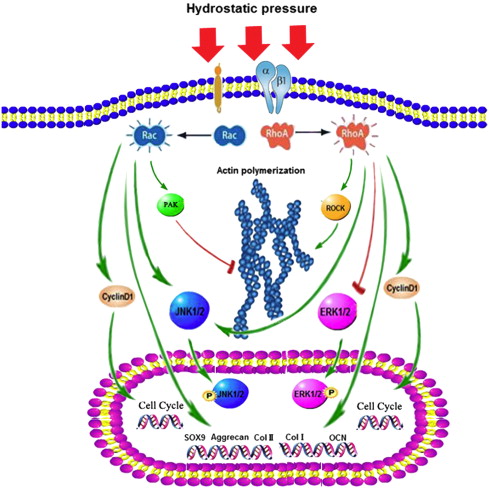
“Hydrostatic pressure can serve as an active regulator for bone marrow mesenchymal stem cells (BMSCs). [We investigate the roles] of cytoskeletal regulatory proteins Ras homolog gene family member A (RhoA) and Ras-related C3 botulinum toxin substrate 1 (Rac1) in hydrostatic pressure-related effects on BMSCs. Hydrostatic pressure promoted cell cycle initiation in a RhoA- and Rac1-dependent manner. RhoA played a positive and Rac1 displayed a negative role in the hydrostatic pressure-induced F-actin stress fiber assembly. RhoA and Rac1 play central roles in the pressure-inhibited ERK phosphorylation, and Rac1 but not RhoA was involved in the pressure-promoted JNK phosphorylation. Pressure promoted the expression of osteogenic marker genes in BMSCs at an early stage of osteogenic differentiation through the up-regulation of RhoA activity. Pressure enhanced the expression of chondrogenic marker genes in BMSCs during chondrogenic differentiation via the up-regulation of Rac1 activity. RhoA and Rac1 are critical to the pressure-induced proliferation and differentiation, the stress fiber assembly, and MAPK activation in BMSCs.”

Hydrostatic pressure applications are methods of applying mechanical loading that mimics the compressive forces borne by cartilage in a joint cavity“<-So we should be able to apply such mechanical load ourselves but in the bone rather than the cartilage to encourage a neo-ectopic growth plate.

“the activities of Rho GTPase signaling molecules are closely related to the differentiation and the fate of BMSCs.”

“the activation of the RhoA pathway and the inhibition of the Rac1 pathway under hydrostatic pressure promoted the assembly of the F-actin cytoskeleton, whereas the inhibition of the RhoA pathway and the activation of the Rac1 pathway blocked the F-actin cytoskeleton assembly.”

“the down-regulation of RhoA activity and/or pressure significantly blocked the phosphorylation of ERK1/2″<-LSJL increases ERK1/2 phosphorylation.

“RhoA activation inhibited the pressure-induced down-regulation of P-ERK1/2 expression and that RhoA played an important role in the regulation of ERK1/2 phosphorylation upon pressure stimulation.”

A combination of decreased RhoA activity and pressure stimulation (P/RhoA − group) achieved the maximum expression of the chondrogenic marker genes in BMSCs{So we have to find a way to decrease RhoA levels in the bone}. After two weeks of chondrogenic induction, the expression levels of the chondrogenic genes in the P/RhoA + group were significantly reduced compared with those of the P group. The up-regulation of RhoA antagonized the promoting effect of pressure on the chondrogenic differentiation of the BMSCs{So how do we downregulate RhoA?}. After 4 weeks of chondrogenic induction, the expression levels of Sox-9, Aggrecan and Col II in the RhoA −, P and P/RhoA − groups were significantly higher than those of the control group ”

” pressure inhibited ERK phosphorylation, suggesting that the induction of cell cycle initiation by pressure may not require a modulation of the cyclin D concentration, which involved the regulation of ERK activity through the LIMK protein.”

“pressure promoted the phosphorylation of JNKs but not ERKs in the BMSCs.”

the damage to the cytoskeletal structure and the inhibition of ROCK activity induced rounded cell morphologies and promoted the expression of chondrogenic marker genes in the BMSCs“<-Maybe hydrostatic pressure will induce more cytoskeletal damage over time and more chondrogenic differentiation will be induced naturally.

“Hydrostatic pressure regulates cell cycle initiation through both the RhoA/Rock and the Rac1 signaling pathways. At the same time, the mechanical stimulation promoted cytoskeletal assembly in BMSCs through the up-regulation of RhoA/ROCK activities, and activation of the JNK1/2 pathway by down-regulation of Rac1 activity. Hydrostatic pressure could also enhance expression of marker genes for early osteogenic differentiation through the up-regulation of RhoA activation or enhance the expression of chondrogenic marker genes in BMSCs during chondrogenic differentiation via the up-regulation of Rac1 activity ”

hydrostatic pressure effect

According to Flavoprotions: Advances in Research and Application: 2011, egf is a stimulator of Rac1.

There are several skin applicators of EGF but I couldn’t find any oral.
I’m not sure if applying it to the skin near the bone/joint region would work in stimulating Rac1 and I’m not sure if ingesting a product meant for the skin is safe nor if it will stimulate any desired region.  But the study suggests Rac1 versus RhoA is a key cellular distinction for osteoblasts versus chondrocytes.  And since there is as yet no RhoA inhibitor(but it’s being investigated due to cancer applications), stimulation of Rac1 via EGF is a worthwhile path to go down.  By encouraging chondrogenesis versus osteogenesis in the bone, it would be easier to great a neo-growth plate.

Rac1 promotes chondrogenesis by regulating STAT3 signaling pathway. has a possible suggestion of how STAT3 could also promote chondrogenesis,

“The small GTPase protein Rac1 is involved in a wide range of biological processes including cell differentiation. Previously, Rac1 was shown to promote chondrogenesis in micromass cultures of limb mesenchyme. However, the pathways mediating Rac1’s role in chondrogenesis are not fully understood. This study aimed to explore the molecular mechanisms by which Rac1 regulates chondrogenic differentiation. Phosphorylation of signal transducer and activator of transcription 3 (STAT3) was increased as chondrogenesis proceeded in micromass cultures of chick wing bud mesenchyme. Inhibition of Rac1 with NSC23766, janus kinase 2 (JAK2) with AG490, or STAT3 with stattic inhibited chondrogenesis and reduced phosphorylation of STAT3. Conversely, overexpression of constitutively active Rac1 (Rac L61) increased phosphorylation of STAT3. Rac L61 expression resulted in increased expression of interleukin 6 (IL-6), and treatment with IL-6 increased phosphorylation of STAT3. NSC23766, AG490, and stattic prohibited cell aggregation, whereas expression of Rac L61 increased cell aggregation, which was reduced by stattic treatment. Rac1 induces STAT3 activation through expression and action of IL-6. Overexpression of Rac L61 increased expression of bone morphogenic protein 4 (BMP4). BMP4 promoted chondrogenesis, which was inhibited by K02288, an activin receptor-like kinase-2 inhibitor, and increased phosphorylation of p38 MAP kinase. Overexpression of Rac L61 also increased phosphorylation of p38 MAPK, which was reduced by K02288. These results suggest that Rac1 activates STAT3 by expression of IL-6, which in turn increases expression and activity of BMP4, leading to the promotion of chondrogenesis.”

So BMP4, STAT3, and IL6 are all potential targets to induce chondrogenesis.

Contribution of the Interleukin-6/STAT-3 Signaling Pathway to Chondrogenic Differentiation of Human Mesenchymal Stem Cells.

“Mesenchymal stem cells (MSCs) are multipotent cells that can differentiate into chondrocytes. Articular cartilage contains MSC-like chondroprogenitor cells, which suggests their involvement in the maintenance of cartilage homeostasis by a self-repair mechanism. Interleukin-6 (IL-6) is a cytokine [which is] produced by MSCs in a steady manner and in large quantities. The purpose of this study was to investigate the involvement of IL-6 signaling in MSC differentiation into chondrocytes.
Human bone marrow-derived MSCs were cultured using a pellet culture system in medium containing transforming growth factor β3. Chondrogenic differentiation was detected by cartilage matrix accumulation and chondrogenic marker gene expression.
IL-6 was detected at a high concentration in culture supernatants during chondrogenic differentiation. The expression of the IL-6 receptor (IL-6R) was significantly increased, accompanied by markedly increased phosphorylation and expression of STAT-3. Addition of IL-6 and soluble IL-6R (sIL-6R) to the chondrogenic culture resulted in concentration-dependent increases in cartilage matrix accumulation and cartilage marker gene expression (type II collagen/aggrecan/type X collagen). Phosphorylation of the master transcription factor SOX9 was enhanced upon addition of IL-6 and sIL-6R. STAT-3 knockdown suppressed chondrogenic differentiation. IL-6 and the MSC markers CD166 and nestin were colocalized in macroscopically normal human cartilage taken from the lateral femoral compartment of knees with medial tibiofemoral osteoarthritis.
During differentiation of human MSCs into chondrocytes, the activation of IL-6/STAT-3 signaling positively regulated chondrogenic differentiation. The presence of IL-6 around MSC-like cells in the cartilage tissue was identified, suggesting that IL-6 contributes to homeostasis and cartilage self-repair by promoting chondrogenic differentiation.”

“cartilage contains chondrogenic progenitor cells with mesenchymal stem cell (MSC)–like characteristics”<-possibly actually more like epithelial cell characteristics.

“MSCs exhibit immunosuppressive activity and inhibitory effects on osteoclast differentiation via trophic effects, by releasing various humoral factors”

“MSCs produced high levels of IL-6 during chondrogenic differentiation”

“Chondrocytes are also capable of producing IL-6 upon stimulation, although under physiologic conditions (i.e., embedded in the cartilage matrix), chondrocytes are reported to have little ability to produce IL-6”

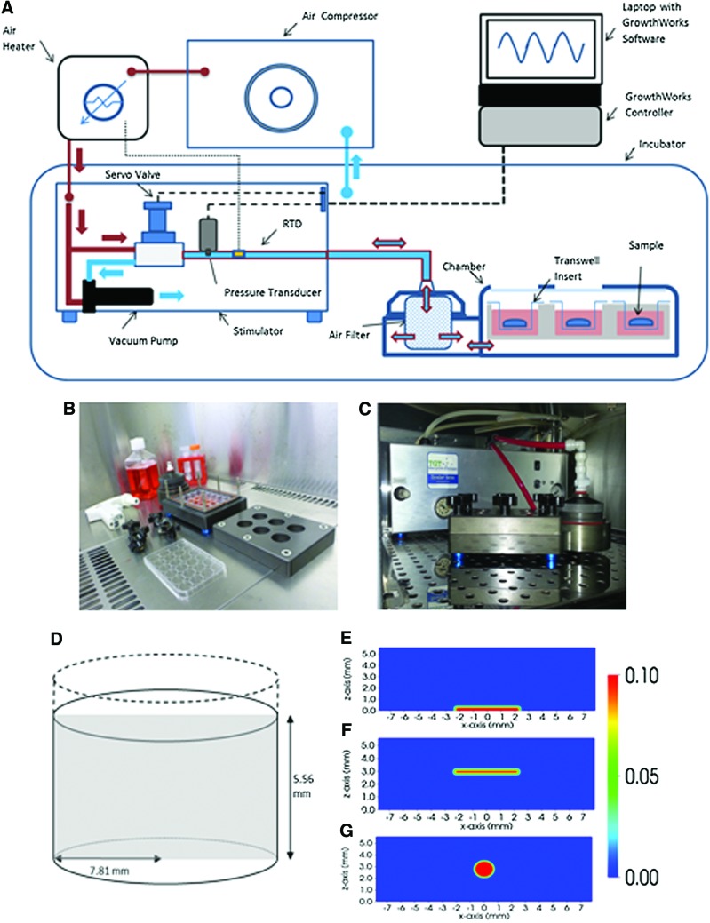
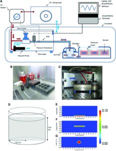
Evaluation of the Growth Environment of a Hydrostatic Force Bioreactor for Preconditioning of Tissue-Engineered Constructs

“To determine the effect of hydrostatic pressure on bone formation, chick femur skeletal cell-seeded hydrogels were subjected to cyclic hydrostatic pressure at 0-270 kPa and 1 Hz for 1 h daily (5 days per week) over a period of 14 days. At the start of mechanical stimulation, dissolved O2 and CO2 in the medium increased and the pH of the medium decreased, but remained within human physiological ranges.”

“Hydrostatic pressure has been shown to be an important mechanical stimulus for the direction of cell fate in various tissues, including articular cartilage, the intervertebral disc, bone, and the vascular system”<-We want to induce the cell fate of chondrogenesis.

“Osteocytes in the canalicula-lacuna network of load-bearing bones are subjected to physiological pressures of approximately 270 kPa”

“he heartbeat of chick embryos delivers a dynamic pressure of 4 kPa, the blood pressure is usually between 8–24 kPa, the hydrostatic pressure in the cerebrospinal fluid is around 1.2 kPa and the interstital fluid pressure is around 0.27 kPa”

“The application of hydrostatic pressure during tissue formation could result in enhanced transfer of small molecules, such as oxygen and CO2, into the tissue matrix and provide physical forces to cells and tissues

hydrostatic pressure machine

Biomechanics-driven chondrogenesis: from embryo to adult.

“Cartilage is relatively acellular, with chondrocytes only comprising 1–5% of the tissue by volume”

“Joint loading results in direct compression of chondrocytes inside a relatively impermeable matrix. Following tissue loading, hydrostatic pressure initially develops in the interstitial fluid, which is followed by fluid flow-induced shear. However, in time scales > 10 μs, the solid matrix begins to bear the applied load, resulting in deformation. Consequently, the cells residing in the matrix experience hydrostatic pressure, shear, compression, and, to a lesser extent, tension. This mechanical stimulation produces a signaling cascade, resulting in increased gene expression, matrix protein production, and intracellular ion influx”<-Our goal though is to induce hydrostatic pressure within the bone to induce chondrogeneic differentiation.

Spatiotemporal changes in progenitor cell adhesion molecule expression cause similar cells to transiently associate during chondrogenesis. However, cell-cell adhesion strength correlates linearly with cellular surface tension, irrespective of a homogeneous or heterogeneous interaction, suggesting surface tension as the primary driver of differential adhesion. Therefore, precartilaginous condensation may be the result of mesenchymal progenitor cells exhibiting similar surface tensions rather than similar biomarkers. Furthermore, disruption of surface tension inhibits differential adhesion“<-so altering surface tension may be key to inducing chondrogenesis and cellular biomarkers may not necessarily be a limiting factor on chondrogenic induction.

“the absence of gravitational force reduces precartilaginous condensations in mesenchymal limb bud cells”

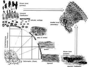
“Biomechanics-driven development of cartilage from embryo to fetal stages and beyond. A, B) Progenitor cells migrate from the early mesoderm to sites of skeletogenesis (A), where they undergo precartilaginous condensations (B). C) Chondrocyte progenitors secrete cartilage-specific matrix and decrease expression of cell-cell interaction proteins. D) Proliferation continues at the subchondral growth front, while endochondral ossification occurs throughout the juvenile stages to transform cartilage into bone. E) Ends of long bones remain capped with a layer of articular cartilage throughout adulthood.”

” Intermittent and cyclic hydrostatic pressure and strain both help regulate matrix protein synthesis to affect macromolecular organization of collagen fibers, which, in turn, leads to changes in the mechanical properties of the tissue ”

“models predict that intermittent hydrostatic pressure inhibits degeneration and ossification of cartilage, while intermittent strain or shear stresses accelerate ossification and degeneration”

“HP does not result in deformation of incompressible media, so it is not expected to deform cells. Direct compression results in deformation of matrix and cells, which will also create fluid flow that is not observed with HP.”

Response to mechanical loading may be differentiation dependent.

bonemarrowchondrodifferentiation

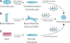
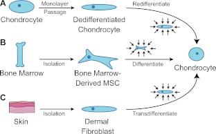
The above image is our intent where we intent to get bone marrow MSC’s to differentiate into chondrocytes.  However, we cannot isolate them in culture.

“Harnessing biomechanics to drive adult cell chondrogenesis. A) Following monolayer culture, chondrocytes rapidly dedifferentiate. Biomechanical stimuli, such as HP, can promote redifferentiation. B) Under mechanical stimulation, mesenchymal stem cells migrate and chondrodifferentiate. C) Mechanical stimulation can be used to induce transdifferentiation into chondrocytes.”

 Human mesenchymal stem cell responses to hydrostatic pressure and shear stress.

“the present study investigated the early responses of human mesenchymal stem cells (hMSCs) to intermittent shear stress (ISS) and to cyclic hydrostatic pressure (CHP) simulating some aspects of the biological milieu in which these cells exist in vivo. Production of nitric oxide (NO) and mRNA expression of several known mechanosensitive genes as well as ERK1/2 activation in the hMSC response to the two mechanical stimuli tested were monitored and compared. NO production depended on the type of the mechanical stimulus to which the hMSCs were exposed and was significantly higher after exposure to ISS than to CHP. At the conditions of NO peak release (i.e., at 0.7 Pa for ISS and 50,000 Pa for CHP), ISS was more effective than CHP in up-regulating mechanosensitive genes. ERK1/2 was activated by ISS but not by CHP. The present study is the first to report that PGTS2, IER3, EGR1, IGF1, IGFBP1, ITGB1, VEGFA and FGF2 are involved in the response of hMSCs to ISS. These findings establish that, of the two mechanical stimuli tested, ISS is more effective than CHP in triggering expression of genes from hMSCs which are bioactive and pertinent to several cell functions (such as cell differentiation and release of specific growth factors and cytokines) and also to tissue-related processes such as wound healing.”

“Exposure of the cell distal membranes to shear stress was achieved by flowing the cell-culture liquid medium through low-wall microchannels (μ-Slide) on whose surfaces (2.5 cm2) the cells were cultured; during these experiments, the medium flow was in the axial direction of the microchannels (μ-Slide).”

“PTGS2, IER3, EGR1, IGF1, IGFBP1, ITGB1, VEGFA and FGF2 genes are expressed in the response of human MSCs exposed to ISS, and that (ii) the FGF2 gene (but neither PTGES, EGR1 or VEGFA genes) are involved in the response of hMSCs to CHP. Last, but not least, the observed expression of mechano-sensitive genes (specifically, PTGS2, IER3, EGR1, IGF1, IGFBP1, ITGB1, FGF2 and VEGFA) returned to basal levels within 6 to 24 h after exposure of the hMSCs to the two mechanical stimuli tested”