Author Archives: Tyler

Evidence that foot length may increase after epiphyseal fusion

If the feet bones can increase in length after epiphyseal fusion perhaps we can use the mechanisms which increase foot length on the long bones.

Gender and age related differences in foot morphology

“Anatomical parameters such as foot length, circumference and height and ankle length, circumference and height were assessed in a sample of males and females divided into three age groups. The groups included young-adult, aged between 20 and 25 years; adult, aged between 35 and 55 years; and old, aged between 65 and 70 years individuals.”

“Comparative analysis of morphometric data between young-adult and adult individuals revealed that the instep length was smaller in adults. The opposite was observed for the great toe and medial foot arch height. Length of ankle was higher in adult than in young-adult individuals, whereas ankle circumference and height were smaller.”<-I don’t know what conclusions to possibly draw from this but there could be something to gather here.

“From a topographic anatomy point of view, foot is in general divided into three parts, namely forefoot, midfoot and hindfoot. The forefoot includes the five toes (phalanges) and the five longer bones (metatarsals). The midfoot represents a pyramid-like association of bones (cuneiform, cuboid and navicular bones) forming the arches of the feet. The hindfoot includes the heel (calcaneus) and the ankle. The heel bone is the largest bone of the foot, whereas the talus bone supports the leg bones (tibia and fibula), forming the ankle”

The elderly is reported to have flatter, longer, and wider feet than younger adults“<-Taken from Age-related differences in foot structure and function.  Although I could not find the information that elderly feet tended to be longer from that study.  They state in regards to this study “Other investigations enlarging their analysis to old people aged 80.2 ± 5.7 years noticed in the elderly group compared with a younger one of the mean age of 20.6 ± 2.6 years flatter, longer, and wider feet than younger adults”

The other citation they use for this is: Foot Health and Shoewear for Women.  And I can’t find anything compelling here other than a statement that a woman’s foot grows larger with age.

“The foot length was measured between the extreme point of heel (foot end) and the extreme point of the longest toe (either first or second toe).”

“The ankle height was considered as the distance between the heel of foot and the line right above the medial malleolus.”

Average foot length of age 35-55 was 269.2 cm and 261.7cm for age 20-25 cm male group.  Foot length was similarly longer for the female middle age versus young adults group.  In males age 65-70 Foot Length was 264.4 cm which is a decrease for middle age but the female group continued to increase in foot length.

So we can’t really conclude that foot length continues to grow with age but this indicates that it may be possible that the feet do grow.

HUGE LSJL update-x-ray proof of LSJL

I loaded my right index finger and my left thumb.  This post isn’t finished because I still have to break things down via gimp and it’s a lot harder to measure the bones than I thought.   But I wanted it up right away to get feedback right away. Maybe someone could help measure the bones of the right and left index finger and the right and left thumb.  I need someone to measure the bones of the right and left index finger to confirm that I’ve grown.  The x-ray’s are standardized as you can see by the centimeter mark on the bottom of the page.  Help me with this and we can prove LSJL.

One thing to note about the x-rays.  Is that the whiter something is on an x-ray the denser it is while the blacker it is the less dense it is.  So darker areas of the bone could be some indication of the possibility of a micro growth plate in that region.

X-Ray left hand:

img007

img009

X-Ray right hand:

img006

img008

No visible growth plates but that does not rule out microgrowth plates.  You’d need a lot more than a an x-ray to detect micro growth plates.  The difference in finger length does not seem to be due to swelling nor are there signs of osteoarthritic degeneration.  The bumps seem to be due ganglion cysts(see below).  The epiphysis on the right clamped finger is noticeably thicker.  If you look at the lateral view of the right versus the left finger then the difference is extreme and noticeable.

Here’s a side to side comparison between the two fingers:

20140829_143800The right index finger is longer than the left although I will have to do further analysis with GIMP or have Michael or someone do independent analysis.  Same with the left thumb(clamped thumb being longer than the right).

Turns out that I don’t really have osteophytes on my finger but really it’s closer to a ganglion cyst.  See this image here of this photo with someone with ganglion cysts and it’s very similar to how my finger looks:

1547906sThe lump is completely in the soft tissue and cannot contribute to height (see the x-rays)

—————————

Michael: This is what I can say currently after studying the X-ray of the left & right index fingers placed right next to each other. I stared for a full 10 minutes at the picture that Tyler uploaded but could not tell any differences in in the distance between the interphylangeal joints of the right hand compared to the left hand, at least for the index fingers. This is from his own comparison pictures.

However, when I took his pictures and put the pictures together (like below), after carefully looking at the X-ray, there does seem to be a noticeable long bone length difference, where the most proximal bone of the right index finger was just SLIGHTLY longer than the one on the left hand. You have to really look at the specific bone very closely but the difference is noticeable, when viewing the left-right hand X-Ray pictures below from a higher up perspective. When they are put to side to side, there is a difference. Of course, then the question would be, was the right index finger always longer than the leftindex  finger to begin with, from natural bone growth, before ever starting LSJL clamping?

When I looked at the left and right thumb bone X-Rays of the lateral perspective, the difference in thickness of the X-Rays is very noticeable. However, to me, it sort of looks like that the RIGHT thumb is thicker laterally than the LEFT one.

The Problem: I am now in a hotel for a conference in San Francisco and I don’t have an exact ruler on hand. All the measurements are done by eye. To be absolutely sure, I would need to find a ruler though, and maybe even go to a Kinkos or FedEx to blow up the X-ray pictures so that a ruler can be used to really do the most exact measurement possible.

What is obvious though is that the overall right index finger is longer. When you compare the skin edge of the right hand index finger to the one on the left hand, it is very noticeably longer.

X Rays of Metacarpal

LSJL may inhibit Src activation in chondrocytes

This isn’t a breakthrough study but it’s always nice to see a new LSJL study coming out.

Distinctive Subcellular Inhibition of Cytokine-Induced Src by Salubrinal and Fluid Flow.

“A non-receptor protein kinase Src plays a crucial role in fundamental cell functions such as proliferation, migration, and differentiation. Inhibition of Src is reported to contribute to chondrocyte homeostasis. In response to inflammatory cytokines and stress to the endoplasmic reticulum (ER) that increase proteolytic activities in chondrocytes, we addressed two questions: Do cytokines such as interleukin 1 beta (IL1β) and tumor necrosis factor alpha (TNFα) induce location-dependent Src activation? Can cytokine-induced Src activation be suppressed by chemically alleviating ER stress or by applying fluid flow? Using live cell imaging with two Src biosensors (i.e., cytosolic, and plasma membrane-bound biosensors) for a fluorescence resonance energy transfer (FRET) technique, we determined cytosolic Src activity as well as membrane-bound Src activity in C28/I2 human chondrocytes. In response to TNFα and IL1β, both cytosolic and plasma membrane-bound Src proteins were activated, but activation in the cytosol occurred earlier than that in the plasma membrane. Treatment with salubrinal or guanabenz, two chemical agents that attenuate ER stress, significantly decreased cytokine-induced Src activities in the cytosol, but not in the plasma membrane. fluid flow reduced Src activities in the plasma membrane, but not in the cytosol. Src activity is differentially regulated by salubrinal/guanabenz and fluid flow in the cytosol and plasma membrane.”

Interesting that fluid flow reduced Src activation in the plasma membrane but the Cytosol and for Salubrinal it was vice versa.  The plasma membrane is the outside covering of the cell so it makes sense to be more affect by fluid flow as it would be in direct contact with it.  Whereas cytosol is the fluid on the inside of the cell. It would be highly significant if we could identify that LSJL mainly affected proteins in the plasma membrane but not so much the cytosol.

“Src is one of the integrin-dependent signaling proteins involved in mechanotransduction, and it plays critical roles in various cellular processes including proliferation, apoptosis, migration, adhesion, and differentiation. To mediate such a variety of cellular processes, Src’s distinct subcellular activation pattern is required. Src is mainly stationed in the cytosol near the endosomes, and activation of Src requires its translocation to the plasma membrane through the cytoskeleton”

“the shear stress of 2–10 dynes/cm2 has been shown to affect chondrocyte signaling and metabolism either positively or negatively”

“Cells were pretreated with Cytochalasin D (CytoD) for 1 h to disrupt the actin cytoskeleton or with MβCD for 1 h to extract cholesterol from the plasma membrane. CytoD partially blocked Cyto-Src activation, and it completely inhibited Lyn-Src activation. MβCD reduced both Cyto-Src and Lyn-Src activations, although to a lesser degree to Cyto-Src. Collectively, these data suggest that the actin cytoskeleton and lipid rafts are essential components for cytokine-induced Lyn-Src activation”

“Lyn-Src was responsive to fluid flow in a magnitude-dependent manner. In response to shear stress at 5 dynes/cm2, a rapid inhibition of Lyn-Src activity was observed (9.7% decrease). In contrast, shear stress at 10 dynes/cm2 led to its activation (14.9% increase). However, Cyto-Src activity was not altered at any magnitude of shear stress”

According to one article, inhibition of Src kinase activates the chondrocyte phenotype.  The article however did not distinguish between Src activities in the cytoplasma or plasma membrane.  It’s possible for instance that only Src activities in the cytoplasm discourage the chondrogenic phenotype and fluid flow would therefore have no effect.

LSJL Progress Update 8-26-14

In my previous progress post I continued to establish that I had in fact grown due to finger clamping.  I have been using the new LSJL method which emphasizes clamping force rather than duration.  I’ve been clamping right index finger, left thumb, elbows, wrists, knee, ankle, calcaneous, and experimentally toes. Michael suggested using two clamps at the same time and I’ll have to try that.  I’ve been trying to find alternative ways to get X-rays but looks like Michael was right and the best bet is Urgent Care.  It’s going to be a pain in the butt but I don’t think I have a choice.

I’ve been gradually increasing the clamping force.  I think it’s key to be clamping on the synovial joint.  Since the bone is so strong, it’s likely that a lot of the LSJL affects are due to clamping the synovial joint.  If a clamp only the epiphysis of one bone than I can seemingly clamp forever but if I clamp more at the synovial joint I can tolerate much less.  It’s interesting to note that I did grow in the arms from a wingspan of 72.5″ to 74.4″ and the way I’m been clamping the elbow is more on the humerus side because the bone structure is so awkward.  So I can’t clamp exactly in the the middle of the humerus and ulna.  However, I have still gotten results there.   So I think it’s more important to clamp in an area where you get some sort of feedback from your body rather than to clamp in some specific spot.  If you can clamp an area forever, you’re doing it wrong.   Right now I’m clamping areas between 100 to 140 seconds and that’s after working up to it.

20140826_123611Here’s another finger angle with the tips aligned.  You can see that the knuckle is higher on the right finger.  But yeah, yeah, yeah,  you have to accept X-rays.  And I have to make sure I get both hands in the x-ray.  Although if there are neo-growth plates in the right finger that would be enough regardless of a comparison.  Although it’s possible that the growth was due to another mechanism say fibrocartilage, etc.

As for my legs and arms, there are signs that I am growing.  Last measured wingspan was 74.5″ but since arms can be stretched a little bit it’s not enough.

As for legs, they seem longer and my jeans and shorts seem to be a little higher but nothing definitive yet.  Now when I have a chance I’m clamping twice a day.  At least once a day seven days a week.

LSJL Update-8-18-2014

So I’m still working on getting a hand x-ray.  I’m contacting facilities to see if they take walk-ins.  No replies so far. I also read you may be able to get an x-ray at the airport(but that was in context of a full body scan)?  I’m looking for alternative medicine facilities that do x-rays but I’ve read they need a doctors script too.  I don’t really want to go do the Urgent Care but it looks like I may not have a choice.  If anyone has any experience let me know. Here’s the last update.

20140817_150801The right finger could be longer since last update but not sure.

Here’s a thumb pic:

20140817_150737I’ve been clamping my left thumb but not my right and from this picture you can see the left thumb is slightly but significantly longer.  The osteophytes are much less noticeable in my left thumb than my right index finger but they are there if I feel around the clamping region.

Interestingly, I clamp each of the three joints of the finger.

Here’s an image of the finger bones:

027 Bones of the hand (dorsal view)I clamp the two interphalangeal joints from side to side  but at the metacarpophalangeal joint I clamp from top to bottom.   The epiphysis near the MP on the index proximal phalanx of my right (clamped) finger is thicker than my left finger but not as enlarged as the epiphysis near the IPs of the distal and middle phalanx.  The IPs of the clamped joints are also rougher than the MP which is clamped overhead rather than side to side.  So some of the enhanced epiphyseal thickness may be due to LSJL growth stimulation and other effects may be due to irritation from the clamping surface on the joint.  If I can get an x-ray it will be interesting to compare the two IPs to the MPs.  For a period of time, I tried clamping the two IPs from top and bottom but didn’t get noticeable results from that.  Top to bottom clamping may be more effective in the MP than the IPs due to differences in joint structure.  You can feel much more pressure generated by top to bottom clamping of the MP than you can the IPs.

As for leg clamping, I am increasing the duration and intensity of the clamping.  I may be getting taller but I cannot yet rule out placebo(like I have with my thumb and index finger which are too noticeable and significant to be placebo) or measurement error.  I grew about 1 1/2 inches in wingspan for sure but I don’t have photographic evidence.  I’ll have to keep monitoring there.  In a week if I don’t have noticeable leg progress, I’ll bite the bullet and go to urgent care to get the x-rays.

Pressure plasma for height

Non-Thermal plasma is plasma that is not in thermodynamic equilibrium.  Atmospheric pressure plasma matches the pressure of the surrounding atmosphere.

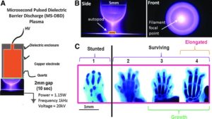
Non-Thermal Atmospheric Pressure Plasma Enhances Mouse Limb Bud Survival, Growth and Elongation.

“appendage regeneration is dependent on reactive oxygen species (ROS) production and signaling. Mesenchymal cell stimulation by non-thermal (NT)-plasma, which produces and induces ROS, would (1) promote skeletal cell differentiation and (2) limb autopod development. Stimulation with a single treatment of NT-plasma enhanced survival, growth and elongation of mouse limb autopods in an in vitro organ culture system. Noticeable changes included enhanced development of digit length and definition of digit separation. These changes were coordinate with enhanced Wnt signaling in the distal apical epidermal ridge (AER) and presumptive joint regions. Autopod development continued to advance up to 144 hr in culture, seemingly overcoming the negative culture environment normally observed in this in vitro system. Real time qPCR analysis confirmed the up-regulation of chondrogenic transcripts. Mechanistically, NT-plasma increased the number of ROS positive cells in the dorsal epithelium, mesenchyme and the distal tip of each phalange behind the AER, determined using dihydrorhodamine. The importance of ROS production/signaling during development was further demonstrated by the stunting of digital outgrowth when anti-oxidants were applied. NT-plasma initiated and amplified ROS intracellular signaling to enhance development of the autopod. Parallels between development and regeneration, suggest the potential use of NT-plasma could extend to both tissue engineering and clinical applications to enhance fracture healing, trauma repair and bone fusion.”

“Vertebrate limbs grow through the coordinated activity of numerous growth factor signaling pathways, including the Wnt, fibroblast growth factor (FGF), bone morphogenetic protein (BMP), Notch, and transforming growth factor-β pathways. ROS play a primary role in mediating the activity of these factors and/or are integrally involved in regulating downstream signaling pathways. For example, a sustained increase in ROS levels is required for Wnt β-catenin signaling and the activation of one of its main downstream targets, fgf20. In addition, naturally occurring oxidative spikes are observed in most of the transition stages throughout developmental and regenerative processes. Furthermore, there is precedence for ROS production to both direct and enhance mesenchymal cell differentiation into either chondrocyte or osteoblast lineages”

“Since the NT-plasma discharge occurs in air, it is a highly complex mix of reactive chemical species (i.e., nitric oxide [NO], ROS, oxygen singles, and ozone), free ions, visible, ultraviolet, and near-infrared light, electric fields, and electromagnetic (EM) radiation.”

“one study showed that a 2 h treatment with EM field stimulated limb growth through increased proliferation and chondrocyte differentiation”

That is an absolutely huge elongation.

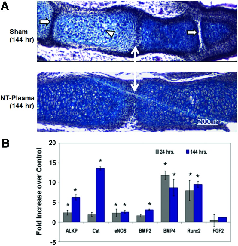
“Morphological formation of the joint region and alcian blue staining of proteoglycan appeared normal in sham and NT-plasma-treated digits. Chondrocytes in the proximal segment of the sham-treated digit were entering the prehypertrophic stage, based on the larger size, greater cellular spacing, and decrease in proteoglycan (white arrowhead). This typically precedes the formation of the primary center of ossification in the diaphysis. In contrast, chondrocytes from the NT-plasma-treated digit were immature and contained large amounts of proteoglycan. Even more striking was the much larger cartilage phalangeal segments in the NT-plasma-treated digit. A white line with double arrows marks the joint between the first and second phalanges in both sections. In the sham-treated digit, the adjacent joint spaces (white arrow) can be clearly observed; whereas in the NT-plasma-treated digits, there is no evidence of the next joint space, due to the increased segment length.”<-So maybe the plasma treatment increases stem cell differentiation to chondrocytes which is what we’d be looking for.

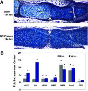
“Histology and quantitative real time-polymerase chain reaction were used to assess chondrogenesis and molecular events associated with the accelerated development. (A) A representative, hematoxylin and eosin (H&E) and alcian blue stained sections of middle phalanges from sham and NT-plasma-treated limb autopods. A white double arrow line marks the first joint space. The white arrows mark adjacent joint spaces, and the white arrowhead marks prehypertrophic chondrocytes in the sham control. (B) At 24 h post-NT-plasma treatment, alkaline phosphate (ALKP), catalase (CAT), endothelial nitric oxide synthase (eNOS), and bone morphogenetic protein 2 (BMP2) expression increased twofold above control. BMP4 and runt-related transcription factor 2 (Runx2) increased 12-fold and 8-fold, respectively, and fibroblast growth factor (FGF)-2 showed no increase. One hundred forty-four hours later, increases for ALKP (6-fold), CAT (14-fold), and BMP2 (3.5-fold). eNOS remained increased twofold above control at 144 h. BMP4 and Runx2 showed a relatively consistent elevated expression at 24 and 144 h. The results are expressed as the mean±standard deviation (n=3 [two limbs pooled per sample], *p<0.05). ”

“NT-plasma treatment promoted both growth and differentiation of cartilaginous elements within the embryonic mouse autopod. Based on the lack of limb growth after NT-plasma treatment in an antioxidant environment, and the increase in H2O2-positive cells in and under the epithelia throughout the 6 day culture period, we propose that NT-plasma induction of intracellular ROS also plays a role in the growth and differentiation observed in the autopod. In addition, H2O2-positive cells were observed in the distal digit tips behind the AER, concurrent with enhanced Wnt signaling. These findings are consistent with the ROS dependence of signaling factors (e.g., Wnt, BMP, and FGF) required for autopod development.”

“ROS production may not be the sole mechanism driving enhanced autopod growth. A second possible reason that NT-plasma enhanced autopod growth was its inhibition of epithelial overgrowth. ”

“In limb regeneration, a thickening of the epidermis has been shown to inhibit signaling to the blastema, halting the regeneration process.”

Hard to determine causality as there are so many potential causal factors as there are so many confounding variables in plasma.