Author Archives: Tyler

Breakthrough study about microgrowth plates

This study is important as although the scientists conclude that interstitial growth of bone is not possible(which would be a huge breakthrough for height growth) it provides a key term to describe the micro-growth plates that are the goal to be formed by LSJL(pseudoarthrosis).  Although this term refers to a fracture case by definition it does show that a microgrowth plate can make a bone longer.  The scientists suggest two possibilities as to how a bone can grow longer after spinal fusion:  either institial growth of the bone itself or psuedoarthrosis(microgrowth plates).  The scientists even state that microscopic areas of pseudoarthrosis may be be responsible for the lengthening in the conclusion.

Thus this study provides additional evidence that microgrowth plates can lengthen bone(as any amount of cartilage is more capable of interstitial growth(which increases bone length) unlike bone) and gives a new term to search for as a key to growing taller: pseudoarthrosis.  The key is to find instances of psuedoarthrosis in adults that are spontaneous and not a result of surgery.  If you want to help find a way to grow taller, helping find such studies would be a great boon.

Bone Growth after Spine Fusion A Clinical Survey

Full study is at the link below.  It should be noted that this study is quite old but it’s still important today because now the focus is more on the gene expression rather than the mechanics as in the old days.  And it’s easier for us to manipulate the mechanics rather than the gene expression.

bone growth after spinal fusion

“When a spine fusion is unquestionably solid and fairly massive, there is little increase in length of the fused area. The small increase that most of the cases we studied showed could be accounted for by magnification and other technical factors, but it is impossible to rule out a small amount of growth. The slight decrease in the kyphos in two of our cases suggests some bending of the fusion mass. No definite pseudarthroses could be seen on the roentgenograms but the presence of one or more pseudarthroses could not be ruled out. Microscopic and transient pseudarthroses are considered by us to be the most likely mechanism by which any real increase in length or any true change in angulation occurs. In our experimental studies, microscopic losses of bone continuity in transepiphyseal bone grafts in the distal femora of young rabbits were demonstrated. These defects were of such a nature that they could not be demonstrated by standard clinical or roentgenographic methods.

In our opinion, the end-result study of Hallock and his associates is valuable in that it indicates, from the practical, clinical point of view, what will happen to the average patient after spine fusion in early childhood. However, their data and the observations of the other authors previously mentioned convey an inaccurate and perhaps unintentional impression that considerable growth occurs in a solidly fused spine segment. It would be unfortunate, we believe, to allow this impression to persist since surgeons not familiar with all that is known about growth of the spine after spine fusion might be falsely encouraged on the basis of published data to perform longer and more massive spine fusions in young children. Our study suggests that a long, massive, and completely solid fusion in early childhood will impair spine growth to a significant degree.

We believe that growth of a fused segment of the spine can occur only at the ends of the segment or at the site of gross or microscopic defects in the fusion plate. Pseudarthroses in spine fusions in children are much more frequent, in our opinion, than is generally suspected because of the tension forces exerted by the growing epiphyseal cartilages, as well as the usual stresses caused by motion. These pseudarthroses or stress fractures may be microscopic or grossly visible; they may occur spontaneously at any time and heal spontaneously. The more massive the fusion plate, the less chance there will be that it will break down under the stress of growth and motion. Finally, we believe that the laws that govern bone growth in general apply to the bone of spine fusions. There is in our opinion no such thing as interstitial growth of bone.”

“all growth in [bone] length of the diaphyses of long boneses takes place at the epiphyseal cartilages, whereas growth of bones its other dimensions occurs through hyaline-cartilage proliferation as in the epiphysis or  through fibrous-tissue proliferation-as in the periosteum and flat bones.”

Scientists reported the apparent growth of fused spinal bones.  If this were to happen in the solidly fused spine two conclusions could be made: either the fusion plate broken down or there was interstitial growth of bone in the fusion plate.

Scientists also observed that  when interbody fusion was performed on the spine.  The bony bridge that was formed elongated in response to vertebral growth.  However, another study found that the fused area remained firm and did not increase in length in response to overall vertebral lengthening.

Another study with spinal fusion found that psuedoarthrosis occured at any interspace[Small hole surronded by bone].  An example of the interspace is perhaps the trabeculae.  Pseudoarthrosis occurs at fracture areas.  The scientists theorized that in their study the longitudinal bone growth was due to these areas of pseudoarhtrosis.

The study mentions Sincher’s law which states that: “increase of pressure or tension beyond the limits of tolerance leads to destruction of bone by resorption.”<-Perhaps LSJL needs to cause this destruction of bone in order to allow for microgrowth plates or psuedoarthrosis and that’s why so far LSJL on the finger has been more successful due to greater ability to increase pressure.

Next steps for proving LSJL

In my last LSJL progress update, the conclusion most people seemed to feel was that people tended to agree that the right index finger was indeed longer than the left index finger but that they weren’t positive that it was due to LSJL and that it wasn’t due to my right index finger being naturally longer than the other.

So here’s some things I’m going to try going further.  I’m going to continue to clamp my right index finger to try to increase the difference from the left index finger.  I’ve also been clamping my left thumb to see if I can get a difference there and there does seem to be a progression.

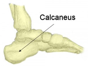
But this does not increase height which has a lot of social value.  For ankle clamping, I was clamping between the tibia and the talus.  Well, I realized that my talus grew bigger.  What if the calcaneus grew bigger than the whole body would be elevated higher increasing height in a manner akin to high heels or shoe lifts.


feet bones

The bone above the calcaneus is the talus(which has grown bigger and may in fact contribute to some of my height gains).  Getting enough force on the calcaneus to induce bone growth is easier as it doesn’t necessarily need to grow by the growth plate as there are no constraining factors on appositional bone growth like the joint does for the long bones.

I’ve done a few clamping sessions aiming for clamping the point between the talus and calcaneus and things seems to be going well.  It’s not as smooth as clamping the fingers as the bones are oddly shaped but I seem to be generating pressure and mechanical strain on the calcaneus.

I think showing calcaneus growth will be a way to inspire people to believe that growing taller is possible and it will also increase the height measurement too.  I’m still working on growing the legs evidence just points to that growth will be slow until a better clamp is developed that can generate more pressure.

Ischemia

Michael talked about Ischemia in the past here.  I found a study earlier that suggested that ischemia of cartilage canals may reduce height growth.  Since intense clamping transiently affects the blood vessels to the cartilage it is important to study this.

Chondro-osseous growth abnormalities after meningococcemia. A clinical and histopathological study.

“The cases of nine children who survived the acute stage of meningococcal septicemia and secondary disseminated intravascular coagulation were reviewed. All of the children had major orthopaedic problems as a result of the acute disease. Detailed histological studies were performed on specimens of bone and cartilage, obtained when these patients had either acute amputation for gangrene or subsequent revision for a chondro-osseous deformity. In the specimens that were obtained from the children who had acute gangrene, the histological changes included small-vessel thrombi[blood vessel clot], osteonecrosis, subperiosteal new-bone formation, cortical disruption, cellular disorganization in the physis, and medullary inflammation. These findings were compatible with a combination of inflammation (acute osteomyelitis) and ischemia. In the specimens that were obtained during revision of the amputation, three years or more after the initial infectious or ischemic process, the clinically relevant findings involved the epiphyses and physes. The growth plates showed variable permanent ischemic damage. Bone bridges connecting the epiphysis and metaphysis were observed in various stages of formation, including several early bridges with involvement of only the physis and metaphysis. Endosteal and cortical bone, in contrast, showed complete recovery with no evidence of permanent ischemic damage. We concluded that children who survive meningococcal septicemia are at high risk for complex orthopaedic problems, both acute and chronic. The disseminated intravascular coagulation and focal infections of the acute phase are primarily responsible for the vascular injuries to the growing chondro-osseous tissues. Ischemic changes also selectively involve the physeal circulation, but may take several years to adversely affect longitudinal and transverse growth of bone.”

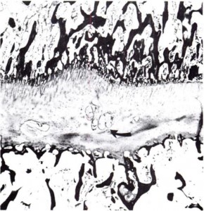
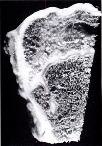
Image of a growth plate clot:

clot in the growth plategrowth plate clot

Growth plate with abnormal “micro” growth plate like structure:

enchondroma

ATP oscillations and Height Growth

I wrote some about calcium secretions(which relate to ATP oscillations) hereTGF-Beta forms pre-chondrogenic mesenchymal condensation(which is what creates the growth plate) via ATP oscillationsATP oscillations also play a role in FGF and Shh mesenchymal condensations.

Analysis of proteins showing differential changes during ATP oscillations in chondrogenesis.

“Prechondrogenic condensation is a critical step for skeletal pattern formation{ie form growth plates}. ATP oscillations play an essential role in prechondrogenic condensation because they induce oscillatory secretion. We examined how differential changes in proteins are implicated in ATP oscillations during chondrogenesis by using liquid chromatography/mass spectrometry. A number of proteins involved in ATP synthesis/consumption, catabolic/anabolic processes, actin dynamics, cell migration and adhesion were detected at either the peak or the trough of ATP oscillations, which implies that these proteins have oscillatory expression patterns that are coupled to ATP oscillations. On the basis of the results, we suggest that (1) the oscillatory expression of proteins involved in ATP synthesis/consumption and catabolic/anabolic processes can contribute to the generation or maintenance of ATP oscillations and that (2) the oscillatory expression of proteins involved in actin dynamics, cell migration and adhesion plays key roles in prechondrogenic condensation by inducing collective adhesion and migration in cooperation with ATP oscillations.”

So we can compare the proteins altered in ATP oscillations to those in LSJL to help see if LSJL induces similar ATP oscillations as those in growth plate chondrogenesis.

“ATP oscillations depend on Ca2+ dynamics.”

“We used the prechondrogenic ATDC5 cell line”<-It would be more ideal if they used normal mesenchymal stem cells as that’s what we’re trying to use to create new growth plates rather than the ATDC5 pre-chondrogenic cells that are like the pre-cursor cells in the Ring of LaCroix.

Peak versus Trough of ATP oscillations.  Genes up and downregulated in LSJL are mentioned in {}

No Peak Trough
1 Obg-like ATPase 1 Suppression of tumorigenicity 5 protein
2 Proteasome subunit alpha type-7 Glucosylceramidase
3 Serine/threonine-protein phosphatase PP1-beta catalytic subunit Alpha-crystallin B chain
4 Hydroxymethylglutaryl-CoA lyase Cytochrome c, somatic
5 Paired box protein Pax-7{down as Pax7a} Arylsulfatase B
6 Arf-GAP with SH3 domain, ANK repeat and PH domain-containing protein 2 Translationally controlled tumour protein
7 Lysosomal protective protein Actin-related protein 2/3 complex subunit 4
8 Protein canopy homolog 4 StAR-related lipid transfer protein 3
9 GRB2-associated-binding protein 3 Transmembrane protein 5
10 Platelet-activating factor acetylhydrolase IB subunit beta Proteolipid protein 2
11 Proteasome subunit alpha type-6 Ribonuclease inhibitor
12 60S ribosomal protein L30 Glycyl-tRNA synthetase
13 26S proteasome non-ATPase regulatory subunit 12 Oxysterol-binding protein-related protein 3
14 Protein CREG1 Forkhead box protein N3
15 Inorganic pyrophosphatase Nuclear transport factor 2
16 Myeloid cell nuclear differentiation antigen-like protein LIM and SH3 domain protein 1
17 Sorting nexin-3 Serine/arginine-rich splicing factor 3
18 Developmental pluripotency-associated protein 4 SAP domain-containing ribonucleoprotein
19 BUD13 homolog Growth/differentiation factor 7
20 cAMP-responsive element-binding protein-like 2 Prostaglandin E synthase 3
21 Tau-tubulin kinase 2 Ornithine aminotransferase
22 Thymosin beta-10 Protein DGCR14
23 40S ribosomal protein S20 Vang-like protein 2
24 Copper transport protein ATOX1 Mitochondrial import inner membrane translocase subunit Tim13
25 Pulmonary surfactant-associated protein D Zinc transporter ZIP8
26 40S ribosomal protein S11 Thrombospondin-3
27 26S protease regulatory subunit 10B Kinase suppressor of Ras 1
28 Haematological and neurological expressed 1-like protein Protein KIAA0284
29 Beta-2-microglobulin 40S ribosomal protein S4, X isoform
30 Vasohibin-2 Galactokinase
31 Ubiquilin-1 Scavenger mRNA-decapping enzyme DcpS
32 Nucleolysin TIAR PHD and RING finger domain-containing protein 1
33 Cleavage stimulation factor subunit 2 Acyl-coenzyme A thioesterase 1
34 Heparanase Aspartyl-tRNA synthetase, cytoplasmic
35 Drebrin-like protein TOM1-like protein 1
36 Interferon regulatory factor 2-binding protein-like Zinc finger CCHC domain-containing protein 14
37 Golgin subfamily A member 2 Anaphase-promoting complex subunit 5
38 Potassium-transporting ATPase alpha chain 1 Armadillo repeat-containing protein 8
39 Collagen alpha-1(XVI) chain{up Col16a1} Coiled-coil-helix-coiled-coil-helix domain-containing protein 2
40 SWI/SNF complex subunit SMARCC1 UPF0568 protein C14orf166 homolog
41 Cleavage stimulation factor subunit 3 Putative potassium channel regulatory protein
42 Centrosomal protein of 170 kDa MAM domain-containing glycosylphosphatidyl -nositol anchor protein 1
43 Zinc finger protein 609 UPF0160 protein MYG1
44 CD44 antigen Nucleoredoxin-like protein 1
45 Transcription factor COE2 Osteoclast-stimulating factor 1
46 Heterogeneous nuclear ribonucleoproteins C1/C2 Pyrroline-5-carboxylate reductase 1
47 Inosine triphosphate pyrophosphatase 40S ribosomal protein S5
48 Latexin Protein SET
49 DNA replication licencing factor MCM2 Succinyl-CoA ligase [GDP-forming] subunit beta, mitochondrial
50 Motile sperm domain-containing protein 2 Anoctamin-1
51 Group XVI phospholipase A2 Annexin A7
52 26S protease regulatory subunit 7 Fibrillin-1
53 Sodium- and chloride-dependent betaine transporter GDP-mannose 4,6 dehydratase
54 Sorcin Homogentisate 1,2-dioxygenase
55 Small ubiquitin-related modifier 2 BTB/POZ domain-containing protein KCTD12
56 Dihydropyrimidinase-related protein 3 Mitogen-activated protein kinase kinase kinase MLK4
57 ATP-dependent RNA helicase DDX39A NEDD4 family-interacting protein 1
58 Eukaryotic translation initiation factor 3 subunit L Secretory carrier-associated membrane protein 1
59 Far upstream element-binding protein 2 Alpha-2,8-sialyltransferase 8E
60 Glypican-5 Cytochrome b-c1 complex subunit Rieske, mitochondrial
61 Intraflagellar transport protein 74 homolog Serine/threonine-protein phosphatase 6 regulatory subunit 2
62 Uncharacterized protein KIAA0141 Interleukin-15 receptor subunit alpha
63 Paralemmin-3 Cell cycle progression protein 1
64 Peroxiredoxin-6 Uncharacterized protein C4orf36 homolog
65 Proteasome subunit alpha type-2 Leukocyte surface antigen CD47
66 Proteasome subunit beta type-3 Cyclic nucleotide-gated cation channel beta-3
67 Reticulocalbin-2 Fibroblast growth factor 14
68 TAR DNA-binding protein 43 Hypoxanthine-guanine phosphoribosyltransferase
69 Ubiquitin-conjugating enzyme E2 K Interferon alpha-12
70 ATP-binding cassette subfamily D member 4 Leucine-rich repeat and IQ domain-containing protein 3
71 Atlastin-3 Neuroplastin
72 Voltage-dependent l-type calcium channel subunit beta-4 Rab proteins geranylgeranyltransferase component A 1
73 Coronin-6 SHC-transforming protein 4
74 Endonuclease/exonuclease/phosphatase family domain-containing protein 1 Vacuolar protein sorting-associated protein 29
75 Exocyst complex component 1 Plakophilin-3
76 Insulin-like growth factor-binding protein 5 Transmembrane protein 223
77 Lysosomal alpha-mannosidase Clathrin light chain A
78 Nucleus accumbens-associated protein 1 Apoptosis-inducing factor 1, mitochondrial
79 Phosphoprotein associated with glycosphingolipid-enriched microdomains 1 Protein-S-isoprenylcysteine O-methyltransferase
80 26S proteasome non-ATPase regulatory subunit 7 Aspartyl aminopeptidase
81 SH3 domain-binding glutamic acid-rich-like protein 3 Tumour protein p53-inducible nuclear protein 1
82 Sepiapterin reductase Nuclear pore complex protein Nup54
83 Tripeptidyl-peptidase 1 ETS domain-containing protein Elk-4
84 V-type proton ATPase subunit d 1 Sideroflexin-3
85 V-type proton ATPase subunit G 1 Ferric-chelate reductase 1
86 Xaa-Pro aminopeptidase 1 Williams–Beuren syndrome chromosomal region 14 protein homolog
87 4F2 cell-surface antigen heavy chain ER degradation-enhancing alpha-mannosidase-like 1
88 Disrupted in schizophrenia 1 homolog Pumilio domain-containing protein KIAA0020
89 Biglycan 25-Hydroxyvitamin d-1 alpha hydroxylase, mitochondrial
90 Apolipoprotein A-I-binding protein Uncharacterized protein C8orf42 homolog
91 Biliverdin reductase A Solute carrier family 12 member 5
92 RNA/RNP complex-1-interacting phosphatase Inosine-5′-monophosphate dehydrogenase 2
93 Homeobox protein Hox-A5 BRCA1-associated RING domain protein 1
94 Dynein light chain 1, cytoplasmic Growth hormone-regulated TBC protein 1
95 Beta-1,3-galactosyl-O-glycosyl-glycoprotein beta-1,6-N-acetylglucosaminyltransferase Calcitonin gene-related peptide 2
96 Arf-GAP with dual PH domain-containing protein 2 ATP-dependent RNA helicase DDX1
97 ATP-binding cassette subfamily A member 8-B Trans-2,3-enoyl-CoA reductase
98 RING finger protein unkempt-like Laminin subunit gamma-2
99 Ankyrin-2
100 Complement C1q subcomponent subunit C
101 Probable ATP-dependent RNA helicase DDX59
102 Homeobox protein Nkx-2.2
103 Heterogeneous nuclear ribonucleoprotein M
104 FtsJ methyltransferase domain-containing protein 1
105 Uncharacterized protein C1orf141 homolog
106 Cell division protein kinase 5
107 Protein tyrosine phosphatase type IVA 1
108 Uncharacterized protein C4orf34 homolog
109 Ephrin-A5
110 Cytochrome b-c1 complex subunit 7
111 Endoplasmic reticulum lectin 1
112 Bifunctional apoptosis regulator
113 Coiled-coil domain-containing protein 27
114 Long-chain specific acyl-CoA dehydrogenase, mitochondrial
115 Iroquois-class homeodomain protein IRX-5
116 Endoplasmic reticulum mannosyl- oligosaccharide 1,2-alpha-mannosidase
117 Cysteine protease ATG4B
118 Galactoside 2-alpha-l-fucosyltransferase 3
119 Apolipoprotein A-II
120 Kelch-like protein 24
121 Serine/threonine-protein kinase Nek5
122 Phosphatidylinositol-4,5-bisphosphate 3-kinase catalytic subunit gamma isoform

With categories

Function Peak Trough
ATP synthesis V-type proton ATPase subunit d 1 Cytochrome c
V-type proton ATPase subunit G 1 Cytochrome b-c1 complex subunit Rieske
Potassium-transporting ATPase alpha chain 1
Cytochrome b-c1 complex subunit 7
Phosphorylation Tau-tubulin kinase 2 Galactokinase
Cell division protein kinase 5 Mitogen-activated protein kinase kinase kinase MLK4
Serine/threonine-protein kinase Nek5
Phosphatidylinositol-4,5-bisphosphate 3-kinase catalytic subunit gamma isoform
Dephosphorylation Serine/threonine-protein phosphatase PP1-beta catalytic subunit Serine/threonine-protein phosphatase 6 regulatory subunit 2
Inorganic pyrophosphatase,
Inosine triphosphate pyrophosphatase
Protein tyrosine phosphatase type IVA 1
RNA/RNP complex-1-interacting phosphatase
Biosynthetic process StAR-related lipid transfer protein 3
Prostaglandin E synthase 3
Pyrroline-5-carboxylate reductase 1
Inosine-5′-monophosphate dehydrogenase 2
Trans-2,3-enoyl-CoA reductase
Catabolic process Proteasome subunit alpha type-7 Ferric-chelate reductase 1
Proteasome subunit alpha type-6 ER degradation-enhancing alpha-mannosidase-like 1
26S protease regulatory subunit 10B 25-Hydroxyvitamin d-1 alpha hydroxylase
26S protease regulatory subunit 7
Proteasome subunit alpha type-2
Proteasome subunit beta type-3
Endoplasmic reticulum lectin 1
Long-chain specific acyl-CoA dehydrogenase
Endoplasmic reticulum mannosyl-oligosaccharide 1,2-alpha-mannosidase
Biliverdin reductase A
Tripeptidyl-peptidase 1
Xaa-Pro aminopeptidase 1
Heparanase
Actin dynamics Thymosin beta-10 Actin-related protein 2/3 complex subunit 4
Alpha-crystallin B chain (stabilize)
LIM and SH3 domain protein 1 (stabilize)
Cell adhesion Collagen alpha-1(XVI) chain Laminin subunit gamma-2
Plakophilin-3
Neuroplastin, leukocyte surface antigen CD47
Thrombospondin-3
Cell migration Platelet-activating factor acetylhydrolase IB subunit beta MAM domain-containing glycosylphosphatidylinositol anchor protein 1
CD44 antigen
Homeobox protein Hox-A5
Cell division protein kinase 5
Ephrin-A5

Comparison to LSJL genes to be done.

” it was found that many proteins involved in cell adhesion, such as laminin subunit gamma-2, plakophilin-3, neuroplastin, thrombospondin-3 and leukocyte surface antigen CD47, were detected only at the troughs of ATP oscillations whereas many proteins involved in cell migration, such as platelet-activating factor acetylhydrolase IB subunit beta, CD44 antigen, homeobox protein Hox-A5, cell division protein kinase 5, and ephrin-A5, were detected only at the peaks of ATP oscillations”

” cell–cell adhesion and cell movement are stimulated at the troughs and peaks of ATP oscillations, respectively, leading to synchronized oscillations of cellular migration during chondrogenesis”

” A nucleation and growth mechanism in which a critical-size aggregate (nuclei) is required for subsequent growth can be applicable to the cellular aggregation process. Therefore, we propose that prechondrogenic condensation proceeds by a nucleation-growth mechanism. This could explain the reason why the oscillatory expression patterns of proteins that are involved in actin dynamics, cell migration and adhesion are required for prechondrogenic condensation. Thermodynamically, a barrier exists in free energy; this barrier occurs at a certain critical size at the initial stage of condensation. Thus, aggregates smaller than the critical size are unstable owing to energy loss due to surface tension, whereas aggregates larger than the critical size grow irreversibly owing to the energy gain due to cellular adhesions surpassing the energy loss, thereby ultimately forming large cellular aggregates. Therefore, the oscillatory expression patterns of proteins involved in actin dynamics, cell migration and adhesion result in collective migration and adhesion, which aggregates cells collectively within a limited time and then drives more efficient formation of the nuclei than random migration and adhesion. Therefore, coordinated oscillatory expression of the proteins is crucial during the initial step of prechondrogenic condensation.”

“platelets are aggregate into clusters at the site of an injury to the skin or blood vessels.”<-Note that blood is involved in distraction osteogenesis

CEBP-Beta

CCAAT/Enhancer Binding Protein β Regulates the Repression of Type II Collagen Expression during the Differentiation from Proliferative to Hypertrophic Chondrocytes.

“we investigated whether C/EBPβ represses type II collagen (COL2A1) expression and is involved in the regulation of sex-determining region Y-type high mobility group box 9 (SOX9), a crucial factor for transactivation of Col2a1. Endogenous expression of C/EBPβ in the embryonic growth plate and differentiated ATDC5 cells were opposite to those of COL2A1 and SOX9. Overexpression of C/EBPβ by adenovirus vector in ATDC5 cells caused marked repression of Col2a1. The expression of Sox9 mRNA and nuclear protein was also repressed, resulting in decreased binding of SOX9 to the Col2a1 enhancer. Knockdown of C/EBPβ by lentivirus expressing shRNA caused significant stimulation of these genes in ATDC5 cells. Reporter assays demonstrated that C/EBPβ repressed transcriptional activity of Col2a1. Deletion and mutation analysis showed that the C/EBPβ core responsive element was located between +2144 and +2152 bp within the Col2a1 enhancer. EMSA and ChIP assays also revealed that C/EBPβ directly bound to this region. Ex vivo organ cultures of mouse limbs transfected with C/EBPβ showed that the expression of COL2A1 and SOX9 were reduced upon ectopic C/EBPβ expression. Together, these results indicated that C/EBPβ represses the transcriptional activity of Col2a1 both directly, and indirectly through modulation of Sox9 expression. This consequently promotes the phenotypic conversion from proliferative to hypertrophic chondrocytes during chondrocyte differentiation.”

So maybe knockdown of CEBP-Beta could increase height?  It upregulates chondrogenic genes but we’d have to see the effects on the growth plate to be sure.

“The expression of COL10A1, RUNX2, and MMP13 was misexpressed through the tibias that were transfected with C/EBPβ, compared with LacZ control. Forced expression of C/EBPβ may lead the ectopic expression of these genes even in the regions that do not show the morphological hypertrophy because C/EBPβ is reported as a direct regulator of them. Moreover, the expression of SOX9 was also decreased and restricted to a small upper area of the growth plate by overexpression of C/EBPβ, similar to the expression of COL2A1. Together, these results further confirmed that C/EBPβ could be involved in regulation of phenotypic conversion from proliferative to hypertrophic chondrocytes by repressing the genes characteristic of proliferative chondrocytes during chondrocyte differentiation.”

Isopsoralen for height growth?

Isopsoralen is also known as Angelicin and is found in Bituminaria bituminosa.  This plant does not seem to be currently available in supplement form.

Isopsoralen Induces Differentiation of Prechondrogenic ATDC5 Cells via Activation of MAP Kinases and BMP-2 Signaling Pathways.

[ATDC5 cells are chondrogenic progenitor cells so it’s much easier to get them to differentiate into chondrocytes than Mesenchymal Stem Cells.  But ATDC5 cells are like prechondrogenic growth plate cells so it may have applications to people with growth plates]

“Endochondral bone formation is the process by which mesenchymal cells condense to become chondrocytes, which ultimately form new bone.  We investigated the possible role of isopsoralen in induction of chondrogenic differentiation in clonal mouse chondrogenic ATDC5 cells. Isopsoralen treatment stimulated the accumulation of cartilage nodules in a dose-dependent manner. Further, ATDC5 cells treated with isopsoralen were stained more intensely with Alcian blue than control cells, suggesting that isopsoralen increases the synthesis of matrix proteoglycans. Similarly, isopsoralen markedly induced the activation of alkaline phosphatase activity compared with control cells. Isopsoralen enhanced the expressions of chondrogenic marker genes such as collagen II, collagen X, OCN, Smad4 and Sox9{all upregulated in LSJL except for Smad4} in a time-dependent manner. Furthermore, isopsoralen induced the activation of extracellular signal-regulated kinase (ERK){stimulated by LSJL} and p38 MAP kinase{LSJL likely upregulates p38}, but not that of c-jun N-terminal kinase (JNK). Isopsoralen significantly enhanced the protein expression of BMP-2 in a time-dependent manner. PD98059 and SB 203580, inhibitors of ERK and p38 MAPK, respectively, decreased the number of stained cells treated with isopsoralen.  Isopsoralen mediates a chondromodulating effect by BMP-2 or MAPK signaling pathways.”

“the upregulation of BMP-2 causes cells to skip cellular condensation stages in early-phase chondrogenic differentiation and also markedly up-regulates the expression of type X collagen mRNA in late-phase differentiation”