Author Archives: Tyler

Need your feedback: 1/4″ finger length increase and new clamping device is key to leg growth

Here’s the before progress with the initial pic of the finger.  Here’s the last pic of the finger

20140429_12175020140429_12173020140429_121710

You can see a comparison of the left and right fingers and see that the right finger is longer than the left finger and much thicker.

Here’s a side to side comparison of the left and right fingers:

20140501_080453

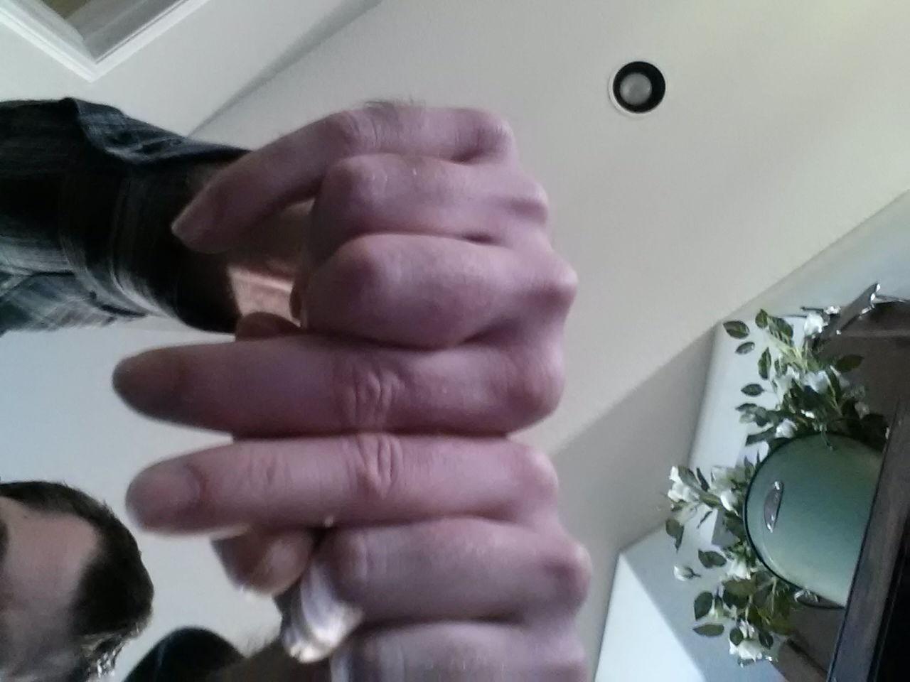
You’ll notice that the right finger is longer than the left finger and not that the right knuckle is higher than the left knuckle so there’s no measurement shennanigans.  Is this pic enough to push as proof that LSJL finger growth is a success?  Let me know if you’d suggest any adjustments.

Here’s another image:

20140502_101938

Both the right finger is longer than the left finger at the tip and the right knucle is higher than the left knuckle.  Nails on the bother inner most fingers are trimmed.

Here’s some pictures of my right hand from four years ago.  Note that the method is obsolete but you can see a finger length increase between then and today.

Here’s a video with the right hand as well:

I found this video about performing LSJL:

http://youtu.be/sWlxaXfUeqM

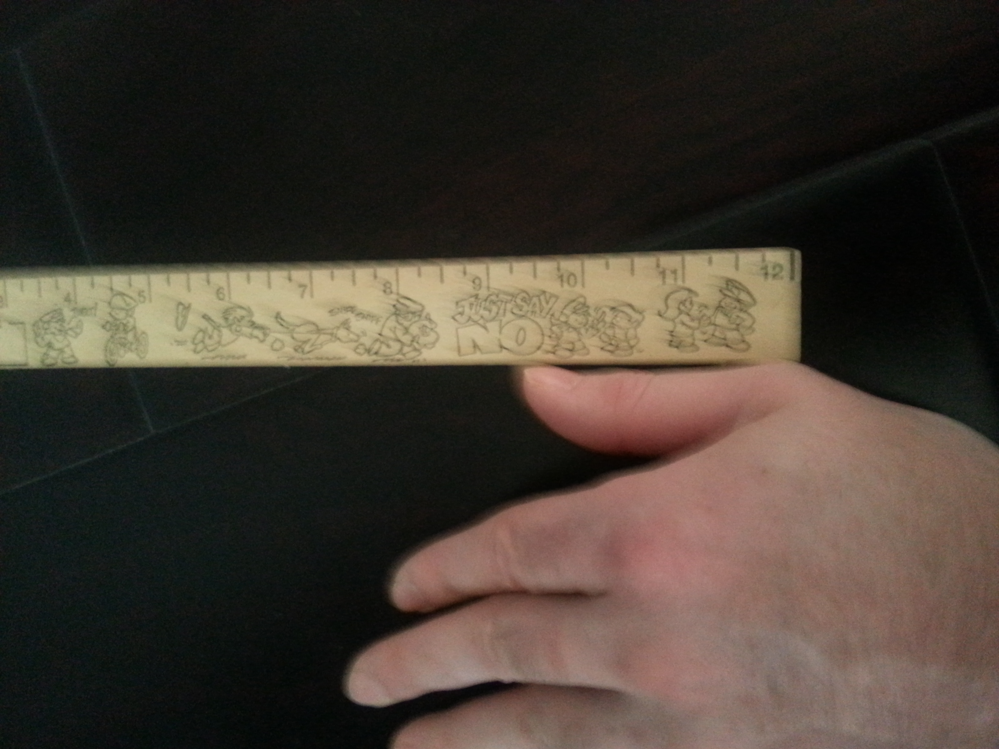
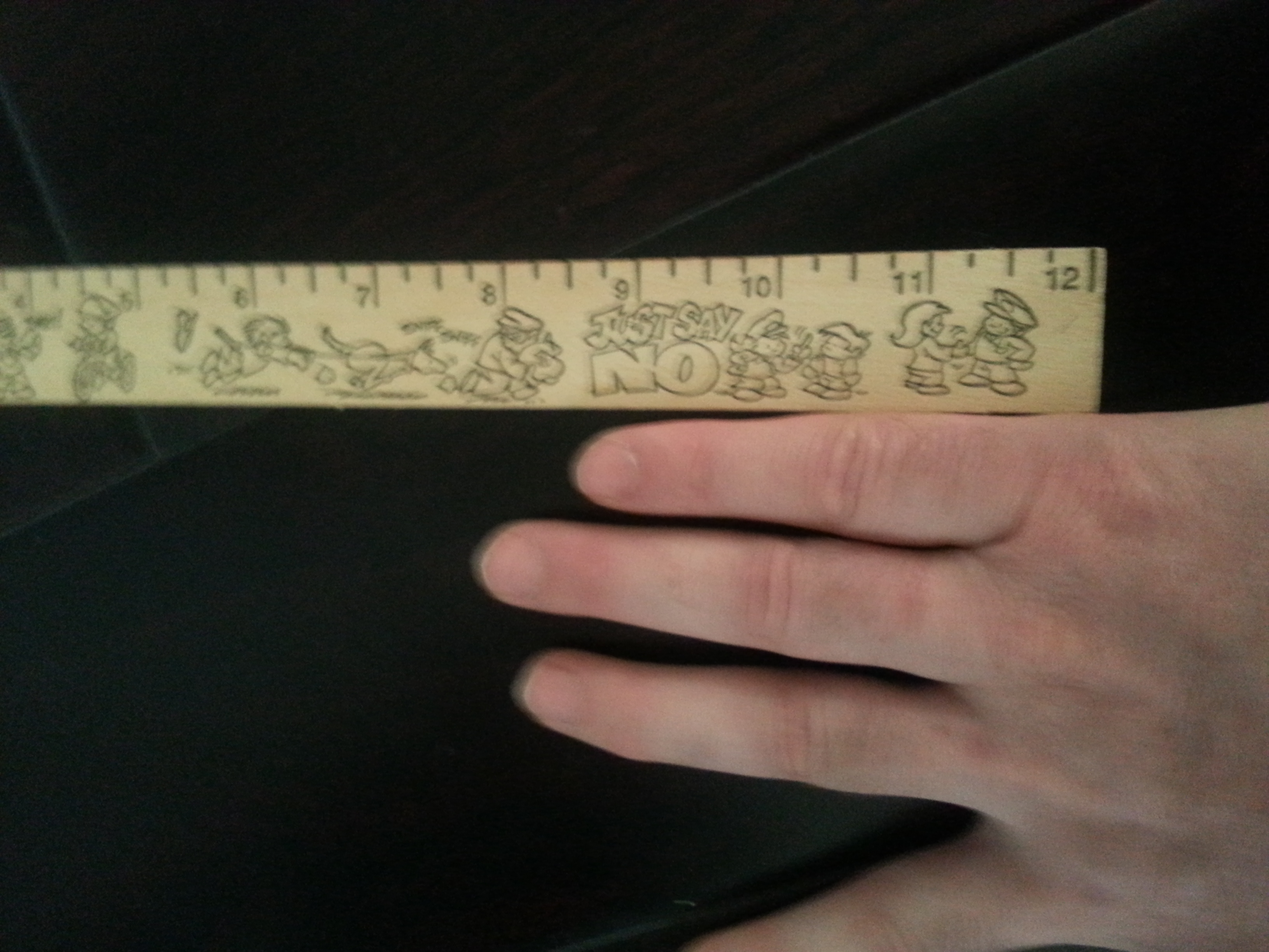
This individual got results after 20 days which is likely due to the superior clamping device.   And is more in line with the finger results I’ve gotten.  I’ve gained about a 1/4 of inch in finger length and a drastic increase in finger width.  Comparing the thumb pics it looks like there may be some length gain there but the thumb is farther along the ruler in the before pic but a side to side comparison of right to left thumb reveals that a possible increase of 1/8th of an inch or so.

So it’s been about 1 3/4 year loading my finger and I’ve gained say 1/4″.  Since my finger is around 3 1/2″ long that’s about a 7% increase.  Now a 7% increase in the tibia bone of say 16 inches would be 17.02 inches which would be a full inch of height.  Now other than the initial gain in height at the beginning I have not gained significantly over time.

Now the key thing is despite my initial gain I’m not gaining length as rapidly in the legs as in the fingers and I’m not getting the drastic increase in width in the legs as in the fingers.  This could be due to physiological differences between the leg and finger bones.  The finger bones do not have periosteum for example or it could be due to the existing clamping device not being sufficient enough load.  The load I’ve been using for 500 counts on the finger is quite extreme relative load.

I’ve been tweaking the load of the legs.  Changing clamp position and bending the knee while loading the knee to get more leverage but we need to find a way to get the same kind of loads that can be done with the finger.

Importantly, we have finger proof.  Longer fingers could be valuable to piano players and guitar players and possibly some other athletes.  So how can we use this finger technique to raise funds to develop a more effective leg clamping device.  I think we can go even further than this gentleman here in developing a better clamping device.

So:

What kind of evidence and documentation do I need to present to the layperson to show the finger proof of concept to generate interest and funding in developing a leg loading device?

New Jeffrey Baron study about Growth Plate Regulation

Jeffrey Baron is one of the scientists studying growth plate senescence so his insights could be very helpful in stopping growth plate closure and forming new growth plates.

Recent insights into the regulation of the growth plate.

“Expression in the resting and the proliferative zone was compared to identify pathways involved in the differentiation of resting zone to proliferative zone chondrocytes. This analysis implicated vitamin D receptor / retinoid x receptor (VDR/RXR) activation, platelet-derived growth factor (PDGF) signaling, BMP signaling, and notch signaling. Similar analysis of the proliferative to hypertrophic differentiation step implicated p53 signaling, ephrin receptor signaling, oncostatin M signaling, and BMP signaling”

“evidence for a BMP signaling gradient across the growth plate with the greatest BMP signaling occurring in the hypertrophic zone and the least in the resting zone”<-Maybe this BMP signaling gradient is caused by some other gradient like a pressure gradient.   We know that LSJL induces a gradient so this could be related to LSJL methodology.

“immunolocalization of phosphorylated SMAD-1, – 5, and -8 in the growth plate increases with increasing distance from the epiphysis”

“Low levels of BMP signaling in the resting zone may help maintain the progenitor cell state. Farther from the epiphysis, greater BMP signaling may induce differentiation to proliferative chondrocytes and, even farther from the epiphysis, yet greater BMP signaling may induce terminal differentiation to hypertrophic chondrocytes.”

The BMP gradient described in figure 2 of the study:

Resting Zone: Bmp-3, Gremlin, Chordin

Proliferative Zone:  GDF10, BMP7

Hypertrophic Zone: Bmp2, BMP6

“In the embryonic skeleton, PTHrP is secreted by periarticular chondrocytes of long bones. PTHrP diffuses across the growth cartilage maintaining chondrocytes in the proliferative state. Cells more distant from the source of PTHrP undergo hypertrophic differentiation.”<-LSJL also puts load on the articular cartilage.  Maybe LSJL induces the release of PTHrP.

“The prehypertrophic and hypertrophic chondrocytes then secrete Indian hedgehog (Ihh), which has a negative-feedback effect on PTHrP production and also independent effects on chondrocyte differentiation. More recent evidence suggests that the Ihh–PTHrP system is maintained in postnatal growth plate but the PTHrP source shifts to the resting zone”<-Some more on PTHrP and chondrocyte differentiation.

Genes involved in human growth plate function:

“the IHH-PTHrP system (GLI2, IHH, HHIP, PTCH1, and PTHLH lie within GWAS loci), BMP/TGF superfamily signaling (TGFB2, BMP6, LTBP3, NOG, BMP2, GDF5), C-type natriuretic peptide signaling (NPPC, PRKG2, NPR3), GH-IGF-I signaling (IGF2BP2, IGF2BP3, IGF1R), and FGF signaling (FGF18).”

“At the molecular level, CNP inhibits the extracellular signal-regulated kinase (ERK) and p38 mitogen activated protein kinase (MAPK) pathways, therefore counteracting the growth-inhibitory downstream signaling of fibroblast growth factor (FGF) in the growth plate”

“BNP [which is involved in growth plate regulation] is transcriptionally regulated by the transcription factor SHOX [which also regulates the growth plate]”

“FGFR1 and FGFR3 signaling are growth-inhibiting, while FGFR2 signaling is growth-promoting. Cartilage-specific (Col2a1-Cre) inactivation of Fgfr1 in mice showed a transient increase height in hypertrophic zone, and delayed terminal differentiation of hypertrophic chondrocytes. However, increase in adult body length has not been reported. In contrast, inactivation of Fgfr2 in the mesenchymal condensations (Dermo1-cre), which affects both the osteoblast and chondrocyte lineages, resulted in mice with skeletal dwarfism”

“transgenic mice with activated Fgfr3 in the growth plate show reduced chondrocyte proliferation, decreased numbers of hypertrophic chondrocytes and decreased height of the hypertrophic zone, while Fgfr3 knockout mice showed increased chondrocyte proliferation, increased height of hypertrophic zone, and increased skeletal growth”

“Fgf9 and Fgf18 promote chondrocyte proliferation during early development of the growth plate, but then function to inhibit chondrocyte proliferation and promote hypertrophic differentiation at later stages of development.”

“FGF21 can activate FGFR1 and FGFR3, both of which elicit growth-inhibitory signaling”

“Fgf21 knockout mice showed no significant difference in body weight and body length as compared to wild type mice”<-FGF21 may play a role in fasting induced growth inhibition.  So FGF21 would be a safe target to inhibit for growth.

“GH has no apparent role in fetal growth, despite the presence of its receptor (GHR) in embryos”

“mice lacking both the GH receptor and IGF-I have shorter bones than mice lacking only IGF-I, suggesting that GH, at least at a super-physiologic circulating concentrations, has an IGF-I-independent effect on bone growth”

“GH-induced Socs2-/-  metatarsal bone growth is not accompanied by increase in Igf1 or Igfbp3 transcript levels, and occurred in the presence of an IGF-I receptor inhibitor”

Manual of Endocrinology and Metabolism(Chapter 4: The growth Plate)

IGF2 is downregulated a thousand fold during senescence.

High levels of estrogen cause cessation of growth due to estrogen receptors.

Environmental Inputs on Longitudinal Bone Growth

Environmental temperature impact on bone and cartilage growth.

“Environmental modulation of tissue temperature can have direct and immediate consequences on cell proliferation, metabolism, matrix production, and mineralization in cartilage. Temperature can also indirectly influence cartilage growth by modulating circulating levels and delivery routes of essential hormones and paracrine regulators.”

environmental inputsThe arrows in white are the functional purposes of the bone.  In red are the factors that can affect bone length.  Bone adapts to the demands placed upon but not always in the way we want.  It doesn’t matter if stretching to us makes sense to use to produce longer bones.  It makes what the bones think.  The bone thinks to think:  I’m being stretched I should grow longer.  We need to recreate specific stimulus in the past that produces the desired results.  For example, the growth plate which results in longitudinal bone growth is a result of mesenchymal condensation of progenitor cells.  The goal of LSJL is to create a stimuli of hydrostatic pressure and shear strain that has been shown to induce mesenchymal condensation in adult mesenchymal stem cells and in turn induce chondrogenesis.

“Chondrocytes differentiate from a reserve pool of round quiescent cells into flattened columns of proliferating cells, which then mature into large terminally differentiated hypertrophic cells, and are ultimately replaced with mineral at the chondro-osseous junction where bone-forming osteoblasts invade from the metaphyseal vasculature”

“limb length correlates with temperature and latitude”

“limb length can be modified within a single generation by rearing young littermate animals at warm and cold temperatures during the postnatal growth period”

“Growth plate morphology was unexpectedly similar at cold and warm temperatures.  There were no major appreciable differences in overall size, shape, or organization of the cartilage.”

“variation in adult long bone length between inbred mouse strains was primarily generated by growth rate differences that occurred during a phase of rapid bone elongation between 3 and 5 weeks age.”<-it could be that lack of enough temperature is a limiting factor on height growth only in the most rapid periods of growth.

Usually these temperature studies are capped at one point in temperature so it’s hard to see if there’s an equilibrium temperature.

“normal capillary blood flow in the skin of the tibia (lower leg) was significantly higher than that in the metatarsal (foot) region, indicating a natural proximal-distal gradient in blood supply.”

” at high temperature extremes outside of a tolerable physiological range, heat does negatively impact body growth.”

“Without countercurrent heat exchangers in a cold environment, blood will rapidly lose temperature as it flows to the distal-most parts of the appendages. Core temperature would consequently drop as cool venous blood returns to the body core. One mechanism to combat a whole body cooling effect is to reduce total outflow of blood flow to the appendages by regulating vasomotor tone”

“This initially perplexing finding (no major differences in morphology of the growth plate when bone lengths did differ) may actually be quite informative as to the mechanism of the growth effect. One possibility is that a complex change in gene expression, cell metabolic activity, and/or maturation rate could contribute to the growth rate differences without changing the superficial appearance of the cartilage. The other corresponding possibility is that the growth rate differences were too slight to detect in the static histology “snapshot.” The total difference in tibia bone length between warm and cold was approximately 370 μm”

The study mentions that heat can induce bone marrow expansion.  Equilibrium temperature can increase calcium uptake.  Cold temperature induces endoplasmic reticulum stress.  HSP70 expression is affected by temperature.  Hydrostatic Pressure also increases HSP70.  HMGB1 is negatively correlated with temperature.  HMGB1 seems to encourage the early stages of chondrogenesis.  This would be a good thing in the formation of the growth but could be a bad thing in the later stages as it may effect the degree of endochondral ossification of the growth plate reducing longitudinal bone growth.  But mice that lack HMGB1 have shorter bones.  Heat also can incude TRPV4 activation.  TRPV4 may modulate bone elongation by affecting cell volume in chondrocytes.  However, continuously active TRPV4 mutants have abnormal endochondral ossification and reduced longitudinal bone growth.

” warm temperature could facilitate bone elongation by increasing the number of cells that could become bone forming osteoblasts at the chondro-osseous junction. There is also evidence that cold could limit bone elongation by directly inhibiting osteoblast activity.”<-Which would mean that heat wouldn’t play a role in neo-growth plate formation.

” leptin deficiency produces a contrasting phenotype in the limb bones and spine (short femurs and long spine)”

“Blood flow could be permanently modified by temperature through the route of new vessel formation, or angiogenesis”

“disrupted blood flow markedly impairs limb growth”

“surgical obstruction of the principal nutrient artery within the tibia marrow cavity of rabbits caused blood to be shunted toward the metaphyseal vessels and increased blood flow to the growth plate. Within three months after the surgery, the operated tibia had grown significantly longer compared to the contralateral side that received the normal blood supply”

So blood flow seems to be the most promising way to increase longitudinal bone growth and inducing new growth plates is not a ruled out effect of increasing blood flow so that LSJL likely increases blood flow is a good sign.

MISC LSJL and Salubrinal studies(but bone effects only)

The LSJL scientists Yokota and Zhang posted another LSJL related study.  Not a lot can be gathered in relation to LSJL on height growth unless Nfatc1 or ATF4 can be found to be chondrogenic inducers or to increase peak chondrocyte hypertrophy.

Evaluating treatment of osteoporosis using particle swarm on a bone remodelling mathematical model.

“The model formulated a temporal BMD change of a mouse’s whole skeleton in response to ovariectomy, mechanical loading[LSJL] and administration of salubrinal.”<-So even though this study is studying mainly BMD we can extrapolate other LSJL effects via LSJL’s affect on gene expression and cells.

“The best treatment was found to start with mechanical loading followed by salubrinal.”

“Ovariectomy (OVX) was modelled through oestrogen deprivation, whereas salubrinal injection and knee loading were modelled by up-regulating p-eIF2α and inhibiting sclerostin, respectively.”<-In studying eif2a, it was unclear whether it could help increase height.

“To model the observed non-responsiveness to salubrinal at normal oestrogen levels in control mice, the model included a range of p-eIF2α values that does not elicit a change in ATF4 and NFATc1. When oestrogen levels decrease as in OVX mice, this range becomes
smaller, and ATF4 and NFATc1 become more responsive to changes in p-eIF2α.”<-ATF4 has some involvement in height but it doesn’t seem to be a powerful effector like say CNP.

Loading BMD

This could connect to cartilage growth via NFATc1 or ATF4.  NFATc’s do affect chondrogenesis.

“Knee loading was applied using a custom piezoelectric loading platform in the lateral-medial direction 3 min/day at 15 Hz, with a peak-to-peak force of 0.5 N”<-LSJL.  Mice were 12 weeks old at time of treatment.

Hydrogel-Based Local Release of Salubrinal Stimulates Healing of Mouse Tibia Fracture

“Salubrinal is a synthetic compound (C21H17Cl3N4OS; 480 Da) which is known to reduce various cellular stresses including stress to the endoplasmic reticulum. It inhibits serine/threonine protein phosphatase 1 alpha (PP1), followed by the elevation of phosphorylated eukaryotic translation initiation factor 2 alpha (eIF2α). Salubrinal is reported to enhance bone formation by stimulating Activating Transcription Factor 4 (ATF4), one of the transcription factors for bone formation, via eIF2α-mediated signaling and stimulating development of bone-forming osteoblasts. It also suppresses nuclear factor of activated T-cells, cytoplasmic 1 (NFATc1), a master transcription factor for osteoclastogenesis, and inhibits development of bone-resorbing osteoclasts. It reduces inflammation and degradation of cartilage tissues{maybe it can delay growth plate cessation?}”

“C57BL/6 female mice (14 weeks, body weight ~20 g”

Here’s how the fracture was induced:

orthopedic-muscular-system-tibia-fracture-study-5-220-g001

“salubrinal can add calcified mass to osteoporotic bone{maybe salubrinal encourages endochondral ossification}?”

orthopedic-muscular-system-micro-ct-images-5-220-g003

In the stump on the left side the salubrinal group looks taller but less wide.

“salubrinal suppresses the proliferation and maturation of osteoclasts by downregulating AP-1 proteins such as c-Fos and JunB, as well as NFATc1”

Salubrinal improves mechanical properties of the femur in osteogenesis imperfecta mice.

“Salubrinal is an agent that reduces the stress to the endoplasmic reticulum by inhibiting de-phosphorylation of eukaryotic translation initiation factor 2 alpha (eIF2α). We and others have previously shown that the elevated phosphorylation of eIF2α stimulates bone formation and attenuates bone resorption. In this study, we applied salubrinal to a mouse model of osteogenesis imperfecta (Oim), and examined whether it would improve Oim’s mechanical property. We conducted in vitro experiments using RAW264.7 pre-osteoclasts and bone marrow derived cells (BMDCs), and performed in vivo administration of salubrinal to Oim (+/-) mice. The animal study included two control groups (wildtype and Oim placebo). The result revealed that salubrinal decreased expression of nuclear factor of activated T cells cytoplasmic 1 (NFATc1) and suppressed osteoclast maturation, and it stimulated mineralization of mesenchymal stem cells from BMDCs. Furthermore, daily injection of salubrinal at 2 mg/kg for 2 months made stiffness (N/mm) and elastic module (GPa) of the femur undistinguishable to those of the wildtype control. Collectively, this study supported salubrinal’s beneficial role to Oim’s femora. Unlike bisphosphonates, salubrinal stimulates bone formation. For juvenile OI patients who may favor strengthening bone without inactivating bone remodeling, salubrinal may present a novel therapeutic option.”

Salubrinal downregulated Nfatc1 in MSC like cells.  If cells are less likely to become osteoclasts it is more likely more them to become cartilage.

New insight on Growth Hormone

The Influence of Growth Hormone on Bone and Adipose Programming.

“In utero growth hormone exposure is associated with distinct immediate growth responses and long term impacts on adult physiological parameters that include obesity, insulin resistance, and bone function. Growth hormone accelerates cellular proliferation in many tissues but is exemplified by increases in the number of cells within the cartilaginous growth plate of bone{can it increase the number of growth plate progenitor cells?}. In some cases growth hormone also potentiates differentiation as seen in the differentiation of adipocytes that rapidly fill upon withdrawal of growth hormone. Growth hormone provokes these changes either by direct action or through intermediaries such as insulin-like growth factor-I and other downstream effector molecules. The specific mechanism used by growth hormone in programming tissues is not yet fully characterized and likely represents a multipronged approach involving DNA modification, altered adult hormonal milieu, and the development of an augmented stem cell pool capable of future engagement as is seen in adipose accrual.”

“Early therapeutic provision of GH to SGA[small for gestational age] neonates having sufficient GH enhances the velocity of bone growth transiently but only for the duration of GH treatment”

“between birth and 28 days is most influential on bone elongation and adult size. Initiating the elevated GH beyond 28 days of age increases growth but not to the
extent realized with earlier exposure despite the presence of a functional growth plate. At the cellular level, GH accelerates bone growth by hyperplasia[an increase in the number of cells] as opposed to growth plate chondrocyte hypertrophy”<-hyperplasia is more powerful than chondrocyte hyertrophy if it increases the amount of growth plate progenitor cells.  If it only increases chondrocyte proliferation then the effect is transient as chondrocytes have a finite proliferative capacity.

Lengthening bone via microcracks, why hasn’t it work?

Now obviously, if walking was able to increase height people would be a lot taller?  But if repetitive stress can induce plastic changes in cortical bone that could possibly contribute to height gain.  More on plastic deformation hereMichael talks about the definition of Plastic deformation here and he talks about the values needed to induce plastic deformation here.

One possibility as to why inducing microcracks to cause plastic(permanent) changes in bone length is that most microcracks such as those induced by walking would be more compressive microcracks rather than tensile strain microcracks which would be what would be needed to make a bone longer.  One study finding longitudinal decrease in height in the lower legs would be consistent with the fact that typical athletic stimuli induces compressive changes rather than lengthening changes. Although another study also mentioned in the same post found that exercise increased height slightly in the spine. Although it would be logical to think that kicking with ankle weights would be one such stimuli to induce longitudinal plastic deformation of the bone.

Effects of fatigue induced damage on the longitudinal fracture resistance of cortical bone.

“As a composite material, cortical bone accumulates fatigue microdamage through the repetitive loading of everyday activity (e.g. walking). Twenty longitudinally orientated compact tension fracture specimens were machined from a single bovine femur, ten specimens were assigned to both the control and fatigue damaged groups. The damaged group underwent a fatigue loading protocol to induce microdamage which was assessed via fluorescent microscopy. Following fatigue loading, non-linear fracture resistance tests were undertaken on both the control and damaged groups using the J-integral method. The interaction of the crack path with the fatigue induced damage and inherent toughening mechanisms were then observed using fluorescent microscopy.  Fatigue induced damage reduces the initiation toughness of cortical bone and the growth toughness within the damage zone by three distinct mechanisms of fatigue-fracture interaction. Further analysis of the J-integral fracture resistance showed both the elastic and plastic component were reduced in the damaged group. For the elastic component this was attributed to a decreased number of ligament bridges in the crack wake while for the plastic component this was attributed to the presence of pre-existing fatigue microcracks preventing energy absorption by the formation of new microcracks.”

“The composite structure of bone consists of three main components, this includes: collagen type I (organic phase), calcium hydroxyapatite (inorganic or mineral phase) and water [8]. These components are arranged hierarchically from macro to nano scale giving bone distinct material behaviour at each characteristic scale. This composite structure causes bone to form microdamage due to fatigue loading. In bone there are two main types of fatigue damage observed (linear microcracks and diffuse damage), which can be linked to different modes of loading. Another consequence of the composite structure of bone is that it exhibits rising fracture toughness with crack extension (i.e. a rising fracture resistance curve). As a macrocrack propagates through bone, microcracks form in the process zone at the tip of the main propagating crack. This damage can act to reduce the driving force available for crack growth by dissipating energy away from the main crack extension and also provides initiation sites for toughening mechanisms, such as uncracked ligament bridges and crack deflection. Pre-existing fatigue microdamage in the crack path can potentially interact with these mechanisms and alter the fracture resistance.”

In the study a microcrack was induced with a drill so it’s hard to extrapolate the results here to what would happen in various of forms of mechanical stimuli.

microcracks

This image shows microcracks in bone.  You can see the drill induced crack on the part of a directly above c.

“a crack path of a typical control specimen showing various toughening mechanisms along the crack path: b microcracking ahead of the tip of the main crack, c several ligament bridges and d deflection and ligament bridge formation.”<-I think this kind of cracking could induce longitudinal bone growth however it would require an initial macrocrack such as that induced by the drill.  If you look at the a image directly above d the ligament bridging covers the entire length of the bone which could potentially result in total longitudinal bone growth.

more microcracks

“Typical damaged specimen following resistance curve testing. The approximate location of the pre-existing fatigue microcracks are marked using white lines. Scale bar is 250 µm”

Exercises like kicking with ankle weights can cause microcracks which can increase the likelihood of getting those macrocracks which can result in plastic deformation like longitudinal bone length increase.  But macrocracks are likely needed to induce that plastic strain in bone and only extreme ankle weight kicking is likely to induce that.

The bone constantly changes in length throughout the day by definition of microstrain.  Just cause a “macrocrack in bone” whole the bone is slightly lengthened and repeat and you should gradually grow taller.