Author Archives: Tyler

Growth plate versus articular cartilage

Regulation of growth plate and articular chondrocyte differentiation : implications for longitudinal bone growth and articular cartilage formation

“the embryonic Ihh/PTHrP feedback system is maintained postnatally except that the source of PTHrP has shifted to a more proximal location in the resting zone.”

“in articular cartilage, superficial chondrocytes differentiate from chondrocytes in the deeper layers following a program that has some similarities to the hypertrophic differentiation program in growth plate cartilage.”

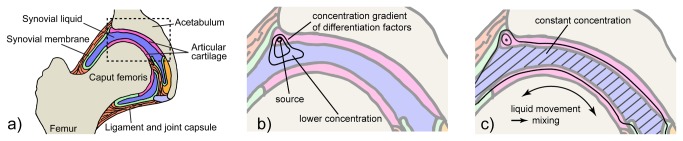
“transplanting growth plate cartilage to the articular surface in an EGFP rat model that enabled cell tracing. We found that hypertrophic differentiation appeared to be inhibited in growth plate cartilage transplanted to the articular surface. The transplanted cartilage also underwent structural remodeling into articular-like cartilage, which suggests that the synovial microenvironment inhibits hypertrophic differentiation and promotes articular cartilage formation.”<-So if we change the bone microenvironment we could encourage growth cartilage.

“An essential genetic switch for patterning of skeletal elements is the expression of Hox genes, which encode a highly conserved family of transcription factors. Mesenchymal condensations are initially uninterrupted and at a later time differentiate into chondrocytes that express type II collagen”

“At the bottom of the proliferative zone, chondrocytes stop proliferating and undergo hypertrophy, a process characterized by gains in cell height, intracellular volume, and organelle size up to 4-, 10-, and 3-fold, respectively, and that also contributes to longitudinal bone growth”

“infection of embryonic chick limbs with retroviruses encoding BMP-2, -4, and GDF-5 increased chondrogenesis and final sizes of skeletal elements”

“in vitro administration of BMP-2 to rat fetal metatarsal bones or mouse embryonic stem cell lines increased chondrocyte proliferation and hypertrophy, whereas addition of Noggin elicited the opposite effect of preventing hypertrophic differentiation, thus indicating endogenous production of BMPs”

“mice deficient in both BMPR-IA and -IB receptors in cartilage lacked most skeletal elements that form by endochondral ossification and those that formed were rudimentary, demonstrating the importance of BMP signaling in early chondrogenesis. Conversely, mice overexpressing the BMPR-IA receptor had shortened proliferative columns and accelerated hypertrophic differentiation in the growth plate, suggesting BMP signaling also stimulates chondrocyte maturation”

“loss of BMP antagonism[BMP inhibitors] in Noggin and Gremlin knock-out mice led to multiple skeletal abnormalities including enlarged growth plates and defective patterning and outgrowth of limbs.”<-So increasing BMP expression does always increase growth but it depends on whether it’s worth the side effects.

“cartilage-specific overexpression of antagonist Smad6 in mice caused significantly delayed chondrocyte hypertrophy, thin trabecular bones, and dwarfism”

“activating mutations of PTHR1 cause Jansen’s metaphyseal chondrodysplasia characterized by short bowed limbs with normal growth plates but disorganized metaphyseal regions, whereas inactivating mutations of PTHR1 cause Blomstrand lethal chondrodysplasia characterized by short limbs, increased bone density, and advanced skeletal maturation”

“As proliferative chondrocytes grow distant from the source of PTHrP they undergo hypertrophy. Ihh is produced by prehypertrophic and hypertrophic chondrocytes and signals by perichondrium dependent and independent pathways to periarticular chondrocytes to express PTHrP, proliferative chondrocytes to increase the rate of cell division, and perichondrial cells to form bone collar”

“Perichondrial cells produce FGF-1, -2, -6, -7, -9, -18, -21, and -22, whereas growth plate chondrocytes only express FGF-2, -7, -18, and -22 at very levels, suggesting that FGFs from the perichondrium are the main regulators of chondrogenesis”

“In the growth plate and surrounding tissues, FGFR1 is expressed by prehypertrophic and hypertrophic chondrocytes, FGFR2 is expressed by the perichondrium and the c isoform by resting chondrocytes, FGFR3 expression has been more controversial being suggested in all zones, and FGFR4 is expressed by resting and proliferative chondrocytes”

“overexpression [of FGF-2] in mice causes shortened body length, expanded resting and
proliferative zones, and reduced hypertrophic zone”

“overexpression of Notch-1 in the ATDC5 chondrogenic cell line inhibited chondrogenesis and expression of Notch markers were shown to decline with the differentiation of human articular chondrocytes in pellet mass cultures”

“decreased Wnt signaling in articular cartilage compared to the growth plate due to
increased expression of Wnt antagonists FRP and Dkk-1″<-inhibitors of Wnt signaling may downregulate hypertrophic differentiation.

“We first found that Ihh, Patched, Smoothened, Gli1, Gli2, Gli3, and PTHR1 were expressed in regions analogous to the expression domains in prenatal epiphyseal
cartilage: Ihh was differentially expressed in the prehypertrophic (pre-HZ) and
hypertrophic (HZ) zones; Patched, the receptor for Ihh, was expressed in the resting
(RZ) and proliferative (PZ) zones and perichondrium; Smoothened, a second messenger
of Ihh signaling, was differentially expressed in RZ, PZ, and perichondrium; Gli1, Gli2,
and Gli3, transcription factors with activity downstream of Ihh, were differentially
expressed in RZ, PZ, and perichondrium; and PTHR1, the receptor for PTHrP, was
differentially expressed in pre-HZ and HZ. Most notable, however, was that PTHrP
was differentially expressed in RZ, which is a site that differs from the prenatal source
of PTHrP, the periarticular cells”

“We found that, at gestational day 16 (E16), lacZ activity was most pronounced in the superficial articular cartilage and perichondrium and gradually dissipated toward a minimum in HZ. At 1 week of age, lacZ activity was high in the articular cartilage, RZ, PZ, and perichondrium, whereas expression was low in HZ and minimal in hypertrophic cells located in the middle of the epiphysis where the secondary ossification later forms. At 4, 8, and 12 weeks of age, the lacZ activity pattern established at 1 week of age was largely maintained with distinct expression in the articular cartilage, RZ, PZ, and perichondrium, except that the superficial chondrocytes in articular cartilage lost detectable lacZ activity.”

“the prenatal Ihh/PTHrP feedback loop is maintained in the postnatal growth plate, except that the source of PTHrP has shifted to the resting zone. Since the number of resting zone chondrocytes decline with age[which produces PTHrP], our finding may explain why the height of proliferative columns shortens with age until the entire growth plate disappears at the end of puberty.”

Characterization of the proliferating layer chondrocytes of growth plate for cartilage regeneration.

“Cell-based therapy is a strategy capable of repair defect cartilage. At present, the articular chodrocytes (ACs) is the cell source for cartilage repair. Problematically, as serial culture, the ACs de-differentiation occurs, may result in graft failure. In present study, we evaluate the chondrogenic capacity and physical characteristics of proliferating layer chondrocytes (PLCs) derivates from growth plate cartilage, clarify its potential capacity for cartilage repair. We found that PLCs preserved more chrondrogenic phenotypes, such as polygonal appearances, whereas ACs appeared fibroblast-like after seventh passage{so growth plate chondrocytes have more differentiation ability}. Profoundly, the ACs expressed higher apoptosis-related proteins, such as cleaved-caspase-9 and cleaved-caspase-3, than PLCs. Also, the PLCs have higher proliferation rate than ACs, the cell-doubling time is 20.9h for PLCs, and 29.5h for ACs. Using flow cytometry, we demonstrated that 26.6% PLCs entered S-phase after 16h serum re-addition to starved cells, compared to 13.3% of ACs. Otherwise, col2a1, aggrecan, sox5, sox6 and sox9 mRNA were significantly increased in PLCs compared to ACs, in contrast, the col1a and col10a1 mRNA expression level in PLCs is less than in ACs. The glycosaminoglycan (GAG) content in PLCs was higher than ACs by the direct 1,9-dimethylmethylene blue (DMB) assay. Histological and immunohistochemical evaluations have demonstrated that significantly more chondrogenic extracellular matrix was detected in PLCs group when compared with ACs group after implantation in nude mice. Taken together, our data indicate the PLCs preserved much more chondrogenic phenotypes than ACs in vitro and in vivo. Those might imply that PLCs as the better cell source for transplantation can effectively repair and regenerate growth plate and articular cartilage.”

Plasma Discharge Treatment for Height

This treatment could potentially increase chondrogenic differentiation.  However, it involves free radicals and too many free radicals can be harmful to height growth.  Too much ROS accumulation can lead to MSC senescence.

Skeletal cell differentiation is enhanced by atmospheric dielectric barrier discharge plasma treatment.

“This study investigated the potential of non-thermal atmospheric dielectric barrier discharge plasma (NT-plasma) to enhance chondrocyte and osteoblast proliferation and differentiation. [The mechanism by which] NT-plasma interacts with cells [involves ionizing the atmosphere] generating extracellular reactive oxygen and nitrogen species (ROS and RNS) and an electric field. Appropriate NT-plasma conditions were determined to evaluate DNA damage and mitochondrial integrity.  Specific NT-plasma conditions were required to prevent cell death{How could we manipulate this in the growth plate?}, and that loss of pre-osteoblastic cell viability was dependent on intracellular ROS and RNS production. To further investigate the involvement of intracellular ROS, fluorescent intracellular dyes Mitosox (superoxide) and dihydrorhodamine (peroxide) were used to assess onset and duration after NT-plasma treatment. Both intracellular superoxide and peroxide were found to increase immediately post NT-plasma treatment. These increases were sustained for one hour but returned to control levels by 24 hr. Using the same treatment conditions, osteogenic differentiation by NT-plasma was assessed and compared to peroxide or osteogenic media containing β-glycerolphosphate. Although both NT-plasma and peroxide induced differentiation-specific gene expression, neither was as effective as the osteogenic media. However, treatment of cells with NT-plasma after 24 hr in osteogenic or chondrogenic media significantly enhanced differentiation as compared to differentiation media alone. NT-plasma can selectively initiate and amplify ROS signaling to enhance differentiation, and suggest this technology could be used to enhance bone fusion and improve healing after skeletal injury{healing after skeletal injury often involves endochondral ossification which is the mechanism by which you become taller}.”

“The NT-plasma discharge is generated by applying a high voltage, time-varying waveform between a dielectric covered electrode and the biological target. To prevent high temperature build-up and transition to arc, high voltage current is alternated between the two electrodes, one of which is covered with a dielectric. Within the NT-plasma discharge, the molecules present in air (O2, N2, H2O, CO2, etc) are ionized resulting in the direct formation of numerous reactive oxygen species (ROS) and reactive nitrogen species (RNS)”

” NT-plasma exposure promoted endothelial cell proliferation, which was abrogated by fibroblast growth factor (FGF)-2 neutralizing antibody. Proliferation and FGF-2 release were blocked by N-acetyl-cysteine (NAC), which prevented changes in intracellular redox. Mechanistically, these studies directly link NT-plasma effects to ROS or RNS generation.”<-FGF2 is involved in height growth.  However, elevated levels of FGFR3 which is an FGF2 receptor decrease height growth. Maybe a mechanism of inhibiting FGFR3, such as an over the counter pharmaceutical supplement, would be a solution to using NT-plasma.

“he Map5kinase Apoptosis signal-regulating kinase 1 (ASK1), is particularly sensitive to ROS as its activity is tightly regulated by four ROS sensitive proteins thioredoxin, glutaredoxin, Akt and 14-3-3. ROS activated ASK1 phosphorylates and activates both p38 and jnk kinases, which play key roles in cellular differentiation”<-LSJL activates Akt as well.

NT-Plasma increased ROS levels by 25-50% for about 1 hour.

“the N1511 chondrocyte cell line was subjected to the same NT-plasma treatment in the presence of BMP2 (200 ng/ml), a known inducer of chondrocyte differentiation. 24 hr after treatment, chondrocyte differentiation markers Runx2, ALKP were increased 3–6-fold above BMP-treated controls. By 56 hr, collagen type X (Col X) and another late marker, matrix metalloprotease 13 (MMP13) were both increased 20 and 4-fold, respectively above BMP-treated control.”<-Given the upregulation of Col X and MMP-13 NT-Plasma seems to encourage enchondral ossification.  Perhaps it could do so for the articular cartilage and we could grow taller that way?

Within the stem cell pool, quiescence and pluripotency is maintained by the repression of ROS generation. As such, mouse and human embryonic stem cells have immature mitochondria, reduced expression of OXPHOS enzymes, low metabolic activity, low oxygen consumption, decreased levels of ATP production, express modest levels of antioxidant enzymes and have a high glycolytic flux”

“chondrocyte differentiation in response to NT-plasma may be directly linked to ROS induced expression of SOX-9 and Runx2”

“laser irradiation induced intracellular ROS production and enhanced SOX-9 expression leading to chondrocyte differentiation and expression of collagen and aggrecan“<-One laser irradiation study to impact longitudinal bone growth was discussed here.

NT-Plasma Treatment was not tested directly on Mesenchymal Stem Cells but still could be useful if other factors are in the right place.

BSP(Bone sialoprotein)

LSJL upregulates Bone sialoprotein.

Loss of bone sialoprotein leads to impaired endochondral bone development and mineralization.

“Bone sialoprotein (BSP) is an anionic phosphoprotein in the extracellular matrix of mineralized tissues, and a promoter of biomineralization and osteoblast development. Previous studies on the Bsp-deficient mouse (Bsp-/-) have demonstrated a significant bone and periodontal tissue phenotype in adulthood.  Early endochondral ossification in the Bsp-/- mouse was studied. Embryonic day 15.5 (E15.5) wild-type (WT) tibiae showed early stages of ossification that were absent in Bsp-/- mice. At E16.5, mineralization had commenced in the Bsp-/- mice, but staining for mineral was less intense and more dispersed compared with that in WT controls. Tibiae from Bsp-/- mice also demonstrated decreased mineralization and shortened length at postnatal day 0.5 (P0.5) compared to WT bones. There was no detectable difference in the number of tartrate-resistant acid phosphatase-positive foci at P0.5, although the P0.5 Bsp-/- tibiae had decreased Vegfα expression compared with WT tissue. Due to the shortened tibiae the growth plates were examined and determined to be of normal overall length. However, the length of the resting zone was increased in P0.5 Bsp-/- tibiae whereas that of the proliferative zone was decreased, with no change in the hypertrophic zone length of Bsp-/- mice{So BSP can possibly aid in the transition of resting zone chondrocytes to proliferative chondrocytes}. A reduction in cells positive for Ki-67, an S-phase cell-cycle marker, was noted in the proliferative zone. Decreased numbers of TUNEL-positive hypertrophic chondrocytes were also apparent in the Bsp-/- tibial growth plates, suggesting decreased apoptosis. Expression of the osteogenic markers Alp1, Col1a1, Sp7, Runx2, and Bglap was reduced in the endochondral bone of the neonatal Bsp-/- compared to WT tibiae. These results suggest that BSP is an important and multifaceted protein that regulates both chondrocyte proliferation and apoptosis as well as transition from cartilage to bone during development of endochondral bone.”

From this information though we can’t be clear whether the loss of BSP slows down growth or results in alteration of adult bone length.

“Overexpression of BSP downstream of a CMV promoter (CMV-BSP) in vivo resulted in mice that are significantly smaller than their wild-type controls. There are also defects in their endochondral bones, with aberrant growth plate formation and development. The growth plates of the CMV-BSP mice have no change in overall length, although there was an increase in hypertrophic zone size and number of hypertrophic chondrocytes. At 8 weeks of age, the mice also show a decrease in the proliferative zone length. This suggests that the overexpression of BSP promotes terminal differentiation in these chondrocytic cells.”<-So mice with overexpression of BSP had shorter bones.  Perhaps lower levels of BSP is beneficial as growth may be slower but take place over a longer period of time.

“these CMV-BSP mice have increased numbers of osteoclasts in their trabecular bone, suggesting that BSP promotes osteoclast formation”

“Mice deficient in BSP (Bsp−/−) also have decreased long bone length and cortical bone thickness relative to wild-type (WT) mice at 4 months of age. Conversely, they have a higher trabecular bone density than WT mice, but an apparent lower rate of bone turnover. This decrease in turnover could be due, in part, to a decrease in osteoclast numbers.”

“lack of BSP results in delayed patterns of bone development, which can be attributed, in part, to the reduction in the proliferation and apoptosis of the chondrocytes within the growth plate.”<-Delayed development not necessarily reduced development.

” The RP fraction was assayed for expression of developmental markers of chondrocyte development Alp1, Col2a1, Acan, Runx2, and Sox9 and no differences were seen between genotypes”

Slc26a2 height increase gene

Slc26a2 is involved in sulfur intake by chondrocytes and mutations in Slc26a2 can cause Diastrophic Dysplasia, which causes chondrocyte undersulfation and reduces height.  Read more about DTD here.  A supplement that upregulates Slc26a2 expression could be an alternative to increasing IGF-1 expression and given that DTD is a disease treatments will be developed that may be applicable to people with operating normal growth plates to grow taller.

Multiple Roles of the SO42-/Cl-/OH- exchanger Slc26a2 in Chondrocyte functions.

“Mutations in the SO42-/Cl-/OH- exchanger Slc26a2 cause the disease diastrophic dysplasia (DTD), resulting in aberrant bone development and therefore skeletal deformities. DTD is commonly attributed to a lack of chondrocyte SO42- uptake and proteoglycan sulfation. However, the skeletal phenotype of patients with DTD is typified by reduction in cartilage and osteoporosis of the long bones. Chondrocytes of patients with DTD are irregular in size and have a reduced capacity for proliferation and terminal differentiation. This raises the possibility of additional roles for Slc26a2 in chondrocyte function. Here, we examined the roles of Slc26a2 in chondrocyte biology using two distinct systems: mouse progenitor mesenchymal cells differentiated to chondrocytes and freshly isolated mouse articular chondrocytes differentiated into hypertrophic chondrocytes. Slc26a2 expression was manipulated acutely by delivery of Slc26a2 or shSlc26a2 with lentiviral vectors. Slc26a2 is essential for chondrocyte proliferation and differentiation, and for proteoglycan synthesis. Slc26a2 also regulates the terminal stage of chondrocyte cell size expansion{thus perhaps extra Slc26a2 could increase height}.”

“Slc26a2 functions as a SO42-/Cl-/OH exchanger that is exquisitely regulated by extracellular Cl-”

“Slc26a2 expression is highly enriched in the long bone proliferating zone.”  Slc26a2 is minimally expressed in the resting zone but moderately expressed in the hypertrophic zone.  Thus, perhaps Slc26a2 plays a role in chondrocyte differentiation.

“Proteoglycan sulfation depends on SO42- uptake by chondrocytes. The SO42- transporter Slc26a2 mediates most SO42- uptake by chondrocytes”

IGF-1 activates Slc26a2.

“Knockdown of Slc26a2 strongly inhibited the effect of IGF-1 on proteoglycan synthesis, and IGF-1 had a very modest effect on proteoglycan synthesis in cells overexpressing Slc26a2.  Inhibition of PI3K by LY294002 inhibited the effects of both Slc26a2 and IGF-1.”<-Thus Slc26a2 is highly responsible for the rate of proteoglycan synthesis.  High IGF-1 can make up for a Slc26a2 deficit and vice versa.

“treatment with IGF-1 and overexpression of Slc26a2 increased proteoglycan synthesis, whereas knockdown of Slc26a2 inhibited synthesis.”

“IGF-1 and Slc26a2 increased chondrocyte size, with a combined effect smaller than the additive effect. PI3K activity was necessary for both IGF-1 mediated and Slc26a2 mediated chondrocyte volume expansion.”

“the function of Slc26a2 in chondrocytes cannot be compensated for by any of the other SLC26 SO42- transporters”

“Slc26a2 is constitutively phosphorylated by PI3K”

Chondrogenic Induction with only Mechanical Stimulation

The following study provides a new paradigm in inducing longitudinal bone growth in adults.  Cartilage versus Bone cell differentiation is movement based and bone is stiff.  If bone tissue was exposed to more deformation(like that which occurs at a joint) then cartilage differentiation could occur and neo growth plate formation is one possible result.  LSJL is one possible mechanism for inducing this deformation as bone is more malleable in the lateral direction than the axial direction that occurs more frequently in physiological loading.

This paradigm opens the possibility for other ways to deform the bone to encourage chondrogenic differentiation within the bone to form new growth plates.

Pressureless mechanical induction of stem cell differentiation is dose and frequency dependent.

“Pressureless, soft mechanical stimulation precipitated by the cyclic deformation of soft, magnetic hydrogel scaffolds with an external magnetic field, can induce chondrogenesis in mesenchymal stem cells without any additional chondrogenesis transcription factors (TGF-β1 and dexamethasone). A systematic study on the role of movement frequency revealed a classical dose-response relationship for human mesenchymal stem cells differentiation towards cartilage using mere mechanical stimulation.”

“Diffusion, local release of growth factors and concentration gradients shape an organism’s 3D structure. Unfortunately, liquid filled pockets and particularly movement destroy such local information pattern (e.g. in a joint)

cartilage interface

“Hip joint (anatomy, a) with a local concentration profile of a differentiation factor secreted at the interface of the cartilage if the joint is not moved (hypothetical, b) and under physiologic movement (c). Diffusion alone usually results in rather steep concentration gradients (static situation). Movement induces liquid mixing through convection (synovial fluid) flattening concentration profiles. ”

” Chondrocyte growth has been associated with demanding physical input such as cell deformation, hydrostatic pressure gradients, fluid flow, streaming currents and physicochemical changes”<-LSJL could potentially alter all of these.

“Without adequate traumatic surgery or fixation, pseudoarthrosis may occur at non-fixed bone fracture sites. Continued movement of such non-fixed primary callus then promotes local stem cell differentiation into cartilage and ligament tissue instead of bone tissue.”<-Mimicking this concept without the fracture may enable neo-growth plate formation.

“If a joint is not moved anymore (e.g. due to arthritis), even an otherwise fully functional joint stiffens (ankylosis) and the tissue locally transforms into a bone-type material”

So movement tends to encourage cartilage growth whereas lack of movement encourages bone growth.  The bone changes in length constantly as noted by the definition of microstrain(which measures strain as change in bone length).  So natural movement is not enough.  However, something like LSJL may mimic such movement via lateral bone deformation as bone is more malleable from a lateral direction than an axial direction.

Bone bending and twisting does occur in a natural environment.  It’s just that the natural loads that occur are not enough movement to encourage cartilage over bone stem cell differentiation.

“For the impact of mechanical stimulation at different intensities regarding to differentiation, the stimulus was performed in 30 min cycles every 1.5 h for 8 h per day during 3 weeks (daytime activity, total number of cycles = 12600, 2016 and 672 respectively) for each group (n = 3). Cell culture samples were taken after 1, 2 and 3 weeks, respectively.”

“The only difference between differentiating and non-differentiating hMSC seeded on the scaffolds used here, was the application of the external magnetic field and the resulting reversible, soft deformation of the scaffold. “

ERR-lambda

Cartilage-Specific Overexpression of ERRγ Results in Chondrodysplasia and Reduced Chondrocyte Proliferation.

“To assess the effect of increased ERRγ activity on cartilage development in vivo, we generated two transgenic (Tg) lines overexpressing ERRγ2 via a chondrocyte-specific promoter; the two lines exhibited ∼3 and ∼5 fold increased ERRγ2 protein expression respectively in E14.5 Tg versus wild type (WT) limbs. On postnatal day seven (P7), we observed a 4-10% reduction in the size of the craniofacial, axial and appendicular skeletons in Tg versus WT mice. The reduction in bone length was already present at birth and did not appear to involve bones that are derived via intramembranous bone formation as the bones of the calvaria, clavicle, and the mandible developed normally. Histological analysis of P7 growth plates revealed a reduction in the length of the Tg versus WT growth plate, the majority of which was attributable to a reduced proliferative zone. The reduced proliferative zone paralleled a decrease in the number of Ki67-positive proliferating cells, with no significant change in apoptosis, and was accompanied by large cell-free swaths of cartilage matrix, which extended through multiple zones of the growth plate. We identified known chondrogenesis-associated genes with at least one predicted ERR binding site in their proximal promoters, as well as cell cycle regulators known to be regulated by ERRγ. Of the genes identified, Col2al, Agg, Pth1r, and Cdkn1b (p27) were significantly upregulated, suggesting that ERRγ2 negatively regulates chondrocyte proliferation and positively regulates matrix synthesis to coordinate growth plate height and organization. ”

” in a cartilage-specific ERα-deleted mouse, appendicular bones developed normally, but exposure to high levels of estrogen failed to reduce bone length as it did in wild type (WT) mice, indicating that ERα was required for the natural deceleration of bone growth that occurs in mice upon sexual maturity”

Ironically, ERR-lambda decreased FGFR3 expression despite reducing height.

“Overexpression of ERRγ2 in a cartilage-specific manner leads to dose-dependent abnormalities in the axial and appendicular skeletons due to alterations in Cdkn1b expression and chondrocyte proliferation as well as differentiation-maturation- matrix synthesis”<-This is despite that ERRy2 results in overexpression of many anabolic chondrogenic genes.  Thus, it only takes one misexpressed gene to ruin longitudinal bone growth.

thumbnail