Author Archives: Tyler

(Breakthrough)Direct chondroinduction by ESC implantation

Being able to induce chondrogenesis directly by stem cell implantation would be a huge breakthrough as there are stem cell sources available in breast milk for instance or umbillical cords.

Developmental-like Bone Regeneration By Human Embryonic Stem Cell-derived Mesenchymal Cells.

The in vivo osteogenesis potential of mesenchymal-like cells derived from human embryonic stem cells (hESC-MCs) was evaluated in vivo by implantation on collagen/hydroxyapatite scaffolds into calvarial defects in immunodeficient mice{This is a problem in extrapolating results to humans as humans are not immunodeficient!  The human immune system may reject stem cells}. This study is novel because no osteogenic or chondrogenic differentiation protocols were applied to the cells prior to implantation. After six weeks, x-ray, microCT and histological analysis showed that the hESC-MCs had consistently formed highly vascularized new bone that bridged the bone defect and integrated seamlessly with host bone. The implanted hESC-MCs differentiated in situ to functional hypertrophic chondrocytes, osteoblasts, and osteocytes forming new bone tissue via an endochondral ossification pathway. Evidence for the direct participation of the human cells in bone morphogenesis was verified by two separate assays: with Alu and by human mitochondrial antigen positive staining in conjunction with co-localized expression of human bone sialoprotein in histologically verified regions of new bone. The large volume of new bone in a calvarial defect and the direct participation of the hESC-MCs far exceeds that of previous studies and that of the control adult hMSCs. This study represents a key step forward for bone tissue engineering because of the large volume, vascularity and reproducibility of new bone formation and the discovery that it is advantageous to not over-commit these progenitor cells to a particular lineage prior to implantation. The hESC-MCs were able to recapitulate the mesenchymal developmental pathway and were able to repair the bone defect semi-autonomously without pre-implantation differentiation to osteo- or chondro-progenitors.”

It’s not quite true that the hESCs were implanted as is into the bone as the hESCs were first differentiated into MSC-like cells which requires for instance silencing or activating some genes.

“direct transplantation of undifferentiated hESCs induces uncontrollable spontaneous differentiation and teratoma formation instead of the desired healthy, functional tissue”<-a teratoma is a tumor made of ectopic tissue.

The hESCs were more epithelial cell types whereas the hESCs-MCs were more fibroblastic cell type.

The hESC-MCs have a fibroblastic morphology resembling adult hMSCs“<-So they were like adult MSCs but with some epigenetic modifications.

“Flow cytometry analysis of the hESC-MCs for markers of adult MSCs demonstrated they were positive for CD73 (99.9%), CD90 (85.4%), CD105 (100%), CD146 (99.6%), and CD166 (100%), and were negative for the hematopoietic markers CD34 and CD45. These values were nearly identical to those obtained for the control adult hMSCs, except that the hESC-MCs had lower Stro-1 expression than adult hMSCs (0.3% vs 11%).”

“The percent of cells positive for SSEA-4 was 60% for hESC-MCs vs 35.5 % for adult hMSCs. For Oct4 the percent of hESC-MC cells expressing the marker was 85.8 vs 94.1 % for adult hMSCs, for Nanog: 67.8% positive in hESC-MCs vs 63.7% in adult hMSCs, and lastly 100% of hESC-MCs were positive for Sox 2 vs 99.9% for adult hMSCs.”

Osteogenic differentiation was three times higher for adult MSCs than for ESC-MCs.  Endochondral ossification was observed in the bone defect healing for ESC-MCs but not for the adult MSCs.

“adult bone marrow-MSCs are an adult tissue resident stem cell whose normal function is small scale tissue repair to maintain homeostasis and its own self-renewal. When extracted and cultured, adult bone marrow-MSCs will have a higher tissue specific gene expression because of their developmental lineage in that tissue. However, this also potentially limits their capacity for large-scale tissue regeneration, perhaps because of inherent functionality or even limited proliferation.”

“adult hMSCs from bone marrow are capable of tissue repair, while hESC-MC are capable of induced developmental tissue generation.”

“the hESC-MC cell morphology is similar to that of adult MSCs, although adult MSCs have more elongated filopodia[slender cytoplasmic projections that extend beyond the leading edge of lamellipodia in migrating cells].”

So the breakthrough isn’t that you can grow taller by eating umbillical cords as these cells were pre-differentiated into mesenchymal cells.  The breakthrough is that the limitation on height growth after puberty is based on the characteristics on the cells themselves.  The presence or absence of the growth plate or bone mineralization may not be the limiting factor but rather the cells themselves.

That means that any height increase modality such as LSJL should be ensured to have an effect on the cells themselves to induce them to a more developmental stem cell type.  Now stimuli induced by LSJL like hydrostatic pressure, interstitial fluid flow, and dynamic compression have all been shown to induce changes in cellular gene expression.  MSCs and hESC-MCs were largely similar between pluripotency markers Oct4, SSEA4, Sox2, and Nanog.  Differences lied mainly in the expression of Stro-1 was lower in hESC-MCs than adult MSCs.  Which is odd as Stro-1 positive MSCs tend to decline with age.

The hESC-MCs were also implanted into a defect with a scaffold so it’s unclear whether these implanted cells could generate endochondral ossification on their old without a defect nor scaffold.

Here’s some stuides on how mechanical stimulation can alter the genetic expression of mesenchymal stem cells so we can see whether LSJL does in fact prime adult MSCs to be more chondrogenic.

Gene Expression Responses to Mechanical Stimulation of Mesenchymal Stem Cells Seeded on Calcium Phosphate Cement.

“[We] investigate the molecular responses of human mesenchymal stem cells (MSC) to loading with a model that attempts to closely mimic the physiological mechanical loading of bone, using monetite calcium phosphate (CaP) scaffolds to mimic the biomechanical properties of bone and a bioreactor to induce appropriate load and strain. Methods: Human MSCs were seeded onto CaP scaffolds and subjected to a pulsating compressive force of 5.5±4.5 N at a frequency of 0.1 Hz. Early molecular responses to mechanical loading were assessed by microarray and quantitative reverse transcription-polymerase chain reaction and activation of signal transduction cascades was evaluated by western blotting analysis. The maximum mechanical strain on cell/scaffolds was calculated at around 0.4%. After 2 h of loading, a total of 100 genes were differentially expressed. The largest cluster of genes activated with 2 h stimulation was the regulator of transcription, and it included FOSB{also upregulated by LSJL}. There were changes in genes involved in cell cycle and regulation of protein kinase cascades. When cells were rested for 6 h after mechanical stimulation, gene expression returned to normal. Further resting for a total of 22 h induced upregulation of 63 totally distinct genes that were mainly involved in cell surface receptor signal transduction and regulation of metabolic and cell division processes. In addition, the osteogenic transcription factor RUNX-2 was upregulated. Twenty-four hours of persistent loading also markedly induced osterix expression. Mechanical loading resulted in upregulation of Erk1/2 phosphorylation and the gene expression study identified a number of possible genes (SPRY2, RIPK1, SPRED2, SERTAD1, TRIB1, and RAPGEF2) that may regulate this process.

Mechanical loading activates a small number of immediate-early response genes that are mainly associated with transcriptional regulation, which subsequently results in activation of a wider group of genes including those associated with osteoblast proliferation and differentiation.”

Ability of the MSCs to differentiate into chondrocytes was not tested.  This type of loading increased ERK1/2 phosphorylation but not Akt phosphorylation whereas LSJL increased both levels.

“the cytoskeletal organization of the cells displayed alterations, with MSCs taking a more rounded shape when loaded for 2 h, while cells appeared more flattened with a more prominent filamentous actin network when rested for 22 h.”

Comparison of genes altered to LSJL was not done but no genes altered seemed to be involved in chondrogenesis.

Intermittent traction stretch promotes the osteoblastic differentiation of bone mesenchymal stem cells by the ERK1/2-activated Cbfa1 pathway.

“We investigated the osteoblastic differentiation of bone mesenchymal stem cells (BMSCs) affected by intermittent traction stretch at different time points and explored the mechanism of osteoblastic differentiation under this special mechanical stimulation. The BMSCs and C3H10T1/2 cells were subjected to 10% elongation for 1-7 days using a Flexcell Strain Unit, and then the mRNA levels of osteoblastic genes and the expression of core-binding factor a1 (Cbfa1) were examined. Furthermore, we focused specifically on the role of the extracellular signal-regulated kinases 1/2 (ERK1/2) and Cbfa1 in the osteogenesis of BMSCs stimulated by the stretch. The results of these experiments showed that the stretch induces a time-dependent increase in the expression of osteoblastic genes. The synthesis of osteoblastic genes was downregulated after the knockdown of Cbfa1 expression by short-interfering RNA. Furthermore, the stress-induced increase in the expression of Cbfa1 mRNA and osteoblastic genes was inhibited by U0126, an ERK1/2 inhibitor. These results indicate that long periods of intermittent traction stretch promote osteoblastic differentiation of BMSCs through the ERK1/2-activated Cbfa1 signaling pathway.”<-couldn’t get full study.

Hydrostatic pressure has been shown to induce more chondrogenic expression in MSCs when co-cultured with chondrocytes.

Effect of dynamic loading on MSCs chondrogenic differentiation in 3-D alginate culture.

“Mesenchymal stem cells (MSCs) are regarded as a potential autologous source for cartilage repair, because they can differentiate into chondrocytes by transforming growth factor-beta (TGF-β) treatment under the 3-dimensional (3-D) culture condition. In addition to these molecular and biochemical methods, the mechanical regulation of differentiation and matrix formation by MSCs is only starting to be considered. Recently, mechanical loading has been shown to induce chondrogenesis of MSCs in vitro. In this study, we investigated the effects of a calibrated agitation on the chondrogenesis of human bone MSCs (MSCs) in a 3-D alginate culture (day 28) and on the maintenance of chondrogenic phenotypes. Biomechanical stimulation of MSCs increased: (i) types 1 and 2 collagen formation; (ii) the expression of chondrogenic markers such as COMP and SOX9; and (iii) the capacity to maintain the chondrogenic phenotypes. Notably, these effects were shown without TGF-β treatment. These results suggest that a mechanical stimulation could be an efficient method to induce chondrogenic differentiation of MSCs in vitro for cartilage tissue engineering in a 3-D environment. Additionally, it appears that MSCs and chondrocyte responses to mechanical stimulation are not identical.”<-couldn’t get the full study but this one seems to suggest that adult MSCs can upregulated chondrogenic genes by mechanical stimulation.  The details of the mechanical stimulation are left absent in the abstract unfortunately.

A combination of dynamic and shear stress has been used to induce chondrogenic differentiation in adult MSCs.

One tensile strain study at 3000 microstrain found that it upregulated both chondrogenic and osteogenic genes.

Forces induced by LSJL such as tensile strain, dynamic and shear stress, and hydrostatic pressure can induce chondroinduction of MSCs.  Whether these stimuli induce osteo- or chondro-(the ideal) induction may depend on various concentrations of growth factors in the serum(altered by supplements) and properties of the bone itself.

Great New Free Review Paper on Cartilage

Cartilage to bone transitions in health and disease.

“The apical ectodermal ridge and the zone of polarising activity control proximo-distal and anterior-posterior patterning in the growing limb bud. These two centres are regulated by signalling pathways including Indian hedgehog (Ihh) and Wnt/beta-catenin”

“planar cell polarity (PCP), a non-canonical Wnt pathway involving the cadherins Fat and Dachsous (Fat/Dchs), is also important in embryonic skeletal development. Gradients of Dchs expression appear to regulate cell shape and directional movement during limb morphogenesis and growth”

“movement-induced mechanical bone loading regulates longitudinal growth of skeletal elements [via epigenetic alteration]”

“Endochondral ossification is initiated by embryonic mesenchymal cells migrating to form pre-cartilage condensations, which then undergo differentiation into chondrocytes and secrete an extracellular matrix rich in collagen type II and aggrecan. The chondrocytes of these cartilage condensations undergo an ordered and highly regulated process involving  predominant marginal proliferation and central maturation, hypertrophy and cell death”

Other events are described until eventually a secondray ossifaction center is formed called the epiphyseal plate.  Maybe the formation of the secondary ossification center can give insights into height growth.

Chondrocytes maintain fixed positions while undergoing their various differentiation states.

“The primary zone of the growth plate, often known as the ‘resting’ or ‘germinal’ zone, consists of undifferentiated chondrocyte progenitors. Unlike the remainder of the growth plate, the chondrocytes of the resting zone are distributed sporadically and have a low rate of proliferation”<-Our idea is that any stem cell can function as a chondrocyte progenitor to form a new growth plate.

“As chondrocytes progress from the resting zone, they gain a proliferative phenotype and adopt a flattened, oblate shape, arranging themselves into longitudinal columns. It is proposed that the creation of these highly organised columns is directed by the chondrocytes in the resting zone which have been postulated to produce a growth plate-orientating factor”<-Conceivably a mesenchymal stem cell could acquire the same phenotype.

WISP3 regulates IGF-1 control of chondrocyte hypertrophy which affects longitudinal growth.

The ECM matrix is mineralized during the hypertrophic stage to facilitate vascular invasion.  Since ECM matrix mineralization affects the degree of interstitial growth possible, this could be a key stage for affecting height growth.

“the formation of mineralised tethers[cords] between epiphyseal and diaphyseal bone [promote] the fusion of the primary and secondary ossification centres”<-The authors suggest that longitudinal bone growth cessation occurs at the end of sexual maturity but there is large reason to believe this is not the case.

“The chondrocytes of the growth plate reach a state of senescence as they exhaust their proliferative potential, and longitudinal bone growth is ceased. In humans, oestrogen mediates these effects in both males and females and the processes controlling fusion are relevant to understanding the ‘permanent’ loss of this transient chondrocyte phenotype”

Differences between type IIA and Type IIB collagen characterize the differences between articular and growth cartilage.

“As joint development progresses, the interzone differentiates into three recognisable layers; two chondrogenic layers which cover the articular surfaces of the developing opposed skeletal elements and an intermediate layer which separates them. There is evidence to suggest that the cells derived from this intermediate layer differentiate to become articular chondrocytes,  while the outer layer chondrocytes are incorporated into the growing epiphysis”<-Maybe some outer layer cells are retained that can be used for neo growth plate formation?

“Interzones first appear as densely cellular, homogenous regions with GDF-5, Wnt9a, autotaxin and chordin being known interzone markers”<-These four elements are linked to c-Jun by Wnt9a and c-Jun is upregulated by LSJL.  Maybe LSJL can form these interzones?

Articular chondrocytes tend to be smaller than GP chondrocytes and they express Tenascin C.  Chondrocyte progenitors are present in the superficial zone in mature cartilage.  Perhaps articular chondrocytes can become GP chondrocytes and induce longitudinal growth at the joints?

“The calcified cartilage layer is semipermeable and whilst it acts as a physical barrier for vascular invasion of the overlying articular cartilage, it does permit the passage of small molecules from the underlying subchondral bone”

“there is increasing evidence implicating the re-initiation of the transient chondrocyte phenotype in osteoarthritic aetiology and pathology”

A BMP receptor ALK2 has been implicated in heterotopic ossification.

“in repair of fractured bone tissue in which there is a deliberate re-initiation of the endochondral processes”<-since re-initiation of endochondral ossification can occur in any fractured bone tissue it is likely that any set of mesenchymal cells can be induced to form an ectopic growth plate as well.

Manipulating ECM stiffness for height increase

Edit 8/20/13 by Michael: The acronym ECM stands for Extracellular Matrix. More specifically, we are talking about the extracellular matrix that is inside the bone tissues, between the bone cells, which are composed of both living and non living components, and organic as well as non-organic compounds.

The obstacle to stretching adult bones is ECM stiffness and why bones do not grow longer as adults.  If the ECM is too stiff then interstitial growth, which is the mechanism by which bones grow longer, is not possible.  The cartilagenous growth plate has a less stiff ECM than the typical bone.  If LSJL can induce micro-growth plates such that the whole stiffness of the entire bone ECM is decreased than interstitial and in turn longitudinal growth should be possible.

Given that mechanical loading tends to increase matrix stiffness it is a must that LSJL induce micro-growth plates to decrease the overall matrix stiffness.  According to Effect of high hydrostatic pressure on biological properties of extracellular bone matrix proteins., hydrostatic pressure increased adhesion of osteoblasts to the ECM proteins Col 1, VN, and Fibronectin.

In order to insure that LSJL forms micro-growth plates we must insure that stem cells adhere to a demineralized bone matrix within the epiphyseal bone marrow.  That will insure that the LSJL method decreases matrix stiffness by neo-growth plate formation and does not cause adhesion of osteoblasts to the bone ECM.

Active Manipulation of Uniaxial ECM Stiffness by Magnetic Anchoring of Bio-Conjugated Beads

“by embedding magnetic beads in a ECM through bio-conjugation between the Streptavidin-coated beads and the collagen fibers, the stiffness of the ECM can be actively manipulated by the application of an external magnetic field”

The magnetic field had no effect on ECM stiffness without the presence of the beads.

“embedding 0.1 mg/ml of beads in the pure ECM reduces the difference in stiffness between pure collagen and magnetic bead embedded collagen” 0.5mg/ml on the other hand increased stiffness.

How matrix properties control the self-assembly and maintenance of tissues.

“Tissue formation is regulated, in part, by a balance between cell-cell cohesion and cell-extracellular matrix (ECM) adhesion. Decreasing cell-matrix adhesion by either reducing matrix stiffness or matrix ligand density induces the self-assembly of endothelial cells into network-like structures. These structures are stabilized by the polymerization of the extracellular matrix protein fibronectin. When fibronectin polymerization is inhibited, network formation does not occur. Interestingly, this interplay between substrate mechanics, ECM assembly, and tissue self-assembly is not limited to endothelial cells and has been observed in other cell types as well.”

“Substrates have been made as compliant as 50 Pa and as stiff as 100 kPa moduli which span a large range of physiological mechanical properties. ”

“Pairs of endothelial cells interacting on compliant substrates (E = 500 Pa) tend to remain in contact, while cells on stiffer substrates tend to separate and migrate away from each other. “<-More compliant substrates are likely more pro-chondrogenic.

“If cells are unable to adhere well to a substrate, then cell–cell adhesion is enhanced to enable the cells to assemble their cytoskeleton and spread.”

“fibronectin polymerization stabilizes endothelial cell–cell connections. ”

Influence of stress on extracellular matrix and integrin biology.

“non-lethal stress favors ECM stiffness, integrin activation and enhanced survival.”

” ECM is [composed of] collagens (27 members), glycoproteins (fibronectin, laminin, vitronectin, tenascin, thrombospondin, SPARC for secreted protein acidic and rich in cysteine), proteoglycans (aggrecan, decorin, perlecan, syndecan and versican) and elastin.”

“ECM composition notably influences its mechanical properties such as compliance, which, at least in part, regulates integrin biology. For example, collagen, especially when polymerized, increases the stiffness of the matrix support, compared with fibronectin.”<-Depolymerize collagen to enable bone stretching to grow taller?

“Cells interact physically and functionally with ECM through transmembrane proteins termed integrins, which connect ECM to cell cytoskeleton”

Hypoxia alters the ECM and affects integrin signaling.  It does so to favor ECM-cell and cell-cell contacts.  Mechanical stimulation tends to increase cellular adhesion.  Ultraviolet light also affects ECM.

Elucidating the role of matrix stiffness in 3D cell migration and remodeling.

“in matrices with low stiffness, single cells can overcome the resistance of the matrix by engaging in a degradation-independent three-dimensional migration mode”

“Cells in soft gels quickly adopted a spindle-shaped morphology. With increasing stiffness the morphology became less elongated and reticulate filopodia were formed. In the stiff gels, the cells generally remained round with frayed filopodia.”<-The stiffness of the bone may inhibit hypertrophy which is a key stage for bone elongation.

“the overall mobility of cells entrapped in the stiffest gels was dramatically reduced compared to the intermediate and soft gels”

“With increasing stiffness, the density of these cellular networks decreased, as cells were increasingly hindered from proliferating and penetrating the matrix.”<-This may be way too stiff an ECM inhibits interstitial growth.

Addition of hydroxyapatite improves stiffness, interconnectivity and osteogenic potential of a highly porous collagen-based scaffold for bone tissue regeneration.

Conversely, removal of hydroxyapatite may reduce stiffness.

“e investigated how the addition of discrete quantities of HA affected scaffold porosity, interconnectivity, mechanical properties, in vitro mineralisation and in vivo bone healing potential. The results show that the addition of HA[hydroxyapatite] up to a 200 weight percentage (wt%) relative to collagen content led to significantly increased scaffold stiffness and pore interconnectivity (approximately 10 fold) while achieving a scaffold porosity of 99%. In addition, this biomimetic collagen-HA scaffold exhibited significantly improved bioactivity, in vitro mineralisation after 28 days in culture, and in vivo healing of a critical-sized bone defect.”

Imaging articular cartilage tissue using atomic force microscopy (AFM).

“Cartilage is a complex avascular tissue composed of cells (“chondrocytes”) embedded in an extracellular matrix (ECM) consisting of 70%-80% water. The primary components of the ECM are negatively charged aggrecans and collagen II fibrils, which possess a characteristic, ordered three-dimensional structure. The components interact to ensure that the cartilage is able to absorb shock and can function to protect the bone ends.  mechanical testing of cartilage at the micrometer scale results in unimodal distribution of the stiffness because the bulk elastic property of the ECM is probed. In contrast, bare AFM tips are able to reveal the molecular components of the ECM at the nanometer scale. Mechanical testing at the nanometer scale reveals a bimodal distribution of the stiffness and reflects the distinct stiffness of the collagen network and the proteoglycan moiety.

New insights into adhesion signaling in bone formation.

“The bone matrix is deposited in a cyclic fashion during homeostasis and integrates several environmental cues. These include diffusible elements that would include estrogen or growth factors and physicochemical parameters such as bone matrix composition, stiffness, and mechanical stress.”

Couldn’t get full study.

Matrix mechanics and fluid shear stress control stem cells fate in three dimensional microenvironment.

“matrix mechanics that control stem cells (primarily mesenchymal stem cells (MSCs)) fate in 3D environment, including matrix stiffness and extracellular matrix (ECM) stiffness.”<-couldn’t get full study.

Wild Olives for Height Increase?

Elaengus angustfolia is also known as silver berry or wild olives.

Toxic effects of Elaeagnus angustifolia fruit extract on chondrogenesis and osteogenesis in mouse limb buds.

“We determined the effect of Elaeagnus angustifolia extract on chondrogenesis and osteogenesis in mouse embryo limb buds in vitro and in vivo. Limb bud mesenchyme from day 12.5 embryos were used for high-density micromass cultures. Water/alcohol extract was added to culture media at 10, 100, 1000 and 10000 μg/L. For in vivo experiments, 40 pregnant mice were given 0.5, 5.0 or 50.0 mg/kg of the extract between days 8 and 18 of gestation.

In limb bud cultures 10 μg/mL of extract reduced chondrogenesis but not osteogenesis. Higher concentrations had no effect on chondrogenesis or osteogenesis. In pregnant mice 50 mg/kg of the extract significantly increased fetal femur and ossified zone length, but significantly decreased bone and cartilage volumes{How can something increase femur length but decrease bone and cartilage volume?}.

The extract had no favorable effects on chodrification or ossification and appeared to reduce chondrogenesis. This is in apparent contradiction to its empirical effects in human adults.”

“Elaeagnus angustifolia (Russian olive, Russian silverberry, Oleander), [is] a plant native to Western Asia”  How the authors prepared the extract can be gotten from the Extract Preparation section in the full study.

The authors hypothesize that the effect of the extract is due to flavinoids and antioxidants.

Both 0.5mg/kg and 50mg/kg reduced cartilage volume but only 5mg/kg and 50mg/kg increased embryo femoral bone length.  5mg/kg actually increased cartilage volume in contrast to 0.5mg/kg, 50mg/kg, and control.

“1000 µg/mL of the extract reduced cell survival. High cell density can induce the mesenchymal cells to differentiate into the chondroblasts. At 1000 µg/mL, cell density decreased; therefore the number of differentiated nodules was reduced. Lower concentrations of the extract led to a decrease in the number and the area of the nodules. Stereological study confirmed that bone and cartilage volumes were reduced by feeding the animals with a high dose of the extract. The difference in the effective concentrations in vitro and in vivo may be attributed to differences in the bioavailability of the extract components”

“the extract may exert an effect on the chondrogenic potential of mesenchymal cells through GPR30.”<-Royal jelly is also associated with GPR30.  Also, GPR30KO is also associated with longer femur length.

Wild Olives wouldn’t be the first supplement to inhibit chondrogenesis and yet increase bone length.  It would be nice to compare the ingredients to Royal Jelly to see if there are any similarities and then the direct height increasing materials could be identified.

Given the chondroinhibitory effects, it would only have potential to increase height during development and not post growth plate cessation.

Adipose Tissue, Increasing Bone Size, and Interstitial Growth

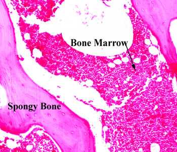
http://bio1151b.nicerweb.com/Locked/media/lab/tissues/SpongyBone.jpg

The White spots within bone marrow are adipose tissue.  There are four above the arrow for instance.

Bone size and bone strength are increased in obese male adolescents.

“We recruited 51 male ObAs (10-19 years) at the entry of a residential weight-loss program and 51 healthy age-matched and 51 bone-age-matched controls. vBMD and geometric bone parameters, as well as muscle and fat area were studied at the forearm and lower leg by peripheral quantitative computed tomography. Muscle force was studied by jumping mechanography. In addition to an advanced bone maturation, differences in trabecular bone parameters (higher vBMD and larger trabecular area) and cortical bone geometry (larger cortical area and periosteal and endosteal circumference) were observed in ObAs both at the radius and tibia at different pubertal stages. After matching for bone age, all differences at the tibia, but only the difference in trabecular vBMD at the radius, remained significant. Larger muscle area and higher maximal force were found in ObAs compared with controls, as well as higher circulating free estrogen, but similar free testosterone and IGF-I levels. ObAs have larger and stronger bones at both the forearm and lower leg. The observed differences in bone parameters can be explained by a combination of advanced bone maturation, higher estrogen exposure, and greater mechanical loading resulting from a higher muscle mass and strength. ”

Obese individuals have a higher bone age than non-obese individuals until age 16.  Obese individuals had taller height than age matched controls but shorter height than bone age matched controls.

“higher values of trabecular vBMD, trabecular area, periosteal circumference, and cortical area at the different pubertal stages in the obese group. ”  So bodyfat causes increase in bone parameters outside of the growth plate.  This includes the bone age matched group and not just the age matched group.

Obese individuals had increased estrogen and leptin but similar levels of free testosterone.

What would be interesting if the fat itself did not increase various hormones and genes to cause bone growth or some kind of loading effect.  But if the fat within the bone itself caused an expansion of bone parameters.

The main difference between a growth plate and adipose tissue is that adipose tissue is disorganized as you can see in the image of the bone marrow however adipose tissue cells are huge.  So is it possible that there could be enough adipose tissue cells to cause an expansion of the bone even if they are not coordinated like a growth plate.

The effect of weight on the femur: a cross-sectional analysis.

if stresses associated with biomechanical modifications of the obese surpass the strain threshold of a bone or bony location, it is possible that discernible differences in long-bone morphology could be observed between different weight categories as a direct result of long-term, abnormal mechanical compensation.”

“The Pearson’s product-moment correlation coefficient results show no correlation between weight and stature. “<-Since only very large amounts of weight would influence stature it’s possible that effect of extreme weights are overlooked.

Mediolateral dimensions of the bone at the midshaft at the bone increased at 4 out of 5 of the sites measured in the bone.  It’s possible that other parameters were increased but not statistically significant.  Anteriorposterior dimensions were increased only at the midshaft.

Is it the adipose tissue cells themselves that increase the bone dimensions or is it a weight loading effect increasing the dimenions.  The question is why would the bone increase in size in only one dimension.  The increase in bone size being mainly in one axis is consistent with it being a weight loaded effect and not a result of internal forces from adipose tissue cells.

research has shown elongation of the proximal ML dimension of the femur in pregnant women

“As ML diameter measures resistance to ML bending, these results suggest that as weight increases, alterations to the femoral angle result in greater ML pressures, forcing the femur to adapt or risk failure.”

Reduced size-independent mechanical properties of cortical bone in high-fat diet-induced obesity.

” femora from C57BL/6 mice fed either a HFD or standard laboratory chow (Chow) were evaluated for structural changes and tested for bending strength, bending stiffness and fracture toughness. Here, we find that in young, obese, high-fat fed mice, all geometric parameters of the femoral bone, except length, are increased, but strength, bending stiffness, and fracture toughness are all reduced. This increased bone size and reduced size-independent mechanical properties suggests that obesity leads to a general reduction in bone quality despite an increase in bone quantity; yield and maximum loads, however, remained unchanged, suggesting compensatory mechanisms. We conclude that diet-induced obesity increases bone size and reduces size-independent mechanical properties of cortical bone in mice.”

Mice were fed high fat diet over 19 weeks.  4 week old mice were used.

“the HFD group showed a 34% increase in serum IGF-I concentration compared to Chow”

Cellular dynamics and tissue interactions of the dura mater during head development

“Morphogenesis of the cranial bones and sutures is dependent on tissue interactions with the dura mater, which control the size and shape of bones as well as sutural patency. Development of the brain also involves interactions with dura mater: secretion of stromal derived factor 1 (SDF-1) is a critical event in directing migration of the external granular layer precursors of the cerebellar cortex and the Cajal-Retzius (CR) cells of the cerebral cortex. The dura mater is also required for growth of the hippocampal dentate gyrus. Wnt1Cre/R26R transgenic reporter mice were used to study the origin and fates of the cells of dura mater during head development. The dura mater of mammals is derived entirely from the cranial neural crest. Beginning around neonatal day 10 (N 10), the dura mater is infiltrated by cells derived from paraxial mesoderm, which later come to predominate. Over the course of infancy, the neural crest–derived cells of the dura mater become sequestered in niche-like distribution characteristic of stem cells. Simultaneously, dura mater cells underlying the sagittal suture migrate upward into the mesodermally-derived mesenchyme separating the parietal bones. Although initially the parietal bones are formed entirely from paraxial mesoderm, the cellular composition gradually becomes chimeric and is populated mainly by neural crest–derived cells by N 30. This occurs as a consequence of osteoblastic differentiation at the dura mater interface and intravasation of neural crest–derived osteoclastic and other hematopoietic precursors. The isolated cells of the dura mater are multipotent in vitro, giving rise to osteoblasts, neuronal cells and other derivatives characteristic of cranial neural crest, possibly reflecting the multipotent nature of dura mater cells in vivo.

” neural crest cells can be found throughout the intrafrontal suture. These cells give rise to fibroblast-like mesenchymal cells in the sutures, as well as chondrocytes, osteoblasts, and osteocytes in developing bones.”

Mineralized bone is incapable of interstitial growth{this would explain why adipose tissue cells don’t cause interstitial growth, however is unmineralized bone capable of interstitial growth; unfortunately there is no citation}, and bones grow at the marginal growth sites—growth plates in long bones and sutures in the skull. ” I also couldn’t find any emails either so I can’t ask where they retrieved that conclusion from.

Further research shows that the possibility of interstitial growth is related to the the rigidity of the ECM.  So adipocytes may be capable of interstitial growth if the ECM is too rigid.

I write about the optimal stiffness of ECM for chondrocyte differentiation here.  However, the stiffness for ECM for chondrocyte differentiation may be different from that for interstital growth.  Here I mention, that the compounds that give the bone ECM it’s stiffness are Calcium, Phosphorus, and Vitmain D.  However, people with deficiencies in those three compounds do not grow taller.  Also, mentioned is that demineralized bone matrix is an effective scaffold for chondroinduction.

In conclusion, the reason that adipose cells do not cause interstitial growth in bone despite being enormous and potentially present in massive quantities is that the ECM of bone is too stiff due to the mineral content.  Although during longitudinal growth, the bone is stiff at the bony area between the top and bottom area of the growth plate.  Thus, a key factor for micro-growth plate success via induction by LSJL is to reduce bone ECM stiffness.  LSJL may do this itself by causing interstitial fluid flow and shear strain.

LSJL dincreasing interstitial fluid flow and shear strain can be supported by the histological slides presented here.  The pink area which represents the bone appears to be much less rigid(compare slides A and B).

Hydroxyapatite crystals and chondrocyte apoptosis

A recent post by Michael stated that one of the obstacles to LSJL success is that hydroxyapatite crystals have sharp edges and that could result in the damage to the cells of any microgrowth plate possibly induced by LSJL.

First, Shear Strain and Fluid Flow induced by LSJL may disrupt the crystals.  However, let’s look at the science of Hydroxyapatite crystals and cell death to chondrocytes.

Intracellular calcium oscillations in articular chondrocytes induced by basic calcium phosphate crystals lead to cartilage degradation.

“Basic calcium phosphate (BCP) crystals, including octacalcium phosphate (OCP), carbonated-apatite (CA) and hydroxyapatite (HA) crystals are associated with destructive forms of osteoarthritis.  We assessed the ability of BCP to induce changes in intracellular calcium (iCa(2+)) content and oscillations and the role of iCa(2+) in BCP-induced cartilage degradation.

Bovine articular chondrocytes (BACs) and bovine cartilage explants (BCEs) were stimulated with BCP or monosodium urate (MSU) crystals. iCa(2+) levels were determined. mRNA expression of matrix metalloproteinase 3 (MMP-3), a disintegrin and metalloprotease with thrombospondin-like motifs 4 (ADAMTS-4) and ADAMTS-5 was assessed. Glycosaminoglycan (GAG) release was measured in the supernatants of BCE cultures.

All three BCP crystals significantly increased iCa(2+) content. OCP also induced iCa(2+) oscillations. Rate of BACs displaying iCa(2+) oscillations increased over time, with a peak after 20 min of stimulation. OCP-induced iCa(2+) oscillations involved both extracellular Ca(2+) (eCa(2+)) influx and iCa(2+) stores. Indeed, OCP-induced iCa(2+) oscillations decreased rapidly in Ca(2+)-free medium. Both voltage- and non-voltage-dependent Ca(2+) channels were involved in eCa(2+) influx. BCP crystal-induced variation in iCa(2+) content was associated with BCP crystal-induced cartilage matrix degradation. iCa²(+) was not associated with OCP crystal-induced mRNA expression of MMP-3, ADAMTS-4 or ADAMTS-5.

BCP crystals can induce variation in iCa(2+) content and oscillations in articular chondrocytes. BCP crystal-induced changes in iCa(2+) content play a pivotal role in BCP catabolic effects on articular cartilage{and potentially possibly LSJL induced micro-growth plates}.”

“BCP crystal deposition in knee articular cartilage is associated with cartilage destruction, more severe clinical symptoms, and chondrocyte phenotype changes towards hypertrophy as suggested by increased expression of type X collagen and greater ability to produce BCPs in vitro”<-So the BCP crystals within the bone will encourage the chondrocytes to hypertrophy.

“BCP crystals may stimulate articular cells through two mechanisms. They can first activate cells as endocytosed or phagocytosed particles leading to intralysosomal crystal dissolution with subsequent elevation of intracellular Ca2+ levels and release of inflammatory cytokines. The other mechanism of cell activation by Ca2+ crystals involves a direct crystal–cell membrane interaction”

“In articular chondrocytes, BCP crystals induce increased DNA synthesis and cell division{this is anabolic}, IL-1β mRNA overexpression, nitric oxide and MMP-13 production, increased caspase-3 activity and chondrocyte apoptosis”

“iCa2+ was involved in OCP-induced proteoglycan degradation but not OCP-induced mRNA expression of MMP-3, ADAMTS-4 or ADAMTS-5.”

This next study relates to HA crystals and causing apoptosis in another type of cell in bone osteoblasts.

Effects of four types of hydroxyapatite nanoparticles with different nanocrystal morphologies and sizes on apoptosis in rat osteoblasts.

“Hydroxyapatite nanoparticles (nano-HAP) have been reported to cause inflammatory reactions. Here, we aimed to compare the effects of four types of nano-HAP with different nanocrystal morphologies (short rod-like, long rod-like, spherical or needle-shaped crystals) and sizes (10-20, 10-30 or 20-40 nm) on growth inhibition and apoptosis in primary cultured rat osteoblasts. The osteoblasts was treated with the four types of nano-HAP at various concentrations (20, 40, 60, 80 or 100 mg/l).  All four types of nano-HAP inhibited the growth of osteoblasts in a dose-dependent manner. These nano-HAP significantly induced apoptosis in osteoblasts. Nano-HAP with smaller specific surface areas induced lower apoptosis rates. The needle-shaped and the short rod-like particles induced greater cellular injury than the spherical and long rod-like particles, respectively. The increased apoptosis rates were accompanied by increased p53 and cytochrome c expression. nano-HAP inhibit the activity of osteoblasts and also induce the apoptosis of osteoblasts in vitro. The nano-HAP-induced apoptotic pathway is mediated by a mitochondrial-dependent pathway. Moreover, the sizes, morphologies and concentrations of nano-HAP have significant effects on the apoptotic level.”

“nano-HAP with diameters less than 100 nm can cause inflammatory reactions, especially when the particles are needle-shaped”

nano-HAP increases caspase 3 and 9 and increases Bax levels while decreasing Bcl2.  The first three being pro-apoptotic proteins and the last being anti-apoptotic.

If HA crystals affect osteoblasts it’s likely they would affect chondrocytes too.  However, osteoblasts manage to survive in bone tissue despite the existence of HA crystals.

Annexin 5 overexpression increased articular chondrocyte apoptosis induced by basic calcium phosphate crystals.

“Basic calcium phosphate (BCP) crystals (octacalcium phosphate (OCP), carbapatite (CA) and hydroxyapatite (HA)) are associated with severe forms of osteoarthritis. In advanced osteoarthritis, cartilage shows chondrocyte apoptosis, overexpression of annexin 5 (A5) and BCP crystal deposition within matrix vesicles.

Apoptosis was induced by BCP crystals, tumour necrosis factor (TNF)-alpha (20 ng/ml) and Fas ligand (20 ng/ml) in normal articular chondrocytes (control) and in A5 overexpressed chondrocytes, performed by adenovirus infection. Apoptosis was assessed by caspase 3 (Cas3) activity, and DNA fragmentation.

All BCP crystals, TNF-alpha and Fas ligand induced chondrocyte apoptosis as demonstrated by decreased cell viability and increased Cas3 activity and DNA fragmentation. TUNEL (terminal deoxyribonucleotide transferase-mediated dUTP nick end-labelling)-positive staining chondrocytes were increased by OCP (12.4 (5.2)%), CA (9.6 (2.6)%) and HA (9.2 (3.0)%) crystals and TNF-alpha (9.6 (2.4)%) stimulation compared with control (3.1 (1.9)%). BCP crystals increased Cas3 activity in a dose-dependent fashion. BCP-crystal-induced chondrocyte apoptosis was independent from TNF-alpha and interleukin-1beta pathways but required cell-crystal contact and intralysosomal crystal dissolution. Indeed, preincubation with ammonium chloride, a lysosomal inhibitor of BCP crystal dissolution[dissolved], significantly decreased BCP-crystal-induced Cas3 activity. Finally, overexpression of A5 enhanced BCP crystal- and TNF-alpha-induced chondrocyte apoptosis.

Overexpression of A5 and the presence of BCP crystals observed in advanced osteoarthritis contributed to chondrocyte apoptosis.”

So, Chondrocyte Apoptosis doesn’t occur unless the crystal is dissolved.  But it’s possible this may occur as a result of shear strain due to LSJL.    However, the apoptosis induced is not complete and wouldn’t totally inhibit micro-growth plate formation due to LSJL.

“Chondrocytes undergo apoptosis after exposure to NO or Fas ligand (Fas-L).”

“Annexins are ubiquitous proteins that can interact with acid phospholipids, membranes and cytoskeleton constituents in the presence of Ca2+. They are involved in regulating intracellular and extracellular activities such as endocytosis and exocytosis and Ca2+ fluxes. Chondrocytes produce annexins 2, 5 and 6 (A2, A5 and A6), whose levels are increased in OA cartilage. A2, A5 and A6 have been identified on matrix vesicles. A5 can form voltage-gated Ca2+ channels and mediates Ca2+ influx into matrix vesicles, which initiates extracellular mineralisation, and into cellular cytoplasm, which induces apoptosis of growth-plate chondrocytes”

” chondrocyte apoptosis induced by BCP crystals was independent from elevations in extracellular calcium and/or phosphate concentrations but required direct cell-crystal contact.”

“[BCP induced chondrocyte apoptosis requires] cell-crystal contact, crystal endocytosis and intralysosomal crystal dissolution responsible for intracellular Ca2+ elevation.

Hydroxapatite crystals require too many things to go wrong to induce apoptosis in cells to likely occur in a normal physiological environment.  And if they did they would cause apoptosis to a variety of cells not just chondrocytes so you would want to eliminate them regardless.

Thus, I do not believe that HA Crystals or BCP crystals are a hindrance of micro-growth plate formation.