Author Archives: Tyler

Bone Adaptation to typical loads

Possible information on how mechanical stimulation can alter the bone.

Increased density and periosteal expansion of the tibia in young adult men following short-term arduous training.

“Tibial bone density and geometry were measured in 90 British Army male recruits (ages 21±3years, height: 1.78±0.06m, body mass: 73.9±9.8kg) in weeks 1 (Baseline) and 10 of initial military training. Scans were performed at the 4%, 14%, 38% and 66% sites, measured from the distal end plate, using pQCT (XCT2000L, Stratec Pforzheim, Germany). Customised software (BAMPack, L-3 ATI) was used to examine whole bone cross-section and regional sectors. T-tests determined significant differences between time points (P<0.05).
Bone density of trabecular and cortical compartments increased significantly at all measured sites. Bone geometry (cortical area and thickness) and bone strength (i, MMi and BSI) at the diaphyseal sites (38 and 66%) were also significantly higher in week 10. Regional changes in density and geometry were largely observed in the anterior, medial-anterior and anterior-posterior sectors. Calf muscle density and area (66% site) increased significantly at week 10 (P<0.01).
In vivo mechanical loading improves bone strength of the human tibia by increased density and periosteal expansion, which varies by site and region of the bone. These changes may occur in response to the nature and distribution of forces originating from bending, torsional and shear stresses of military training. These improvements are observed early in training when the osteogenic stimulus is sufficient, which may be close to the fracture threshold in some individuals.”

” The purpose of this training is to improve physical fitness and teach recruits basic military skills. In a 10-week period, infantry recruits typically undertake 27 periods of military drill, 45 physical training periods comprising of 8 continuous runs, 9 loaded marches (starting at 2 miles carrying a 10 kg backpack building up to 5 miles carrying a 20 kg backpack), 6 swimming sessions and 22 40-min periods of military specific fitness (obstacle course, circuit training, and steeplechase run).”

“There was no significant change in endosteal perimeter at any measured site.”

“Military activities that are not performed often, but likely exceed the strain threshold, include downhill running and/or zigzag motions, which elicit up to 2000 microstrain at the tibial shaft”

The blood supply to the growth plate

growthplatebloodsupply

The blood supply of the growth plate and the epiphysis: a comparative scanning electron microscopy and histological experimental study in growing sheep.

” The vascular supply of growth plate and epiphysis of the proximal tibia was reinvestigated using a modern technique, the Mercox-perfusion method, in six sheep aged 6-24 weeks. A comparison was made among pure perfusion specimens, the corrosion casts, and histological sections. The metaphyseal, epiphyseal, and perichondral blood supply systems were confirmed. However, there was evidence of regular transphyseal anastomoses{reconnection of two systems} between the metaphyseal and epiphyseal system. Based on the histological arrangement of the blood vessels, the arterial blood flow would appear to be from the metaphysis to the epiphysis. The existence of transphyseal arterial vessels originating metaphyseally and seen both in cast preparations and histological sections was added to the present description of the blood supply of the growth plate. Age-related differences in the vascularization of the growth plate were not found. ”

I couldn’t copy and paste from the full study at top.

Some vessels do cross the growth plate.

Longitudinal bone growth may cease if blood supply is cut off.

Here’s an image of a growth plate artery:

growth plate blood vessel

Cartilage canals run parallel to growth plate columns.

These transphyseal vessels may only be present up to 24 weeks of age.

Estrogen’s effect on height may be mediated by chondrogenic effects

The effect of estrogen on the expression of cartilage-specific genes in the chondrogenesis process of adipose-derived stem cells.

“During adolescence, sex hormones play an important role in regulating proliferation, differentiation, maturation, and the scheduled death of chondrocytes.
In the present study, we used adipose-derived stem cells (ADSCs) to differentiate into cartilage. Differentiated cartilage cells were used in the control (without estrogen E2 in the culture medium) and experimental (with estrogen in the culture medium) groups to evaluate the expression of type II collagen and aggrecan as chondrogenic genes markers.
Estrogen leads to inhibition of type II collagen gene expression and reduction of aggrecan gene expression.
Estrogen probably has negative effects on chondrogenesis process of ADSCs”

“17-β estradiol (E2) increased the type X collagen, inhibited the type II collagen, and reduced the proteoglycan content of the cartilage”

Loading effects on bones and possible transdifferentiatiion

Since chondrocytes are what make up growth plates and growth plates are what make you taller.  This is huge.

Molecular events caused by mechanical stress in bone.

“The shape of bone changes as a result of bone remodeling corresponding to physical circumstances such as mechanical stress. The tissue which receives the loaded mechanical stress most efficiently is bone matrix. Recent studies revealed the function of osteocytes as mechanosensors in the early stage of bone remodeling. Loaded mechanical stress is converted to a series of biochemical reactions, and finally activates osteoclasts and osteoblasts to cause bone resorption and formation. Biochemical and molecular biological studies have recently resulted in the identification of the gene of which expression level is changed by mechanical stress. Nitric oxide (NO) and cAMP is secreted in response to mechanical stress in the immediate early stage. Genes encoding enzymes such as glutamate/aspartate transporter (GLAST), nitric oxide synthetase (NOS) and prostaglandin G/H synthetase (PGHS-2) are identified as mechanical stress-responsive. The expression level of IGF-I is enhanced under the control of PTH/PTHrP. The expression of c-fos is increased by loading of mechanical stress[LSJL upregulates C-Fos]. AP1, a heterodimer of c-FOS/c-JUN, functions as a transcription factor of downstream gene(s). Elements including AP1 sites, cyclic AMP response elements (CRE) and shear stress response elements (SSRE) are found in the promoter region of mechanical stress-response genes. The enhanced expression of osteopontin (OPN) in the osteocytes of bone resorption sites was demonstrated by in situ hybridization and immunohistochemistry and transdifferentiation of chondrocytes with the abundant expression of BMP-2 and -4 in the process of distraction osteogenesis was observed.”

“Mechanical stress loaded to bone causes the deformation of bone matrix and generates strain force, which is thought to initiate the mechanotransduction pathway. Since the bone contains a water solution, the strain causes fluid flow in the bone matrix. These represent secondary changes known as streaming potential and shear stress. The activities of ion channels, focal adhesion kinases, as well as the structure of the cytoskeleton of the bone cells, are also modulated by mechanical stress.”<-Wish LSJL we hope that this fluid flow induces differentiation of stem cells into chondrocytes.

“GLAST mRNA expression level in the osteocytes decreased in the loaded ulnae after 6 h of load on two occasions 24 h apart.”

“OPN can inhibit the formation of hydroxyapatite. Immunohistochemical studies demonstrated that OPN was localized only on the surface of matured bone. In contrast, much abundant and broad distribution of OPN was observed in embryonic bone, osteoid and fractured callus”<-OPN is upregulated in LSJL.

“OPN can promote the attachment of osteoclasts possibly via interaction of GRGDS amino acid sequence of OPN and the αvβ3 integrin of the osteoclasts ”

“OPN mRNA was detected predominantly in osteoclasts and osteocytes in the resorption site of the septum.”<-This could suggest that LSJL degrades existing bone to make room for growth plates.

“After 7 days of osteoectomy in the femur of an 11-week-old rat, 0.25 mm of distraction was carried out every 12 h for 28 days. Cartilaginous external callus with endochondral ossification, and endosteal callus formation with membranous ossification were observed at the osteoectomized site. In addition to these typical ossification patterns, we observed the direct bone formation by chondrocyte-like cells with the temporal appearance of fibrous tissue“<-So cells differentiated into chondrocytes with fibrous tissues as an intermediary giving evidence of transdifferentiation.

“The molecular/histological analysis in the process of transchondroid bone formation demonstrated the expression of type II collagen mRNA in the chondrocyte-like cells at the early stage of osteogenesis. With the decrease of type II collagen mRNA, the expression level of type I collagen became enhanced at later stages.”

“the cells present in the transitional region from cartilage to fibrous tissue express alkaline phosphatase mRNA, OPN mRNA and osteocalcin mRNA simultaneously. These results suggest that the differentiation of chondrocyte to hypertrophic chondrocyte is blocked by distraction force, and indicated that chondrocyte may transdifferentiate to the osteoblastic lineage.”

Some insights into the growth plate from fish growth plates

One of the key parts of LSJL is to cause new growth plate formation via inducing differentiation of epiphyseal bone marrow.  This study has insight that epiphyseal bone marrow may participate in the growth plate process and thus in turn may be capable of contributing to neo-growth plate formation.

The humerus of Eusthenopteron: a puzzling organization presaging the establishment of tetrapod limb bone marrow.

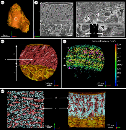
“Because of its close relationship to tetrapods{A tetrapod is a four limbed organism}, Eusthenopteron{a kind of fish} is an important taxon for understanding the establishment of the tetrapod body plan. Notably, it is one of the earliest sarcopterygians in which the humerus of the pectoral fin skeleton is preserved. The microanatomical and histological organization of this humerus provides important data for understanding the evolutionary steps that built up the distinctive architecture of tetrapod limb bones. Previous histological studies showed that Eusthenopteron’s long-bone organization was established through typical tetrapod ossification modalities. Based on a three-dimensional reconstruction of the inner microstructure of Eusthenopteron’s humerus, obtained from propagation phase-contrast X-ray synchrotron microtomography, we are now able to show that, despite ossification mechanisms and growth patterns similar to those of tetrapods, it also retains plesiomorphic{trait that only eusthenopteron’s have but not other tetrapods} characters such as a large medullary cavity, partly resulting from the perichondral ossification around a large cartilaginous bud as in actinopterygians. It also exhibits a distinctive tubular organization of bone-marrow processes. The connection between these processes and epiphyseal structures highlights their close functional relationship, suggesting that either bone marrow played a crucial role in the long-bone elongation processes or that trabecular bone resulting from the erosion of hypertrophied cartilage created a microenvironment for haematopoietic stem cell niches. ”

Now even though these fish and humans are different it suggests still that bone marrow has a capability to form new growth plates.

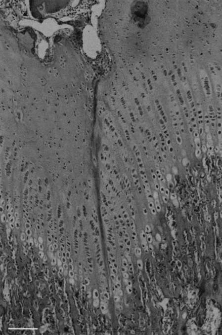
Here’s the anatomy of the mid-bone shaft of this fish(juvenile):

mid bone shaft

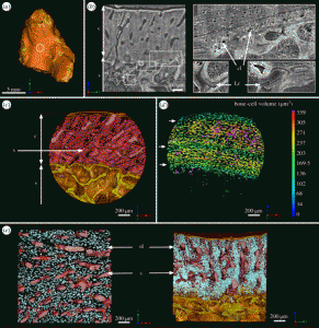
“(b) Virtual thin section (made along the longitudinal axis) showing the primary bone deposit of cortical bone and its connection to the spongiosa. Some remnants of calcified cartilage are still preserved at the location of Katschenko’s line (chondrocyte lacunae) and within the spongiosa (Liesegang rings).

So “micro-growth plates” within the bone marrow.

“At mid-shaft (figure 1a), the inner surface of the cortical bone is delimited by clusters of numerous large globular cell lacunae (cl, figure 1b,d) that can be identified as chondrocyte lacunae of cartilage. This suggests that remnants of Katschenko’s line are still present. Several stacks of Liesegang rings, typical of calcified cartilage, are also notable among the endochondral trabeculae”

“In extant tetrapods, the spongiosa forms when chondroclasts create erosion bays in the cartilage that are then lined with a thin peripheral bone layer, and it is common for small remnants of calcified cartilage to be left behind by the process; spongiosa formation in Eusthenopteron appears to have been similar.”

“The trabeculae in the spongiosa [of adult fish] are greatly remodelled and show no visible remnant of calcified cartilage”

“In extant actinopterygians, the fin endoskeleton develops from a blastema that differentiates into cartilages. Perichondral bone is deposited on the surface of the cartilages and continues to grow centrifugally as periosteal bone”

“a typical metapterygial bone is composed of a cartilaginous rod surrounded by a bony tube with cartilage projecting as condyles”

“In contrast to tetrapods, actinopterygian long bones have no haematopoietic bone marrow but only fatty tissues in the spaces created by the erosion of the cartilaginous rod. Neither do they show any evidence of a growth plate with longitudinally oriented columns of chondrocytes. The large longitudinal tubular mesh observed in Eusthenopteron humerus appears to constitute the earliest and phylogenetically deepest documented occurrence of a complex functional bone marrow in the tetrapod stem group.”

“As the tubular channels in Eusthenopteron are obviously connected to the epiphyses, the appearance of a complex bone marrow seems to be related to the appearance of tetrapod-like epiphyseal structures and elongation growth. Eusthenopteron lacks the comprehensive remodelling and trabecular resorption that creates an open medullary cavity in the majority of extant tetrapods, but the reduction of trabeculae between the longitudinal tubes in the adult compared with the juvenile may represent an evolutionary precursor of this process. ”

“Tetrapod bone marrow has been shown capable of degrading cartilage proteoglycans and inducing the initial stage of endochondral ossification”

Osteoarthritis and Ligaments

Loading ligaments may be able to contribute to height.  Ligaments connect to bone via entheses.  Loading ligaments may be able to alter bone and in our case ideally causing formation of new growth plates.

How does bone and surrounding cartilage affect the surrounding ligaments?

High-resolution ultrasound evaluation of extrinsic wrist ligaments in patients affected by rheumatoid arthritis.

“Twenty-one consecutive patients affected by RA (12 men, 9 women; mean age 57 ± 14.6 years) were compared with 21 controls (12, 9; 54 ± 12.1, respectively). Wrists were evaluated using ultrasound on both palmar and dorsal sides along each ligament, using carpal bones as references. The following ligaments were studied: radioscaphocapitate, radiolunotriquetral, palmar ulnolunate, palmar ulnotriquetral, dorsal radiotriquetral, dorsal ulnotriquetral, and radial collateral ligament. Ligament number and thickness were noted. Echotexture was rated as fibrillar, fragmented, or heterogeneous; the surface was rated as smooth or blurred.

The number of palmar ulnolunate and palmar ulnotriquetral ligaments detected by ultrasound in patients was significantly lower than in controls. All ligaments had significantly more fragmented or heterogeneous echotexture and blurred surface and were significantly thinner in patients than in controls. No correlation was found between ligament thickness and RA duration or clinical parameters.

Extrinsic wrist ligaments were less detectable and thinner in patients affected by RA compared with healthy volunteers matched for age and sex. Ligament thinning did not directly correlate with RA duration and clinical parameters.”

“Damage in RA mainly originates from the synovium in the joints and tendon sheaths. During the course of the disease, inflamed synovial tissue may extend to bone, tendons, capsule, and ligaments”<-And possible the refuse may be true in which stimulus from the ligaments progresses into the bone.