“Tachypleus tridentatus (T. tridentatus) is a marine animal and traditional Chinese medicine. T. tridentatus plasma is a valuable resource for important medical and health-based functions. In this experiment, in order to evaluate the effect and mechanism ofT. tridentatus plasma with respect to the promotion of bone tissue growth in rats, the processes of ultrafiltration and mass spectrometry were first used to separate and identify the components of T. tridentatus plasma. Then, a comparison of the effects of the T. tridentatus plasma samples, which each possessed different molecular weights, regarding the growth of the long bones of rats was conducted. Finally, transcriptomics, proteomics, and bioinformatics were all used to analyze the biological functions and related signaling pathways of the T. tridentatus plasma in order to promote rat bone growth. The results showed that the contents of amino acid residues in peptides are related to the growth promotion that was contained in the 10–30 kDa plasma group. Moreover, the T. tridentatus plasma samples were found to be higher in this respect than those in the whole plasma group. In addition, the 10–30 kDa plasma group could significantly promote bone growth activity in rats. The proteomic analysis showed that the proteins that were differentially expressed in the 10–30 kDa plasma group were mainly enriched in the PI3K-AKT signal pathway. Our study suggested that theT. tridentatus plasma possesses promising potential for the purposes of clinical use, whereby it can serve the role of a growth-promoting agent.“
The fact that they think that horshoe clab has clinical use is promising but we have to see what unique benefits it has over other compounds. Lots of things stimulate the PI3K-AKT pathway. We have to see if there’s anything the plasma does that is unique or superior in promoting growth.
“T. tridentatus possesses anti-inflammatory, analgesic, hemostatic, and antidysentery medicinal effects. It is also worth noting that T. tridentatus blood that contains hemocyanin possesses important medical application value. The blood extract can be made into a Limulus-reagent (LR), which is widely used for the purposes of endotoxin detection and quantification in injections, as well as in radiopharmaceuticals, vaccines, and other biological products”
“T. tridentatus plasma can promote the growth and development of Wistar rats by increasing body weight, body length, and tail length in adolescent Wistar rats; in addition, despite this range of growth, the rats do not appear obese.”
” IGF-1, BMP2, and ALP are applied to monitor bone growth indicators.”<-lots of things can increase those things. The question is does Horshoe Crab does anything that is unique?
“the plasma group showed macroscopic changes in total longitudinal bone length after 14 days of treatment. In addition, morphological changes were observed, such as the increased height of the growth plate area and an increased number of proliferating cells. Furthermore, T. tridentatus plasma treatment significantly increased the total serum levels of BMP-2, IGF-1R, and IGF-1, as well as induced the upregulated expression of IGF-1 and IGF-1R in the growth plate region.”
So the fact that scientists like that this compound can enhance longitudinal bone growth is interesting but the question is is whether it is really better than say other things in the diet?
The below study suggests that growth is manituable not only be nutrition but also by mechanical loading(tool use). It states that growth of some elements of the body is prioritized over others in times of nutrient restriction(“the thrity phenotype” hypothesis). Although this study is more geared to that nutrition can alter body height and skeletal length growth. Proving the plasticity(ability of bone to change in length) of bone is essential in proving that bone can change in response to mechanical loading.
“Plasticity in the growth of body segments between populations has been researched in relation to migration, temporal change and high-altitude studies{height seekers could potentially manipulate altitude or temperature}. We study the within population variation in body segments, thus controlling for some of the environmental and genetic differences that could be at play in between populations studies. We test a version of the thrifty phenotype hypothesis, where the growth of head-trunk and hand are prioritized due to their functional significance over height and leg growth.
A total of 3913 Guatemalan, rural, semi-urban and urban, Maya and Ladino children 6 to 15 years old were studied. Height, sitting height, leg length, and metacarpal length were studied in relation to three proxies for living conditions: height- and leg length-for-age, and maternal education. Estimation statistics and null hypothesis significance testing were used to analyze the data.
Metatarsal length and sitting height values were higher than height and leg length respectively. Relative metacarpal length was conserved across height-for-age groups. Females were less affected than males for metacarpal length and sitting height, but more affected for leg length.
Our results agree with the thrifty phenotype hypothesis, where metacarpal and sitting height growth would be prioritized over height and leg length due to greater functional significance.“
“Plasticity refers to the ability of an organism to modify its biology to respond to changes in the environment, particularly when these changes are physiologically stressful”<-plasticity in height is a very promising idea for height as it means height and bone length can be manipulated.
“in conditions of environmental stress, leg length is often more sensitive than is the head-trunk segment, and that tibia and ulna lengths are also more sensitive than humerus, hand and foot lengths. A proximate reason for this sensitivity is due to the allometry of skeletal growth, where the more proximal body segments grow fastest prenatally and are less exposed to extra-uterine environmental stress, but more distal segments grow most rapidly after birth and are more exposed to environmental stress. The thrifty phenotype hypothesis offers an ultimate evolutionary explanation; the growth of human body segments with greater functional significance, such as “head-brain,” “trunk-major organs,” and “hands-tools” will be prioritized over forearms and legs.”
“the thrifty phenotype hypothesis over the predictions of the distal blood flow and cold adaptation models in the growth of different body segments in living populations “
“According to this hypothesis, the growth of the head-trunk segment would be prioritized due to the important organs that it houses (brain, heart, lungs), and the growth of the hand would be prioritized due to its manipulative function in human tool technology, thus offering an ultimate evolutionary explanation for the differential plasticity in the growth of body segments under different conditions of stress.”
the thrifty phenotype hypothesis is basically that growth is conserved based on nutrients with some bones being prioritized over others in times of nutrient restriction.
Infigatinib is a potential alternative to Vosoritide to increase height during development. FGFR3 reduces growth in everyone it just does so in a greater manner in people with dwarfism as they have a mutation. CNP may have other beneficial effects on top of inhibiting FGFR3 but this just seems to be an FGFR3 inhibitor.
“Fibroblast growth factor receptor 3 (FGFR3) gain-of-function mutations play a crucial role in achondroplasia (ACH), thanatophoric dysplasia (TD), and hypochondroplasia (HCH).{But FGFR3 may decrease height in everyone during development just to a lesser degree if you have the mutation}. HCH is a less severe form of dwarfism than ACH, but similarly is caused by gain-of-function mutations in the FGFR3 gene. HCH is characterized by a disproportionate short stature and a growth deficit affecting both endochondral and intramembranous ossification. While multiple therapeutic strategies are being tested for ACH, currently there are no approved therapeutic options for individuals with HCH. We tested the hypothesis that the oral, selective FGFR tyrosine kinase inhibitor (TKI) infigratinib (BGJ398) could improve the HCH phenotype and improve endochondral and intramembranous ossification in a preclinical mouse model of HCH Fgfr3N534K/+.
The first Hch mouse model studied expresses the most frequent human mutation p.Asn540Lys (Fgfr3Asn534Lys/+), and exhibits a mild dwarfism and most of the hallmarks of the human pathology. Fgfr3N534K/+ mice received subcutaneous injections of infigratinib or vehicle control every 3 days (1 mg/kg) or daily (1 mg/kg) for 15 days (post-natal day [PND] 4–19) or 21 days (PND 3–24), respectively.
Fgfr3N534K/+ mice treated with 1 mg/kg infigratinib every 3 days did not show obvious and significant modification of the dwarf phenotype. In contrast, Fgfr3N534K/+ mice treated with 1 mg/kg infigratinib daily for a total of 21 days showed a statistically significant increase in appendicular and axial skeletal measures. Length of the long bones was statistically significantly increased in Fgfr3N534K/+ mice compared with Fgfr3+/+ mice (tibia +3.18%, femur +3.16%, humerus +3.04%, ulna +2.94%, radius +3.01%). Treatment also modified the skull shape (skull width, skull height, nasal bone length and naso-occipital length), the length of the mandible and skull base, as demonstrated by measurement of the foramen magnum (foramen magnum length +3.72%). Infigratinib treatment modified the cartilage growth plate organization, in particular the hypertrophic chondrocyte area. Finally, the high activation of the MAP kinase pathway due to the HCH missense FGFR3 mutation was reduced by treatment, as revealed by the immunolabelling of phosphorylated Erk1/2 proteins.
Treatment with daily 1 mg/kg infigratinib improved the length and weight of Fgfr3N534K/+ mice and significantly modified the skull and the axial and appendicular skeleton. We demonstrated in Fgfr3N534K/+ mice that infigratinib is able to counteract the constitutive activation of FGFR3 due to the heterozygous N540K mutation localized in the tyrosine kinase 1 domain of the protein. These results provide a rationale for targeting FGFR3 with a specific TKI for the treatment of children with HCH.”
So basically infigratinib works to increase height in people with dwarfism but given everyone has FGFR3 receptors it could help increase height in anyone with growth plates to a lesser degree.
“– In the highest dose level (Cohort 5, 0.25 mg/kg once daily), the mean change from baseline in annualized height velocity (AHV) at six months was +3.03 cm/yr (p = 0.0022) for the first 10 children with at least six months of follow-up in Cohort 5. The two remaining children who have not yet had six months of follow-up have a mean change from baseline in AHV of +8.8 cm/yr based on three months data“<-this is huge but again for normal children the gain will be smaller.
“80% of children at six months were responders, as defined by an increase from baseline AHV of at least 25%. The mean change from baseline in AHV of responders was 3.81 cm/yr“<-but that does not mean the 20% who weren’t responders didn’t get additional height at all.
” Infigratinib is an oral small molecule designed to inhibit FGFR3 and target achondroplasia at its source”
“Combined with the previously reported Cohort 4 change from baseline in AHV value of +1.52 cm/yr, the Cohort 5 data demonstrate a strong dose response for infigratinib”<-meaning the more of it you take the better but there is usually a better of diminishing returns…
I’ve written about heat being used to grow taller before. Heat also increases the rate at which chemical reactions occur which could be part of it. Here’s more on heat to grow taller. Heat could potentially increase solute uptake of the growth plate cartilage. Such things wouldn’t be really helpful to those who are skeletally mature although there is the possibility that heat could provide some kind of stimulus to the bone in which case it would help skeletally mature individuals.
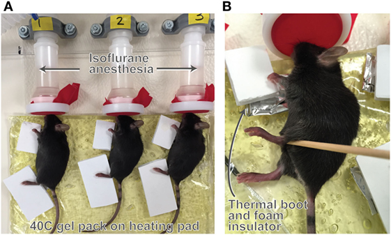
“As a noninvasive alternative, we previously developed a once daily limb-heating model using targeted heat on one side of the body for 2 weeks to unilaterally increase bone length by up to 1.5% in growing mice. In this study, we applied heat for 1 week to determine whether these small differences in limb length are functionally significant, assessed by changes in hindlimb weight bearing. We tested the hypothesis that heat-induced limb length asymmetry has a functional impact on weight bearing in mouse hindlimbs. Female 3-week-old C57BL/6 mice (N = 12 total) were treated with targeted intermittent heat for 7 days (40 C for 40 min/day){most humans have intermittent heat exposure though}. High-resolution x-ray (N = 6) and hindlimb weight bearing data (N = 8) were acquired at the start and end of the experiments. There were no significant left-right differences in starting tibial length or hindlimb weight bearing. After 1-week heat exposure, tibiae and femora were ~1 and 1.4% longer, respectively, on the heat-treated sides (40 C) compared to the non-treated contralateral sides (30 C). Tibial elongation rate was over 6% greater. “
“warm-reared mice had consistently longer ears, limbs, and tails when compared to littermates raised at cooler temperatures”<-studies have found however that humans raised at cooler environments tend to be taller. This finding though also means that it’s possibly that not only is growth rate increased but also height at skeletal maturity.
Here’s the heating device:
“The growth acceleration averaged 10 µm/day”
“heat-induced limb length differences do persist at skeletal maturity in 12-week-old mice that were examined 7 weeks after a juvenile heat-treatment”
” heat-based methods for treating minor limb length discrepancies may ultimately provide alternatives to traditional surgical approaches that can be painful and invasive.”<-so they do think that the method could be applied to humans.
“The increase in bone elongation rate generated by localized heat could help equalize minor limb length and weight bearing asymmetry in children”<-although we’d want something that works in older individuals.
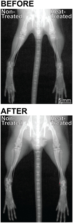
“Limb length inequality results from many types of musculoskeletal disorders. Asymmetric weight bearing from a limb length discrepancy of less than 2% can have debilitating consequences such as back problems and early-onset osteoarthritis. Existing treatments include invasive surgeries and/or drug regimens that are often only partially effective. As a noninvasive alternative, we previously developed a once daily limb-heating model using targeted heat on one side of the body for 2 weeks to unilaterally increase bone length by up to 1.5% in growing mice{1.5% is huge. That’s the difference between 5’9″ and 5’10”}. In this study, we applied heat for 1 week to determine whether these small differences in limb length are functionally significant, assessed by changes in hindlimb weight bearing. We tested the hypothesis that heat-induced limb length asymmetry has a functional impact on weight bearing in mouse hindlimbs. Female 3-week-old C57BL/6 mice (N = 12 total) were treated with targeted intermittent heat for 7 days (40 C for 40 min/day). High-resolution x-ray (N = 6) and hindlimb weight bearing data (N = 8) were acquired at the start and end of the experiments. There were no significant left-right differences in starting tibial length or hindlimb weight bearing. After 1-week heat exposure, tibiae (t = 7.7, p < 0.001) and femora (t = 11.5, p < 0.001) were ~1 and 1.4% longer, respectively, on the heat-treated sides (40 C) compared to the non-treated contralateral sides (30 C). Tibial elongation rate was over 6% greater (t = 5.19, p < 0.001). Hindlimb weight bearing was nearly 20% greater (t = 11.9, p < 0.001) and significantly correlated with the increase in tibial elongation rate on the heat-treated side (R2 = 0.82, p < 0.01). These results support the hypothesis that even a small limb length discrepancy can cause imbalanced weight distribution in healthy mice. The increase in bone elongation rate generated by localized heat could be a way to equalize limb length and weight bearing asymmetry caused by disease or trauma, leading to new approaches with better outcomes by using heat to lengthen limbs and reduce costly side effects of more invasive interventions.”
“warm-reared mice had consistently longer ears, limbs, and tails when compared to littermates raised at cooler temperatures”
Here is the skeletal effects:
This next paper is a review paper that covers everything more thoroughly:
Temperature, heat shock proteins and growth regulation of the bone tissue
“Ambient heat modulates the elongation of bones in mammals, and the mechanism of such a plasticity has not been studied completely. The influence of heat on growth and development of bone depends on its values. Five zones of temperature influence on the bone tissue with different biological effects have been distinguished : a) under-threshold thermal zone < 36.6 ºС, insufficient amount of heat is a limiting factor for osteogenesis; b) normal temperature zone 36.6‒37.5 ºС, the processes of breakdown and development of bone in this temperature range is balanced; b) zone of mild thermal shock 39‒41 ºС, the processes of functioning of osteoblasts, osteocytes and formation of the bone tissue intensify; d) the zone of sublethal thermal shock > 42 ºС, growth of bone slows; e) zone of non-critical shock > 50 ºС, bone tissue cells die.{so seems like heat as a stimulus is biphasic where there’s an optimal zone, but heat can be combined with other stimuli}. We propose a model of the mechanism of influence of heat shock on bone growth. Mild heat shock is a type of stress to which membrane enzymes adenylyl cyclase and cAMP-protein kinase react. Protein kinase A phosphorylates the gene factors of thermal shock proteins, stress proteins and enzymes of energy-generating processes – glycolysis and lipolysis. Heat shock protein HSP70 activates alkaline phosphatase and promotes the process of mineralization of the bone tissue. In the cells, there is intensification in syntheses of insulin-like growth factor-I, factors of mitogenic action, signals of intensification of blood circulation (NO) and synthesis of somatotropin. The affinity between insulin-like growth factor I and its acid-labile subunit decreases, leading to increased free and active insulin-like growth factor I. Against the background of acceleration of the capillarization process, energy generation and the level of stimulators of growth of bone tissue, mitotic and functional activities of producer cells of the bone – osteoblasts and osteocytes – activate{would be interesting to see how osteoclasts are affected as they alter growth}. The generally known Allen’s rule has been developed and expanded: “Warm-blooded animals of different species have longer distal body parts (tails) if after birth the young have developed in the conditions of higher temperature”. The indicated tendency is realized through increased biosynthesis of heat shock proteins and other stimulators of growth processes in the bone tissue.”
“In one experiment, two month-old male mice C57BL/6J were exposed to cold (4 °C) and normal (23 °C) temperatures for 28 days. Cold increased the apoptosis of osteocytes and decreased the length of the canals. Those changes were accompanied by decrease in the number of osteocytes that were positive to Е11 (transmembrane glycoprotein, important for differentiation of osteocytes, first of all prolonging dendrites) and ММР13 (matrix metallopeptidase that breaks down the collagen I in extracellular matrix and potentially plays a role in rotating the articular cartilage) after 14 days. The indicated parameters returned to the initial levels after 28 days. The study revealed that after 14 days of influence of low temperatures the volumetric bone fraction significantly decreased, but recovered after 28 days. Brown adipose tissue affected remodeling of the bone by increasing thermogenesis”
“Mild thermal stress increases the processes of regeneration of blood vessels and bones”
“A study reported that heat stress effectively induced the development of new bone tissue in an experimental model with 58 rats and 10 rabbits. The experimental animals underwent hyperthermia in 45 °С for 15 min once and three times a week, the changes in their bone tissue being determined by x-ray and histological evaluation. After the procedure of thermotherapy (1 time/week over 4 weeks), the experimental groups of rats and rabbits were observed to have a heightened level of osteogenesis compared with the control. The researchers inferred that thermal stimuli with heated materials enhance osteogenesis and increase the area of the development of bones and would be useful in treatment of bone defects in cases of skeletal diseases”<-this could potentially impact height.
Note that just because the muscles are stimulated by exercise to increase temperature does not mean bones do in the same way.
“Heat influence induces HSP expression. They stabilize proteins that strengthen the transmission of the signals of nitrogen oxide (NO), decrease oxidative stress and inflammation of the vessels and improve their function. During passive heat stress, blood circulation in the legs increased ~ 3–4-fold”
“The cartilage has no blood vessels and its nutrition is performed by diffusion of substances. The semi-penetrable “barrier at the border of vessel-cartilage surface” obstructs the molecular transport. To study the peculiarities of overcoming this obstacle, a model of heating the hind legs was used for manipulating the blood circulation in bones in 5-week-old female mice. In the experiment, dextrans were used weighing 10, 40 and 70 kDa, which are close in size to physiological regulators. Increase in the temperature in hind legs from 22 to 34 °C led to increase in vascular access of the abovementioned molecules. In 34 °C, penetration of dextrans 10 kDa into the growth plate increased by > 150%, and 40 and 70 kDa increased only by < 50%“<-so temperature managed to increase nutrient delivery into the growth plate by a significant amount.
“at HSP70 (200 ng/mL) increases the activity of alkaline phosphatase and promotes the mineralization of the bone tissue. In the conditions of osteogenic induction, this heat shock protein increased the expression of osteospecific genes such as transcriptional factors of family runt Runx2 and osterix (OSX). HSP70 promotes osteogenesis and may be a therapeutic mean for treating uncoalesced bones”
“Osteochondral interface between the bone and the cartilage allows these tissues to “communicate” with one another and exchange signal and plastic molecules, thereby providing integrated response to mechanical and thermal irritators”
This age range where height is increased is definitely post fusion I don’t know whether this is torso height or leg height but it’s a pretty big breakthrough. This is a study studying changes in the feet but there is a height increase shown in the paper and the authors do acknowledge it. The reason that the study found height increase in a japanese population but studies have not find height increase in other populations is that japanese tend to be thinner and having access fat plus being sedentary could lead to bad posture.
“The subjects were 135 males and 133 ware 135 males and 133 females born before 1940 (the 1930 group below), and 383 males and 414 females born between 1960 and 1979 (the 1970 group below).”<-so it’s not a longitudinal study unfortunately.
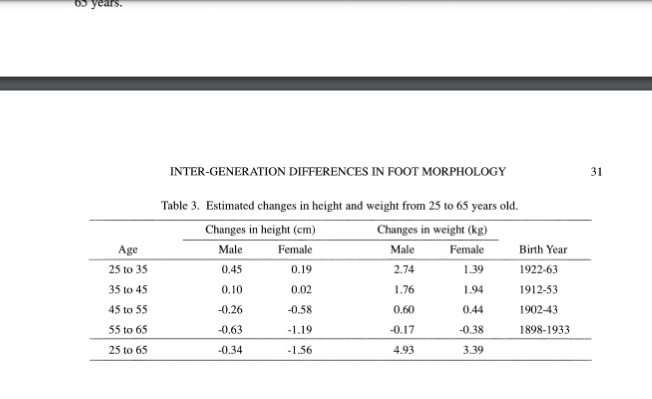
The study may have indications that the feet bones change over time but it’s unclear by the methodology but here’s the interesting part where height gain is shown:
Not only is there a height gain between 25 and 35 but also between 35 and 45. And the gain is greater for males and males tend to exercise more. Leading to a possible correlation with exercise and height.
The authors acknowledge the increase in height “A small increase in height was observed between the ages of 25 and 45 years“
The paper also makes an interesting observation regarding relaxin and feet: “Female feet are thought to become wider after their first pregnancy because the ligaments of the feet loosen due to hormonal effects. If this is true, inter-generation differences in foot dimensions would be larger in females than in males, and the differences between a 18-year-old and a 30-yearold would be much larger than the differences between a 30-year-old and a 50-year-old. Inter-generation differences in females were in fact larger than those in males in the present study (Table 2), but this was due to the exceptionally robust feet of the female 1930 groups in the IPRI and NIBH series. The trend in FB with age or with BY does not support this hypothesis”
This may also suggest too that ultrasound can affect longitudinal bone growth post skeletal maturity via articular cartilage endochondral ossificaiton.
GROWTH MODULATION BY STIMULATING THE GROWTH PLATE:A PILOT STUDY
“This study investigates the ability of low-intensity pulsed ultrasound (LIPUS) or direct injection of recombinant growth hormone (rGH) to stimulate local growth of long bones. In a randomized controlled animal trial, healthy immature rabbits were allocated to 1 of the following 4 conditions: epiphyseal rGH periosteal injection, transdermal LIPUS, saline periosteal injection, or no treatment. New bone deposition was labeled with calcein at days 1 and 18, and microscopic measurements of growth were conducted by blinded observers.
Statistically significant differences in growth were observed between the LIPUS and rGH stimulated legs compared with contralateral control legs (35% p = 0.04 and 41% p = 0.04, respectively);{35% is huge!} whereas no difference was observed between the 4 control groups (p = 0.37). There was no evidence of physeal bar formation, suggesting that direct injection of rGH and application of LIPUS around the distal femoral physis in rabbits may have a positive effect on microscopic growth without short-term adverse sequelae.”
“Low-intensity pulsed ultrasound (LIPUS) has been demonstrated to improve fracture healing in multiple double-blinded controlled trials, as well as stimulate chondrocytes in culture to increase calcium and aggrecan expression”
“that ultrasound stimulation of chondrocytes in vitro can reversibly change the shape and the orientation of the primary cilium dependent on the duration and pressure-amplitude of the signal and can enhance the cyclic Adenosine Monophosphate level in culture media only in ciliated chondrocytes.”
“The primary cilium is a place of high concentration of integrins that are believed to connect the extracellular matrix and the cytoskeleton in mechanotransduction”
“The bone stimulator used was Exogen (Bioventus, Durham, NC, USA). It emits low-frequency (1.5 MHz) ultrasound with a 1-kHz repetition module creating a 200-ms signal with an intensity of 30 mW/cm2″<-theoretically this could be applied to the articular cartilage
“Therefore, a 3-wk period should be equivalent to approximately 2 y of growth. These rabbits were 15 wk of age at the beginning of the study and may have been approaching skeletal maturity at that time.”
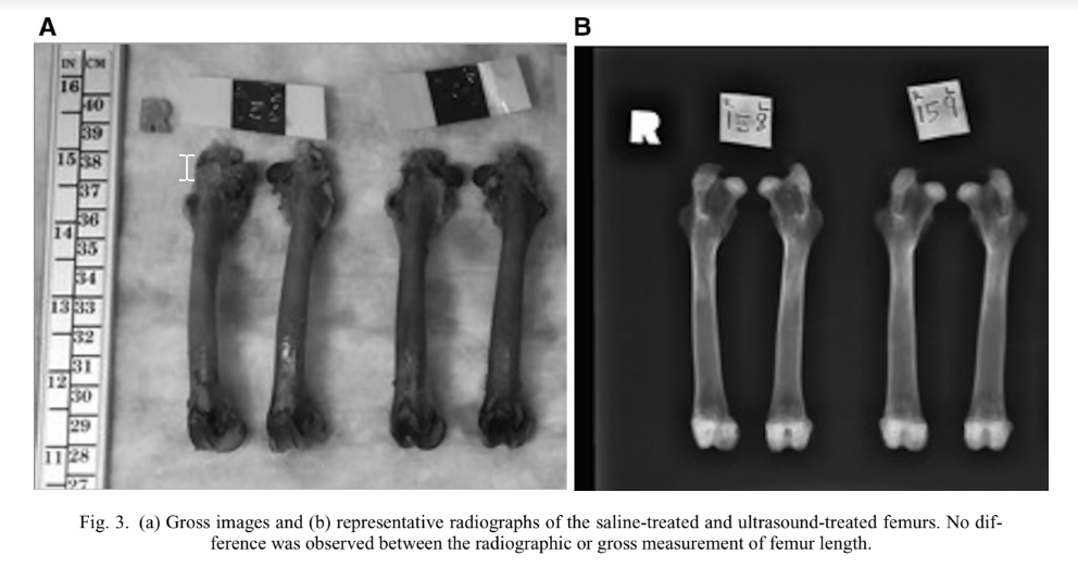
“Direct macroscopic femoral length measurements and radiographic length measurements revealed no statistical difference in growth in animals between any of the treatment arms.”<-So LIPUS didn’t really increase bone length at the macroscopic level? But if you look at 3a I do see a slight difference.
However they directly say “external application of LIPUS and injection of growth hormone does affect the longitudinal growth of bone with no adverse effects such as physeal bar or angular deformity noted.”
“Selective growth stimulation of active growth plates may become a novel noninvasive method to address limb length discrepancies and spine deformities in children.”<-and in adults via articular cartilage endochondral ossification possibly