Category Archives: Uncategorized

Ligaments constrain growth

The reason that the jaw can grow via stretching by forwarding positioning and bite-jumping appliances is that it is not as constrained by ligaments as other bones.  It’s possible that other movable joint regions like the wrists, fingers, and toes would also be stretchable.  The wrists would come under extreme stress due to the farmers walk so if this true there should be anecdotal evidence of longer arms due to the farmers walk.  It is possible that a new height increase method could be invented that causes enough tensile strain in the articular cartilage to activate endochondral ossification in that region.  The ligaments holding the bone together would constrain this.  This would also help explain why people with Marfan’s become taller as they have more flexible joints and therefore their growth is less constrained by ligaments.   Here’s another connective tissue disorder that may affect longitudinal bone growth.  It’s also possible that differences between condylar cartilage and articular cartilage allow this to happen in the mandible but not in other joints.

So if anyone could look for instances of longer arms due to heavy farmers walk.  This would be much easier to do than LSJL.  So any research on this would be a great boon to further the cause of height increase.

The adaptive remodeling of condylar cartilage—a transition from chondrogenesis to osteogenesis.

“Mandibular condylar cartilage is categorized as articular cartilage but markedly distinguishes itself in many biological aspects, such as its embryonic origin, ontogenetic development, post-natal growth mode, and histological structures. The most marked uniqueness of condylar cartilage lies in its capability of adaptive remodeling in response to external stimuli during or after natural growth. The adaptation of condylar cartilage to mandibular forward positioning{basically bringing your jaw forward} constitutes the fundamental rationale for orthodontic functional therapy, which partially contributes to the correction of jaw discrepancies by achieving mandibular growth modification. The adaptive remodeling of condylar cartilage proceeds with the biomolecular pathway initiating from chondrogenesis and finalizing with osteogenesis{so basically by stretching the articular cartilage you activate enchondral ossification enabling you to grow on the longitudinal ends of the bone}. During condylar adaptation, chondrogenesis is activated when the external stimuli, e.g., condylar repositioning, generate the differentiation of mesenchymal cells in the articular layer of cartilage into chondrocytes, which proliferate and then progressively mature into hypertrophic cells. The expression of regulatory growth factors, which govern and control phenotypic conversions of chondrocytes during chondrogenesis, increases during adaptive remodeling to enhance the transition from chondrogenesis into osteogenesis, a process in which hypertrophic chondrocytes and matrices degrade and are replaced by bone. The transition is also sustained by increased neovascularization, which brings in osteoblasts that finally result in new bone formation beneath the degraded cartilage.  The repositioning of the mandibular condyle in adult rats led to a reactivation of chondrogenesis in condylar cartilage which otherwise is at resting status, and finally results in increased bone formation{this is huge if as it means a bigger stronger jaw}

“chondrogenic activity of BMP-2 in vitro involves the action of the cell-cell adhesion protein, N-cadherin, which functionally complexes with beta-catenin the change of condyle position relative to the glenoid fossa constitutes an important trigger for [the endochondral ossification related adaptation of the mandible].”

The deviation of the condyle from the glenoid fossa by mandibular forward translation is the basis for orthodontic functional therapy, which aims to enhance condylar growth and therefore to eliminate the discrepancy between upper and lower jaws.

a decrease in compressive loading enhances condylar growth, whereas an increase in loading inhibits growth”

Note that they do say that condylar cartilage is distinct from articular cartilage.

Here’s another study:

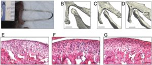
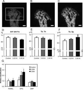
Murine TMJ loading causes increased proliferation and chondrocyte maturation.

“The purpose of this study was to examine the effects of forced mouth opening on murine mandibular condylar head remodeling. We hypothesized that forced mouth opening would cause an anabolic response in the mandibular condylar cartilage. Six-week-old female C57BL/6 mice were divided into 3 groups: (1) control, (2) 0.25 N, and (3) 0.50 N of forced mouth opening. Gene expression, micro-CT, and proliferation were analyzed. 0.5 N of forced mouth opening caused a significant increase in mRNA expression of Pthrp, Sox9, and Collagen2a1, a significant increase in proliferation{These alone will not increase height except maybe the increase in proliferation}, and a significant increase in trabecular spacing in the subchondral bone, whereas 0.25 N of forced mouth opening did not cause any significant changes in any of the parameters examined. Forced mouth opening causes an increase in the expression of chondrocyte maturation markers and an increase in subchondral trabecular spacing.”

“The articulating surfaces of the temporomandibular joint (TMJ) are covered by fibrocartilage, which is composed of chondrocytes at various stages of maturation that are organized into four zones: (1) superior articular zone—cells in this zone express proteoglycan 4 (Prg4); (2) polymorphic zone—cells in this zone express SRY-box 9 (Sox9) and parathyroid-hormone-related protein (Pthrp); (3) flattened zone—the cartilage cells in this layer express collagen type II (Col 2a1); and (4) hypertrophic zone—the chondrocytes express collagen type X (Col 10a1)”

“Growth in the condylar cartilage proceeds from cells exiting the proliferative pool in the polymorphic zone and undergoing endochondral ossification in the inferior cartilage zones. The mandibular condylar cartilage is able to remodel its structure in response to mechanical strains

F and G are both forced mouth opening with F and G to a higher degree.  C and D have longer bones but there is less separation between the two bones with C and D.  The fear with articular cartilage endochondral ossification is that you’ll decrease the joint space.

“Functional appliance therapy is believed to place the mandible in a protrusive position, causing the condyle to be unseated caudally and ventrally from the articular fossa, while forced mouth opening is believed to cause a compressive force between the articular eminence and the mandibular condyle”

“A possible explanation for both functional appliance therapy and forced mouth opening causing similar effects in the mandibular condylar cartilage is that the mandibular condylar cartilage responds similarly to different types of loading compression for forced mouth opening and tension for functional appliance therapy{another possibility is that both activate the lateral pterygoid muscle and that’s what pulls on the cartilage}. Another possible explanation is that forced mouth opening and functional appliance therapy influence the masticatory muscles in such a way that the actual deformational strains within the mandibular condylar cartilage are similar for both.”

Forward mandibular positioning enhances the expression of Ang-1 and Ang-2 in rabbit condylar chondrocytes

“Functional appliances correct dental malocclusion, partly by exerting an indirect mechanical stimulus on the condylar cartilage, initiating novel bone formation in the condyle. Angiopoietin is involved in the angiogenesis associated with novel bone formation. This study aimed to determine the expression of angiopoietin (Ang)‑1 and ‑2 following forward mandibular positioning (FMP) in the condylar chondrocytes of rabbits. Sixty rabbits (age, 8 weeks) were randomly allocated to the experimental and control groups (n=30 per group). In the experimental group, FMP was induced by a functional appliance. Five rabbits from the experimental group and the control group were sacrificed following 3 days and 1, 2, 4, 8 and 12 weeks, respectively. The right temporomandibular joints (TMJs) were collected and the expression of Ang‑1 and -2 was evaluated by immunohistochemical staining. The expression of Ang-1 increased at day 3 and reached a peak at 2 weeks, whereas Ang‑2 reached maximal expression 4 weeks after FMP. Subsequently, the expression of Ang‑1 and ‑2 gradually decreased. Thus, FMP enhanced the expression of Ang‑1 and Ang‑2 in condylar cartilage, which is related to angiogenesis in the process of endochondral ossification.”

“Autocrine Ang-1/Tie-2 modulates blood vessel plasticity and contributes to vascular maintenance. In addition, Ang-1 enhances survival, migration and network formation of endothelial cells in vitro, and induces neovascularization in vivo. Ang-2 is a naturally occurring antagonist of Ang-1 that inhibits Ang-1-induced activation of Tie2. Ang-1 and -2 are located at sites of endochondral bone formation in the growing skeleton”

“The appliances were worn for 24 h to produce a continuous forward and downward positioning of the mandible.”
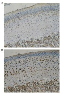
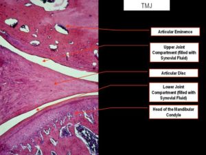
Look at all the cartilage growth.  There seems to be some endochondral ossification going on in the longitudinal ends of bone which is very good for height growth.
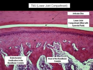
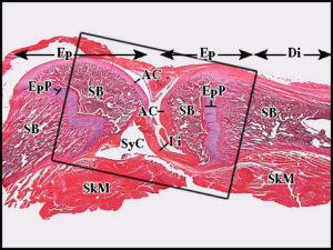
Here’s a normal jointNotice how they are connected strongly by a ligament(Li)
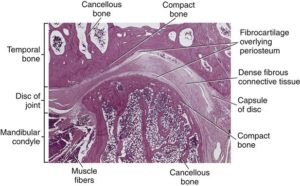
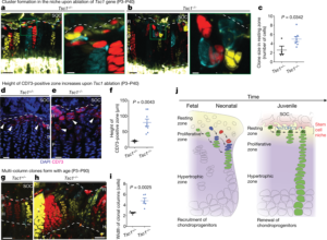
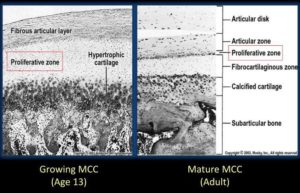
Here’s Mandibular Condylar Cartilage as a Juvenile and as an adult:
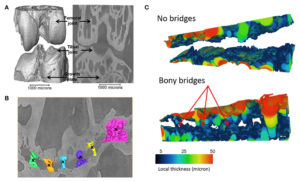
Microcracks effect on fluid flow. Long term loading may be key for LSJL.

This study is important because it indicates that microcracks may be bad for fluid flow which stimulates bone growth but good for cartilage growth as if fluid is not flowing than it is building up pressure and pressure is more conducive to chondrogenesis.  This indicates that a method to induce fluid flow such as  clamping/tapping should be higher duration and more “fatigue loading” based to insure the induction of microcracks.  Fatigue loading is the act of inducing bone damage not by a sudden large damage but by sustained bouts of loading over time.

Influence of interstitial bone microcracks on strain-induced fluid flow.

“microcracks act as a stimulus for bone remodelling, initiating resorption by osteoclasts and new bone formation by osteoblasts.  Microcracks alter the fluid flow and convective transport through the bone tissue. [We evaluate] the strain-induced interstitial fluid velocities developing in osteons in presence of a microcrack in the interstitial bone tissue. Based on Biot theory in the low-frequency range, a poroelastic model is carried out to study the hydro-mechanical behaviour of cracked osteonal tissue. the presence of a microcrack in the interstitial osteonal tissue may drastically reduce the fluid velocity inside the neighbouring osteons{So maybe microcracks will increase hydrostatic pressure as hydrostatic pressure is the pressure exterted by a fluid at rest}. This fluid inactive zone inside osteons can cover up to 10% of their surface. Consequently, the fluid environment of bone mechano-sensitive cells is locally modified.”

“Cortical bone constitutes the outer shell of long bones. This live entity is continuously renewed by bone cells in response due to the loading generated by daily activity”

“microdamage occurring inside the osteonal volume may generate a cell-transducing mechanism based on ruptured osteocyte processes. Concomitantly, microcracks are likely to alter the fluid flow and convective transport through the bone tissue and thus modify the hydraulic vicinity of the sensitive cells”

“the drag force caused by the pericellular fibres is thought to activate the cellular biochemical response through the interactions with the cytoskeleton”

“the pressure inversely increases from its Haversian reference to reach its maximum in the interstitial tissues.”

“the presence of the microcrack strongly modifies the fluid flow velocities in the osteons located in the immediate vicinity of the damage. It may generate an “inactive zone” inside the osteon wherein the fluid velocities are relatively low and thus the osteocytes stimulation too”<-But even though fluid flow may be low hydrostatic pressure may be high which may be better for chondrogenesis.

Fatigue Loading may be important to LSJL

This paper shows that axial loading can induce an almost complete fracture line through the bone through one side to the other.  But the break is on the wrong axis.  If fatigue loading was induced via transverse loading (lsjl or lateral impact loading) it is very likely that the micorodamage would be along the right axis.  But to induce bone fatigue in this way would likely require heavier loads than inducing sufficient hydrostatic pressure to induce chondrogenesis as the goal of increasing fluid forces in the bone is to encourage bone decay and induce differentiation of tissues that are capable of interstitial growth.

Role of Calcitonin Gene-Related Peptide in Bone Repair after Cyclic Fatigue Loading

“We used the rat ulna end-loading model to induce fatigue damage in the ulna unilaterally during cyclic loading. We postulated that CGRP would influence skeletal responses to cyclic fatigue loading. Rats were fatigue loaded and groups of rats were infused systemically with 0.9% saline, CGRP, or the receptor antagonist, CGRP8–37, for a 10 day study period. Ten days after fatigue loading, bone and serum CGRP concentrations, serum tartrate-resistant acid phosphatase 5b (TRAP5b) concentrations, and fatigue-induced skeletal responses were quantified. cyclic fatigue loading led to increased CGRP concentrations in both loaded and contralateral ulnae. Administration of CGRP8–37 was associated with increased targeted remodeling in the fatigue-loaded ulna. Administration of CGRP or CGRP8–37 both increased reparative bone formation over the study period. Plasma concentration of TRAP5b was not significantly influenced by either CGRP or CGRP8–37 administration.”

“sensory innervation of bone may have regulatory effects on skeletal responses to bone loading”

“Periosteum, endosteum, and bone tissue are all innervated by nerve fibers. This innervation exhibits plasticity in response to mechanical loading, in that a single loading event results in persistent changes in neuropeptide concentrations in both loaded and distant long bones, as well as changes in the neural circuits between limbs

Individual bone cells are directly connected to the nervous system via unmyelinated sensory neurons. Bone cells express a range of functional neurotransmitter receptors and transporters, including those for calcitonin gene related peptide (CGRP)”

“12 rats were fatigue loaded until 40% loss of stiffness was attained, using an initial peak strain of −3,000 µε (Fatigue group). ”

“To induce fatigue, the load applied to the ulna was incrementally increased until fatigue was initiated, as indicated by increasing displacement amplitude from a stable baseline. ”

The break extends all along the bone but is on the wrong axis.
“Increased bone blood flow precedes bone repair in response to fatigue loading, and remodeling in response to decreased mechanical loading . Systemic administration of CGRP also decreases blood pressure in a dose-dependent manner””
“”treatment with CGRP8–37 may have increased intraosseus pressure, transcortical interstitial fluid flow, and associated bone formation””

Impact loading on mouse bone length

Normally, I wouldn’t draw attention to this first paper but it draws an interesting thought:  “That long term sustained loading is better for longitudinal bone growth, whereas short term intense loading is better for bone quality.”  This could be due to hydrostatic pressure being good for stimulating a cartilagenous(and thus a pro growth plate micro-environment) whereas rapid changes in fluid flow as induced by short term dynamic loading(like jumping) are better for changes in bone quality.  So I will be changing the way I will be performing LSJL, rather focusing on clamping as hard as possible I will not be clamping as hard but doing longer more sustained clamps.

Enhancement of bone quality and longitudinal growth due to free-fall motion in growing rats

“[We] investigate the synchronous phenomena between bone quality and longitudinal length in a same subject affected by landing exercise. Physical exercise on the ground induces external loading to human body due to resistance from ground which can activate bone generation or remodeling. Especially, when the impact stimulation is applied to bone, it may improve bone quality and lengthening.
6-week-old male Wistar rats were randomly allocated to one of two conditions: free fall from 40 cm-height (I40; n = 7), and control (IC; n = 7). The impact stimulations were administered to the free fall groups, 10 times/day, and 5 days/week for 8 weeks. Structural parameters and longitudinal length of tibia were measured to quantitatively evaluate the variation in morphological characteristics and bone length with maturing.
The landing impact seems to be commonly effective on the enhancement of bone quality as well as longitudinal growth. However, the extent of enhancement may be more dominant in bone quality than longitudinal growth. On the other hand, the ratio of longitudinal growth seems to be dependent on the duration of stimuli whereas the enhancement of bone quality does not.
This study verified that free-falls exercise can be effective on the enhancement of bone qualities and promotion of vertical growth in long bones. We expect that it might be possible for the moderate impact stimulation to be proposed as an aid for prevention of bone loss and promotion of bone lengthening.”

” Bone cells accommodate to a customary mechanical loading environment, making
them less responsive to routine loading “<-thus possibly needing to cycle on and off a method of bone stimulus.  Although our goal is to target stem cells to form new growth plates and not necessarily bone cells.

“In comparison between two groups, there is no significant [differnece] at 4 week, whereas the values in I40 group exhibited slightly but significantly higher than that in IC at 8 weeks”  So there was a change in limb length but it took 8 weeks to notice a difference.  It would be interesting to see if impact loading could have an impact on animals without functioning growth plates.  If it could increase longitudinal bone growth in some other way.

the ratio of longitudinal growth seems to be dependent on the duration of stimuli whereas the enhancement of bone quality does not.”<-this is interesting maybe it’s more important to clamp for a long period of time than intensity of clamping.  This could a lot of sense if longitudinal growth is driven by fluid whereas bone mechanical parameters are driven by stimulation of osteocytes and bone cells.  Longitudinal bone growth could be driven by sustained hydrostatic pressure whereas bone quality could be driven by rapid changes in interstitial fluid flow.

Another study found “. observed significantly greater increase in bone length compared to the sedentary rats when they implemented the similar training 100 times/day in 5 days/week
on Fisher 344 rats of 3-month-old and 6-month-old for 8 weeks”

Here’s that other study:

Effects of Jump Training on Bone Hypertrophy in Young and Old Rats

“The effects of jump training on bone hypertrophy were investigated in 3, 6, 12, 20 and 27 month-old female Fischer 344 rats. The rats of all age groups were divided into jump training
(height: 40 cm, 100 times/day, 5 days/wkfor 8wks), run training (speed: 30 m/min, 1 h/day, 5 days/wk for 8wk) or sedentary group. Fat-free dry weights (FFW) of the femur and the tibia were significantly greater in the jump-trained rats than in the runtrained rats, and were significantly greater in the run-trained rats than in the sedentary rats. jump training significantly increased FNV of the femur and the tibia not only in young rats but also in old rats, while run training did not increase FFW significantly in old rats. In young rats, both jump training and run training significantly increased the length of the femur and the tibia and the diameter of the femur. The diameter of the tibia was greater in the jump-trained rats than in the sedentary and the run-trained rats in all age groups. The results of the present study indicate that jump training was a more effective training mode than run training for bone hypertrophy and that the effects were not limited by age. ”

According to the chart, Jump training increased length of the tibia and femur on rats younger than 12 months but actually decreased tibia and femur length on rats older than that.  So the compressive force actually denatured the tibia and femur such that it was shorter.

But in the study they do say ” In the 3 and 6 month-old rats, both jump training
and run training increased the length of the femur and the tibia and the diameter of the femur.”<-which is contrary to what the graph says.

Key LSJL studies about device design

FINITE ELEMENT ANALYSIS OF AN UNDER-ACTUATED ROBOTIC DEVICE FOR
KNEE LOADING APPLICATIONS

“Knee loading is one form of joint loading modalities, which potentially provides a therapeutic regimen to stimulate bone formation and prevent degradation of joint tissues. Healing of knee injuries is sensitive to many environmental stimuli.  Since mechanical stimuli are crucial for the growth, development, and maintenance of articular cartilage and bone, we have developed an innovative robotic knee loading device to achieve this goal. This device induces mechanical loading to stimulate articular cartilage and bone, and it potentially reduces the healing time of injuries such as bone fractures. The robotic device is an improved version over previous joint loading devices, in which loads are applied at specific points with non-uniform loading around the knee joint. In this paper, finite element analysis (FEA) of this robotic device has been presented that includes static structural analysis and modal frequencies of the device for two different material configurations used in the design. The design with ABS plastic material offers the desired margin of safety while reducing the weight and cost. ”

“The robotic device examined in this paper is designed for small levels of deformation. The intended displacement of the working device is small, a maximum of 13 mm. large
deformations are not recommendable as such deformations will critically affect the effective range of motion of device. “<-we may want larger deformations for our purposes.

” If the stresses are too high for the material selected, the device may yield, resulting in the failure of the device.”<-this can happen with a clamp without enough strength.”

” if very small magnitude of mechanical stimuli is applied fast enough then it may induce a cellular response”<-This is an interesting thought.  It’s very hard to produce a rapid mechanical stimuli physiologically.  How many bicep curls can you perform in one second?

“Osteocytes are the highly mechanosensitive cells which senses the resulting physical stimuli from mechanical forces applied on bones. They constitute of more than 90% of bone cells.
When rapid mechanical loading is applied at the end of long bone, the interstitial fluid present around the osteocytes pressurizes causing the fluid flow which creates hydrostatic
pressure throughout the bone. This hydrostatic pressure excites the osteocytes resulting in enhanced osteogenesis which decreases the healing time of the fractures, increases the bone density and will be helpful in treatment of osteoporosis and osteoarthritis”<-we want additional stimuli from the hydrostatic pressure we want hydrostatic pressure to degrade bone tissue reducing the constraining effect that bone tissue has on growth and we want the stem cells to become more fibrocartilagenous.

“To apply such a load, a device would need to have a means of producing a transverse force directly to the end of a long bone, such as at the knee. A cyclic force applied in such an area would force a slight shift of the fluid within the bone towards the opposite end of the bone in a controlled fashion”<-this is what we do with the clamp.  Produce a transverse(lateral) force directly to the end of a long bone.  It has the potential to be cyclic if you rotate and reverse the rotation of the clamp.  We may want more than a slight shift of fluid and we may not care if it’s in a controlled fashion or not.

“a maximum force of 40 N with the frequency range of 1 Hz to 5 Hz will have a promising effect on a human knee”<-Higher force may be needed for longitudinal bone growth.

” the efficacy of stimulating the osteocytes depended on the stress distribution on the knee. Based on this observation, it is projected that a position specific loading that provides a more
targeted force application on the knee is likely to further improve the efficacy of bone stimulation. It is hypothesized in that this targeted loading would contribute to the
improvement of new bone formation over a distributed loading modality. “<-maybe a smaller clamp pad and instead of clamping the knee as a whole clamping different epiphysis’ separately.  Although we don’t necessarily care about stimulating the osteocytes, we want to degrade bone tissue and stimulate stem cells.  The statement about more targeted loading still applies.

I can’t post an image of the device but look at fig 4 and 5.

” It can be seen that when maximum force of 20 N is applied on human knee, the maximum stress generated is 11.22 MPa. “<-Since around 10MPa is the chondrogenic stimulatory range this is a key pressure however you may need to degrade bone tissue as well.   The study does not indicate exactly where in the knee the stress was generated. This was for alluminum and steel.  Other materials were used and they were all in this same range.

FINITE ELEMENT ANALYSIS OF AN ELECTRO-MECHANICAL KNEE LOADING DEVICE

“When a cyclic but controlled load with a specific frequency is applied to the bones (femur and tibia) and surrounding tissues in the knee, it affects the osseous tissue causing physical deformations.  These deformations result in pressure gradient in the intramedullary cavity of the bone. Due to this pressure difference, there is a fluid flow of molecules and nutrients.
This will result in osteoblast differentiation; a phenomenon will initiate new bone formation or osteogenesis. This can be used as a healing technique in case of bone related injuries
like fractures or diseases like osteoarthritis and osteoporosis.”<-slightly more powerful prediction in this study where they predict osteoblast differentiation and physical deformations.  They mention a specific frequency being needed.

“When the device is loaded with a human knee, the inertial load resisting the driving force is considerable.”

LSJL Update 3-19-17

Here’s the last LSJL update.

I haven’t done experimentation with the rib cage because I’ve focusing on experimentation with a new clamp to get results in the arms and legs which is primarily what people want.

Using a 1500lbs clamp means there’s far less slippage issues.  Finding the key clamping spot is paramount as if you clamp in a spot with a lot of muscle you’ll never be able to generate enough force.  I found this was the case for clamping the shoulder joint as there’s no good place to clamp without getting a lot of deltoid.  But it was sufficient for the wrists, knee, elbow, ankles, and foot.

Here’s another pic of what the clamp looks like:

So hopefully I can have some leg and arm results to start sharing.

Here’s the feet pictures:

Here’s an overhead shot to show that it’s not flattening of the arches:

Here’s a link to when I was performing LSJL solely on my right foot.  Before right was longer.  Now left is longer.

I’m working still on clamping my right hand and right thumb with the new Bessey clamp hopefully I’ll start getting stronger results and get xray results.  There’s a difference but not as strong as I would like.  I’ll keep going to see if the results can get more and the xrays will be more striking.