Category Archives: Uncategorized

Muscle Pump and Hydrostatic Pressure

Getting a pump in your muscles may be a way to induce hydrostatic pressure but it is unlikely as lots of bodybuilders work towards achieving the pump so it’s something that occurs very frequently physiologically.  So if the muscular pump did affect height it would likely be a phenomenon that would be noticed by now.  But the goal of the pump is get the blood to the muscle under target not the bone.  The pump is very localized to the muscle under tension so this may be why the muscular pump does not increase height.  The target area for a hydrostatic pressure increase is the bone and the pump targets the muscle.  This does not mean that an understanding of a muscular pump could help us understand how to increase hydrostatic pressure in the bone.

A bone fluid flow hypothesis for muscle pump-driven capillary filtration: II. Proposed role for exercise in erodible scaffold implant incorporation.

“A model is presented for enhancement of fluid flow through bone matrix and any porous tissue engineering scaffold implanted within it. The mechanism of enhancement is the skeletal muscle pump in compartments adjacent to the bone. Pressure waves from muscle pump contractions aided by increased blood pressure during exercise coupled with temporary occlusion of arteries leading to and veins from the bone, increase hydraulic pressure in cortical bone capillaries so as to amplify capillary filtration. It is proposed that capillary filtration increase is sufficiently convective to contribute to bone fluid flow and associated percolation through tissue engineered scaffold matrix implants. Importance of this contribution is its relative role in maintaining seeded cells in bioreactor scaffolds. Validation of the hypothesis starts at a minimum level of demonstrating that capillary filtration is convective. At a maximum level confirmation of the hypothesis requires demonstration that capillary filtration-based interstitial flow is sufficient to stimulate not only host bone cells (as proposed in part I of the hypothesis) but bioreactor-seeded cells as well. Preliminary data is presented supporting the prediction that skeletal muscle contraction generates convective capillary filtration.”

Although we don’t really want increased hydrostatic pressure in the capillaries that’s more likely to just deliver more nutrients to the bone.  Increased hydrostatic pressure in capillaries increases capillary filtration getting more nutrients to the interstitial fluid.  What we want is increased hydrostatic pressure in the interstitial fluid itself to encourage chondrogenic differenetiation.

“Nutrient exchange is not the sole function of transport in bone. There is increasing evidence that interstitial fluid flow is sensed by and modulates the behavior of bone cells. Percolation through bone matrix and associated implants is referred to as bone interstitial fluid flow
(BIFF). Two mechanisms for bone cell sensing of BIFF have been proposed; one mechanical and the other electrokinetic. The electrokinetic model focuses on streaming potentials that are putatively sensed by electrokinetic receptors in bone cell membranes. The mechanical model focuses on shear stress at the membrane-fluid interface, which is transmitted to second messenger by mechano-receptors”

“osteocytes and their processes are surrounded by relatively thin fluid (not necessarily Newtonian) annuli in the lacunar and canalicular compartments, rather than relatively large volumes of flowing blood.”

“muscle pump and exercise effects combine to increase capillary filtration sufficiently to add a significant component to BIFF.  We reason that skeletal muscle, acting through a muscle pump mechanism, increases the rate of capillary filtration by increasing capillary hydraulic pressure via contraction of skeletal muscle in compartments adjacent to bone. Exercise magnifies the affect by increasing baseline blood pressure through increased heartrate and muscle pump activity. Two anatomical circumstances suggest how the mechanism operates: (1) bone influx and efflux vessels outside bone are contained within fascia bounded compartments, which include skeletal muscle, and (2) efflux vessels (veins) are valved.”

“During the same exercise vascular resistance in bone increases two to fourfold while vasodilation in adjacent muscle increases”<-So blood flow in muscles increase while blood flow in bone decreases.  This could in fact increase hydrostatic pressure in bone as hydrostatic pressure is the force exerted by a fluid at rest and vascular resistance implies that there’s more fluid at rest.

“Solitons in arteries propagate to capillary beds where they increase intravascular hydraulic pressure in fluid unable to escape through veins.  In any given osteon or Haversian canal capillary filtration is increased driving extravascular fluid over perivascular
tissue and through nearest canaliculi. Pressures generated during exercise above heartbeat baselines can be considerable; interstitial values as high as 570 mmHg”

” IMP is a poor indicator of blood flow in bone the blood pressure changes associated with its increase are significant. (2) Blood flow to limb bones increases during exercise. (3) Vascular resistance in limb bones increases during exercise”

“contraction of the quadriceps muscle causes a 30 mmHg or more rise in femur IMP”

The Key Role of the Blood Supply to Bone.

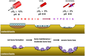
“Blood supplies oxygen, nutrients and regulatory factors to tissues, as well as removing metabolic waste products such as carbon dioxide and acid. Bone receives up to about 10% of cardiac output, and this blood supply permits a much higher degree of cellularity, remodelling and repair than is possible in cartilage, which is avascular. The blood supply to bone is delivered to the endosteal cavity by nutrient arteries, then flows through marrow sinusoids before exiting via numerous small vessels that ramify through the cortex. The marrow cavity affords a range of vascular niches that are thought to regulate the growth and differentiation of hematopoietic and stromal cells, in part via gradients of oxygen tension. The quality of vascular supply to bone tends to decline with age and may be compromised in common pathological settings, including diabetes, anaemias, chronic airway diseases and immobility, as well as by tumours. Reductions in vascular supply are associated with bone loss. This may be due in part to the direct effects of hypoxia, which blocks osteoblast function and bone formation but causes reciprocal increases in osteoclastogenesis and bone resorption. Common regulatory factors such as parathyroid hormone or nitrates, both of which are potent vasodilators, might exert their osteogenic effects on bone via the vasculature. These observations suggest that the bone vasculature will be a fruitful area for future research.”

” impairment of the blood supply is well-known to reduce growth and repair, cause bone loss and, ultimately, necrosis”<-Maybe this could actually be a good thing as hypoxia is often associated with chondrogenesis.

hypoxia

With severe bone loss comes the space for neo-growth plate formation.

“drugs used to treat hypertension[high blood pressure] can increase systemic blood flow”

Mechanotransduction in musculoskeletal tissue regeneration: effects of fluid flow, loading, and cellular-molecular pathways

“Dynamic fluid flow induced by mechanical loading has been shown to have the potential to regulate bone adaptation and mitigate bone loss.”

“High physical activity level has been associated with high bone mass”

“Fluid flow-induced shear stress within bone has been considered as the source of how bone cells sense mechanical stimulation. Bone interstitial fluid is filling a variety of voids and channels within the bone matrix, including lacunae-canaliculi, bone tubules, Haversian canal and Volkmann canal, and osteon. Mechanical loading-induced interstitial bone fluid flow may play a role in mechanical sensing, bone cells response, signal transmission, transfer of nutrients, and so forth.”

“increased venous pressure can promote new bone formation in the periosteum. The data indicated that increased venous pressure will increase blood supply from the capillaries to the bone tissue”

“muscle force alone, if applied at a low rate, such as resistant weight lifting with high intensity, would not be able to generate sufficient strain and fluid pressure in bone. MS with a relatively high rate and a small magnitude, however, can trigger significant fluid pressure in the skeleton.”

“mechanical loading has been shown to reduce sclerostin levels in bone”

My Calling Is Helping The Most Lonely

Sometimes I don’t pay enough attention to this project or the research. I leave for a while to live my life and focus on other areas of it. Then I find something, read something, that makes me remember the real reason why I ever first started this quest. My girlfriend left me, I was devastated, and I believed that one of the reasons she left me was because I felt I was not tall enough. She left me for a guy who was much taller than me. It hurt me at a level which I have never felt before, and I swore on my life that I would find a way to change the situation, not just for me, but for other guys in the world.

I remember once reading this controversial article from some internet website where the author wrote that men in today’s world are no longer needed by women, since women can do almost everything better than men. The way the school system is setup reveals that academics rewards the students based on following orders and being disciplined, which has never been a strong point of young men. While men still need women for the act of reproduction, companionship, intimacy, and sex, women no longer need the skills and qualities within men for survival. They no longer as the males of their family to go out and hunt down a sabretooth tiger for the even meal. As a heterosexual male who have somewhat old-fashion conservative views, the article seemed to push at a pressure point within my psyche that made me feel insecure, sad, and a little bit angry.

It turns out that when the modern young adult female talks about the income inequality among the sexes today, the women are not comparing themselves to the bottom 80% of men in their society, but the top 20% of men. In every society, there is always a heriarchy of men, with some being of higher class, and most men being of the lower class. Throughout the history of the human race, within almost all tribes and groups, it would turn out that the majority of males would never get a chance to have sex, and find a sexual partner or mate in life. Historically, it was the top minority of men in society who get sexual access to the majority of females. Think of the harems of the Emperor of China or the Caliphate of the Ottoman Empire or the Persian Empire, which had up to thousands of young girls who were carefully protected from other men by the army of the male rulers. Being a human guy in this world, in any time frame, has always been hard. It is just that hundreds of years ago, being born as a female was also very hard, with the constant threat of kidnapping, assault, rape, and forced marriages. Now that the world has become more peaceful and most men in the developed world no longer view females as property, the females of our species don’t feel that type of threat from male strangers that they were taught hundreds of years ago. The main point is, in this modern age, it is much harder to be a guy than a girl. When we really, objectively look at the overall condition of the human race, it has been the males who have suffered the most throughout history.

I refer to the readers this amazing book “Is there anything good about men? How Cultures Flourish by Exploiting Men” by the professor Roy F. Baumeister. This book will shake the very core belief system of most young men who were born and/or raised in one of the developed western nations. 

Of all the types of men who would most likely fail with females, I believe that it is men who are short who have it worst. The only exception may be being disabled, and that can be up for debate. The fact that short men are so disrespected and treated badly by society, and looked down upon by girls as unworthy of companionship shows that this type of discrimination is too pervasive in the minds of females. Being short is often the kiss of death.

The silent pain and suffering that this certain group of guys go through in life is felt. I am not God, and I am not a savior. I am trying my best to help a minority group of men in this world who are prevented from finding companionship because they did were not lucky at birth and ended up short. Being short and ending up a certain height is something that is almost completely out of our control. However, we should have that type of power and control, if we really wanted it.

This world is really hard for the short men. Maybe we can find some solution to make it so that their problem of short stature can be solved. It might not come about tomorrow, or even a year from now. However, I believe that one day we will find multiple solutions to solve the problem of being short.

Rejection

From Reddit/r/ForeverAlone – Tried to get a female friend to set me up with someone. Her response: “sorry, you’re too short”  – (https://www.reddit.com/r/ForeverAlone/comments/3hu8el/tried_to_get_a_female_friend_to_set_me_up_with/)


CRISPR-Cas9 Gene Editing Tool Hints At Pre-Programmed Babies With Superhuman Intelligence and Height – Huge Breakthrough!

Most people who are in the online community who discuss the issues and problems related to being short might sometimes wish that their own children (or future children) will be taller than them so that they don’t have to go through the same kind of ostracizing that the parents got when they were younger due to their short stature.

There was even one thread on an old reddit post which turned viral because a wife suggested to her husband (immediately after sex) that she would prefer to have a child with another man instead of him because her husband was short statured. She was afraid to make her future sons or daughters short because they would have it harder in life. Based on what I have read from the evolutionary biology books like The Selfish Gene by Professor Richard Dawkins and The Red Queen by Matt Ridley and Sperm Wars by by Robin Baker, that action is basically the worst suggestion a heterosexual female can tell her chosen lifelong male partner. Genetic infidelity or the idea of using one’s hard earned resources to take care the genetic offspring of another man is probably the worst offense a normal male could ever be asked to do. Humans as animals are evolutionary programmed to resist this idea on the most instinctual level.

Well, there is a type of genetic engineering revolution that is going on in the biology, genetic, molecular biology, and biomedical research labs around the world right now that would signal the possibility that we may one day be able to design a person from birth to give them the qualities that we desire. No more can a woman (or man) complain to the world that the reason they don’t want to mate and be with a potential romantic partner for the long term is because of a perceived genetic lottery loss for their future offspring. When we think about all the times when a short female refused to date, marry, or mate with a short male and used the excuse that she just didn’t want to have short children, we realize that this extremely primal/instinctual desire will no longer be worried over.

This gene editing technique is going to change everything. It is a disruptive technology (as coined by Clayton Christensen) in every sense of the word. It is not going to be immediate and you won’t find super-babies next year, but it has made the idea of “super-babies” an almost guaranteed certainty.

This CRISPR-Cas9 gene editing tool has been so revolutionary that the use of it within the labs around the world has spread like wild-fire. It is probably the biggest Biotech Disruptive Innovation that will come about this century. Where gene editing was extremely hard and inaccurate before, this tool which was only developed in the beginning of this decade has made gene editing extremely easy.

This tool is going to change the field of biology, genetics, and agriculture completely. And that is not an exaggeration. Most researchers from MIT to Caltech are talking about the implications of this technique, which is so simple and non-technical that a BioHacker in his DIY Lab in his/her garage can use it.

There was news that came out about this team in China which has been using the technique already on embryonic cells and the news has shocked quite a people in the biology/genetics world.

If the readers can remember the basic premise of the movie Gattaca, with Ethan Hawke, and one of the few instances where we get a glimpse of a person having limb lengthening surgery performed on them in a blockbuster movie, this technique is basically the key that could very well unlock the door to that world. I am not here to judge whether a potential future where people will be judged solely on their genes is a dystopia or not.

This technique is going to let expectant parents choose the features of their child that they want. We are reaching the level of Science Fiction now.

While genetic manipulation is not going to work on an adult, unless they are going for a radical total cell transplantation with genetically modified cells ala artificial dialysis machine style (where you pump the ordinary cells out of the body and have new gene therapy treated cells flushed into the body), this technology in a few decades will change everything on how we view natural human babies.

How would the average couple in the year 2050 feel knowing that their more well-off neighbor couple can afford the extra $200K treatment to change the phenotypically traits of their unborn child to be 3-4 inches taller and 20-30 IQ points higher? That type of advantage leads to so many bioethical questions for the researcher scientists today.

Of course, we should maybe take a step back and remember the fact that height is not one of those traits that is controlled by one gene. It is not something like the color of one’s eyes or whether one has a widow’s peak or not. Height is quite complex.

It turns out that height is controlled by at least hundreds, if not thousands of genes. The closest we have come to finding one gene that has a profound effect on height was the HMGA2 gene (HMGA2 is confirmed to be associated with human adult height.) From another source, we find out that “…Adults with two copies of the height-increasing version of the HMGA2 variant are on average 0.8 centimeters taller than adults carrying two copies of the other version”. ) Sure, 0.8 cm does not seem like that much, but it is almost 1 cm, and 1 cm will over time become noticeable, when you then combine the effects of HMGA2 with the GDF-5 Gene. From the same source, we learn that “…pointed to a second gene, GDF5, of which the average height difference between genotypes is 0.4 centimeters.” Since most of the world is based on the metric system, and most people state their height to the 3rd digit, that extra centimeter can mean quite a bit.

I personally have heard enough stories of people in China and India being rejected from their dream jobs because they were off the job height requirement by 1 cm.

We know based on just looking at some common organizations like the US based NBA basketball organization that there are families which have the gene for tall stature. There are many brothers, twins, and father-son combos we find in the NBA. Clearly someone like Yao Ming has that genetic trait. It would not take that much to get blood samples from those who have familial/genetic propensity for tall height, and slowly over even a decade figure out what are the other 500+ genes which when expressed properly, would cause a genetically engineered baby to be 4-5 inches taller than through the natural path.

So is it that big of a deal that some potential mother or father would want to spend hundreds of dollars to alter the genetic material of their baby to be taller? I believe that some people will care that much about it. They would gladly open their wallets and pay for that type of treatment. The world is becoming more and more competitive, especially for the younger generation of kids, who are now competing on the global level. The young Chinese prodigy will be competing against the young Indian prodigy in 30 years – a billion vs another billion. People will do what they can to give their offspring as many advantages as they can for later life success.

I wrote about this technology before in the post “CRISPR Technique with Cas9 Enzyme To Alter Hereditary Traits Easily” back in 2013 but in the span of 2 years, everything has changed.

For the love of god, I am begging the readers of this website to read this article from the MIT Technology Review magazine – “Who Owns the Biggest Biotech Discovery of the Century? – There’s a bitter fight over the patents for CRISPR, a breakthrough new form of DNA editing.”

How Close Are We Towards Growth Plate Regeneration To Grow Taller?

This seems like a reasonable question to ask when a person who is past bone maturity and physeal ossification is interested in learning how they can grow taller.

Well, to answer this question, there are many factors and variables we have to take into consideration. It is really hard to give a definite answer, although I have stated before that we might be as close as 15 years away from some commercial company developing the technology to do that. On the other end, it could be as far away as 50-70 years, assuming that the rate of progress continues for the fields of tissue engineering, regenerative medicine, 3D Bio-printing, and stem cell R&D. Let’s remember that the rate of scientific/technological progress for the biological sciences and biomedical application does not follow the trajectory of Moore’s Law, unlike electronics and Computer Science. To make progress and breakthroughs in BIology is extremely resource/financially intensive, unlike CS, which often just requires a programming wunderkid sitting in his underwear in his dorm room eating Cheetos.

I revealed in a post a month ago about the company EpiBone who is developing osteochondral grafts as implants which has as an advisor Dr. Warren Grayson, who I have said since 2013 is one of maybe 6-7 researchers in the world we should be following. Refer to the post EpiBone Company To Engineer Osteochondral Grafts – Research Breakthrough!. The company reveals that there are definitely plenty of people who realizes that there is a huge financial incentive to get this technological problem to work out. There has already been at least 1 patent filed by the research group at EpiBone on how to use a bioreactor to build bone tissue. Refer to “Methods, Devices and Systems for Bone Tissue Engineering Using a Bioreactor – US20120035742 A1”

Let’s now look at the research of Dr. Robert Tracy Ballock, who I have said is the other main researcher we should be following. His work has been on the Growth Plate for the last 15 years or so. He has been working with Dr. Eben Alsberg, who showed that it was possible to growth a functional growth plate. Refer to his grant on Growth Plate Regeneration available here. Further searching on this grant and his work shows that there was 2 grants, one for the 2012-2013 period, and a 2nd grant for the 2013-2014 time frame. (2012-04-11 – 2014-09-30). If you search around the internet for any new academic papers published under Dr. Ballock’s name, he has not published anything for this year, or even late 2014. I would guess that he is finished with his research from the grant which lasted 2 years and plans to write something up. There might even be a Patent application that is filed from his research soon. Whatever he has found, he is not revealing it yet, at least not to the general public.

3 months ago there was a biomedical conference where a university researcher named Dr. Juan Taboas (Ph. D) presented his work called “Repair and Regeneration of the Physis” at the Houston Methodist Research Institute. Just two months ago, the video of his presentation was available for the general public to watch, but now that video has been set to private and one requires a password to watch the video. I did watch maybe the first 5 minutes of the presentation, which was over 1 hour long. There was a video/audio syncing problem so I was not able to record the presentation back then. The part I watched was not that informative and did not reveal too much about his current research.

The abstract of his talk is below…

Physeal regeneration poses a considerable challenge in orthopaedic regenerative medicine. The physis is the cartilaginous interfacial structure at the ends of the long bones that produces appendicular skeleton growth. Fracture, infection, and cancer can result in limb deformity and loss with significant morbidity and medical cost despite their status as rare disorders. Dr. Taboas and his research group are developing hydrogel and stem cell based point-of-care therapies to prevent and repair limb growth disruption in pediatric patients, and for endochondral repair of large boney defects in children and adults. They are currently evaluating the effect of hydrogel composition and zonal patterning on construct growth and architecture maintenance in an in vivo murine subcutaneous implantation model. These techniques may be applied to regenerate other skeletal tissues that require appropriate interfaces with bone for proper function, such as articular cartilage and ligament.

Here is something that the average reader needs to understand. There is a branch (or maybe sub-branch) in the medical research fields known as Orthopaedic Research. Technically, orthopaedic research refers to diseases and injuries of bones, joints, nerves, and muscles. However, there what is not written, but also implied is the cartilage and the tendons as well. Keep this note in mind as we go further along.

When I was at the 2nd Organ-On-A-Chip and 3D Bioprinting Conference in Boston a month ago, there was a presenter at the conference who revealed that she and her group was working on getting stem cells to build tendon tissue. Tendon tissue is what connects muscle tissue to bone tissue. It is similar to cartilage, because of the high level of collagen, although not the same type of collagen. Her presentation was ” Engineering Tissue Microenvironment Informed by Development, Healing and Disease”. Refer to Catherine Kuo, Assistant Professor at Tufts University. Apparently, she got her postdoctoral training in the Cartilage Biology and Orthopaedics Branch at the NIAMS of the NIH. The last I heard, she was changing positions to a different university. This shows that the field of stem cell technology is being applied to form every type of tissue.

There was a paper that was being passed around at the conference which asked all the people at the conference what type of tissue they planned to use the 3D Bioprinter on. The type of tissue that was most often formed from the 3D Printers was cartilage. Not bone, not vascular tissue (blood vessels like capillaries), not tendons, but cartilage. This reveals something critical. It seems that all of the major researchers understand what would be the easiest type of tissue one can print using this new form of technology. Everyone is thinking the same thing, and we are almost all pursueing the same idea: Cartilage.

I would guess that a large percentage of the research done in this well known field known as orthopaedics is on cartilage research, specifically cartilage growth and regeneration.

So what does Dr. Grayson, and Taboas have in common?

It turns out that they both were all at the 2011 Termis Regenerative Medicine Conference in Houston. Their names was on the list of Attendees. I was supposed to go to the 2015 Termis Conference in Boston, but the cost was just too high for me. Dr. Atala of Wake Forest was going to be there. If you plan to take this type of research seriously, you have to spend the money (about $2,000) to attend these types of exclusive conferences to learn what the real academic researchers are working on.

What about Dr. Ballock?

He is a medical doctor as well as a researcher since he has both a Ph. D.and M.D. (Smart guy). Like all academic researchers, he is involved in a lot of societies and academic groups. I am sure he flies around a lot to give presentations and talks in various conferences and conventions. Back in 1998 he was at this conference called “Bioengineering & Orthopedic Science – Gordon Research Conference” which is now known as “Musculoskeletal Biology & Bioengineering”. It was held from July 26-31 in Andover, New Hampshire. He gave a talk called “”Control of Cell Proliferation in the Growth Plate”. It seems that for almost 2 decades this guy has been working on getting the growth plate to work out. He was involved in this multidisciplinary conference back in 2008 called the “Cleveland Clinic Cartilage Innovation Summit”. The conference was basically a group of researchers gathering to talk about the subject of “The Clinical Science and Outcomes of Cartilage Repair, Maintenance, Regeneration and Replacement”.

So What Am I Trying To Conclude From All This Information?

Some people who are in industry who focus mainly on earning income and profit for their employers and no longer in academics (who focus mainly on the research, discovery, and learning more) would say that something along the lines that all the breakthrough research discoveries one finds in the university lab is still useless if one chooses not to bring their findings into real application and shared with the rest of the world.

This applies to the field of Orthopedics (and most any other type of medical research). What I have found in the last 6 months from attending the conferences which talk about the rising field of 3D Printing, and specifically 3D Printing applied to tissue formation is that the researchers in the university labs are trying their hardest to bring their research to the public.

Dr. Grayson is going to help EpiBone succeed in getting bone implants to work. There is already too many companies who have gotten the theory and science down on getting lab grown stem cell derived bone tissue to be reimplanted into the subjects body with complete success. The 1st step in getting bone tissue to be grown from stem cells or mesenchymal stem cells has been multiple times with the academic researchers forming companies to get their research out to possibly make them millionaires. There is no problem with this since the academics see a way to make personal gain from their years of hard work and research.

When we then move towards the 2nd/next step of getting fibrocartilage to be printed from lab grown pieces of cartilage or chondrocyte, there are labs like Dr. Atala or Dr. Lawrence Bonassar who have already achieved that too. You have seen many pictures of the outer ear lobe being grown in the labs in cultures and even on the backs of lab mice. The same can be said about nose tips, which are also fibrocartilage.

The 3rd step is to get Hyaline Cartilage to be printed or grown in the lab. We are getting super close to getting Hyaline cartilage to be printed. There were 2 exhibitors at the Organ-On-A-Chip Conference, (Cellink and RoosterBio) who would be able to provide the raw materials, cell culture medium and seed mesenchymal stem cells, to let any professional lab to play around to eventually get the lamellar structure of hyaline structure to work out. I would be willing to guess that by the time the 2020 Termis World Conference comes around, we would have succeeded there.

Should we try to the traditional seed stem cell into scaffold method or should we try the more revolutionary way of growing cartilage tissue from scratch using the 3D Bioprinter? – We would have to make that decision eventually.

The 4th step would be to get Epiphyseal Hyaline Cartilage to be grown/printed. Ballock has been working on this problem for 20 years. He got a grant for the 2012-2014 time frame to go research on growth plate regeneration. It is not just him who is working on getting this thing to work out. In the grant, he wrote that if he is successful, the growth plate regeneration technology can be used by people who have already reached bone maturity. He knows what his research would mean and how it would effect the public.

Of course, orthopaedic research and development is something that thousands of people around the world are working on. He is not the only one trying to get growth plate regeneration to work out.

  • There has been teams in Hong Kong who got the chondrocyte implantation idea to work back in the early 2000s. Based on certain Non-Disclosure Agreements (NDAs) I signed early on, I can not reveal the exact details of those studies, only to say that the experiments were done and were successful.
  • I revealed early on, in the late 2012 that there were military hospitals in China who have gotten growth plate transplantations to be successful. Where did they get the growth plate in the first place is still suspicious though.
  • I wrote about the fact that even plastic surgeons in Russia, like Dr. Teplyashin have gotten the scaffold technique to be successful but this controversial technique is probably only offered to Russian millionaires in secret for maybe $200 K a pop. They got it to work out in sheep, but that doesn’t mean they have ever performed the same surgery on humans.

The truth is that we are getting quite close, with the best option being people like Ballock. If he is willing to write up his results from his work for the last 3 years, and take those results and form a company around it to develop growth plates grown out of scratch (ie 3D Printed) or based on the traditional tissue engineering method using scaffolds and growth factors, he would have something working in 10-15 years. Of course I could be way too optimistic.

To answer the original question, I would make a very tentative guess based on the idea of Kurzweil’s Law of Exponential Growth, if someone like Ballock is willing to form a company around his discoveries and try to bring it to public, we would have a limb lengthening alternative based on 3D Bioprinting, Stem Cells and Scaffolds, and tissue Engineering within 10-15 years with enough funding. When it comes to funding, I would say that biomedical engineers and researchers would need maybe at least $100-$300 Mil to get this procedure out to public. So who is willing to fund this type of project?

Girl Uses Cosmetic Limb Lengthening Surgery To Increase Height By 8 Inches

Alexandra TranserThis is sort of old news but it is sometimes still worth mentioning. Back in March of this year there was another story that came out of a person who got cosmetic limb lengthening surgery. Unlike most of the people you hear about, this person was a girl.

While it is not very often we find girls who decide to go through with the surgery, there have been enough cases of girls who do it for the prospect of becoming a model. This girl fits that type of case study.

We refer to this article from Mirror, which is a UK based type media outlet/source. – http://www.mirror.co.uk/news/world-news/aspiring-model-spends-thousands-surgery-5115453

A 5′ 4″ tall aspiring model Alexandra Transer got the surgery done for a supposed 6,000 pounds and she has already gotten 6 cms out of it. She comes from a family of people who are below statured. Her mother is 5′ 2″ and her father is 5′ 5″. She chose not to go to University but work for her father in Engineering and saved up the 6,000 UK pounds to get the surgery.

She had wanted to be a model for most of her life, but when she contacted a modeling agency, they told her that she was below the height requirement and that the only way for her to get to that strict modeling height requirement was to extend her legs. She decided to do just that and went through with it, which we found very impressive and brave.

This first surgery she has decided to do is one of a total of 3 planned. There is supposed to be 2 more surgeries she will go through which will eventually put her at 6 feet. Her estimated time frame is that the 3rd surgery will be done by 2018. She will be in her 30s by then and may not get any type of modeling work but she did say that she plans to dye her hair blonde and then feel like a model being tall and blonde.

It seems that maybe what she really wants is to feel beautiful. On this website, we don’t judge. Vanity is something we all go through.

Some new information we learned is that she is actually from Russia herself, Volgograd. For anyone familiar with the LLS community, Volgograd is actually really famous because of the Centre of Anthropometrical (Orthopaedical) Cosmetology and Correction. Refer to Dr. Alexander Barinov and Dr. Viktor Shatov, of the center RUCOSM. Dr. Alexander Barinov is famous for being a protege and student to the more legendary Dr. Yegorov.

She mentioned that a former Soviet Athlete, a Valery Brumel, got this type of surgery done on them in 1968. Why did this olympic athlete get it done back in the 60s? We don’t know about that. I personally suspect that one of the reasons she was willing to go through with the surgery unlike so many other people who wish to be taller but are too afraid of surgery is because she comes from Russia, and specifically her home town has one of the most popular and recognizable centers for height increase surgery in the world. I am sure her parents at first was very reserved in her desires but they probably had a better understanding for why she would go through with it than other parents. Since they are from Russia originally, going a center like RUCOSM would not be as difficult of a transition as other people from other countries who might know any of the local Russian customs or language. Maybe for this girl, going to get limb lengthening surgery is as simple as driving the 3 miles to one’s local gym center to exercise.

Based on current conversion rates, it seems like she paid about $9,400 USD to get the first surgery.

We here have done our own level of research on LLS, and the cost of it. Most people have said that the cost of getting LLS is usually about $50,000 with a trip to one of the more well known, reputable surgeons costing upwards of $100,000 with everything covered, for maybe 10 cm or 4 Inches of increase in the leg bones. The lowest cost I personally have heard of was around $10,000 for the cost of surgery from a former Chinese surgeon. The reduced price of getting one’s leg extended seems to suggest that the cosmetic procedure may be becoming much more prevalant. That may be a good thing for some people who really want to go through with it and are looking for a cheaper alternative.

When we looked at the cost for LLS back in 2011, it seems that the cost was around 12,000 Euros, at least in the RUCOSM center, in Volgograd. Our source was from the Short Support Group website. The 12,000 euros cost was supposed to cover pretty much everything, including a 3 month stay. Of course, from the RUCOSM website, they state that for each surgery, they can only increase one’s bone length by 7-9 cm maximum. This is why Alexandra needed 3 total surgeries, to gain 8 inches or 20 cms of height to reach her desired goal of 6 feet tall from her original 5′ 4″. For her, she was very happy with the results, and there were no complications.

Refer to here for an interview with Dr. Barinov.

Note what he claimed for the cost of the surgery

  • How much does the height increasing procedure cost? – Dr.Yegorov works with different clinics, both state and private. The total cost of the procedure including all necessary expenses is from $15,000 to $30,000 US dollars depending on the clinic and the accommodation and service conditions.

We would say that $10,000 USD is a little unrealistic of cost. A more reasonable cost would be probably $25,000 and that would just be for 7-8 cms of increase, assuming that no complications are developed.  Of course those will be 2011 prices. Since it is 2015, We should expect maybe $30-35 K in fees in total. How Alexandra was able to just pay $10,000 we are not sure. Maybe she got her surgery done in the UK, and not Russia. Maybe instead of paying the thousands of dollars in fee for staying at the hospital facility to recuperate, she was able to have someone take her to her home in Volgograd (which might just be a few blocks away) during that time, allowing her to save thousands in hospital fees.


How bone knows how to grow

How does bone know how to be the proper shape and size for development?  Can we manipulate this to grow taller?

I received this email from the author regarding how distraction osteogenesis would affect how bone manipulates growth in regards to maintaining placement of superstructures:

“It is indeed an interesting question as it challenges the system with an unnatural manipulation – i.e. interstitial growth.

The simple answer is: we haven’t tried, so I can’t say for sure.

If the relative locations of ligament and tendon insertions are what you are interested in, then previous works show that the periosteum is involved in regulation of their positions (see list below). Moreover, if the balance between proximal and distal growth rates is what you are interested in, then other works show that cross-sectional cutting and stripping of the periosteum can cause temporal acceleration in overall growth rate of the bone (also in humans, if I remember correctly), followed by a potential change in proximal to distal growth balance (I don’t think that these works test how these influence the positioning of superstructures in the bone; see list below).
Therefore, if the operation you are applying includes anchoring of the periosteum to the bone or its cutting and stripping, this is something that may influence the scaling of the bones.”

Isometric Scaling in Developing Long Bones Is Achieved by an Optimal Epiphyseal Growth Balance.

“One of the major challenges that developing organs face is scaling, that is, the adjustment of physical proportions during the massive increase in size. Although organ scaling is fundamental for development and function, little is known about the mechanisms that regulate it. Bone superstructures are projections that typically serve for tendon and ligament insertion or articulation and, therefore, their position along the bone is crucial for musculoskeletal functionality. As bones are rigid structures that elongate only from their ends, it is unclear how superstructure positions are regulated during growth to end up in the right locations. Here, we document the process of longitudinal scaling in developing mouse long bones and uncover the mechanism that regulates it. To that end, we performed a computational analysis of hundreds of three-dimensional micro-CT images, using a newly developed method for recovering the morphogenetic sequence of developing bones. the relative position of all superstructures along the bone is highly preserved during more than a 5-fold increase in length, indicating isometric scaling. It has been suggested that during development, bone superstructures are continuously reconstructed and relocated along the shaft, a process known as drift.  most superstructures did not drift at all. Instead, we identified a novel mechanism for bone scaling, whereby each bone exhibits a specific and unique balance between proximal and distal growth rates, which accurately maintains the relative position of its superstructures. Moreover, we show mathematically that this mechanism minimizes the cumulative drift of all superstructures, thereby optimizing the scaling process. [There’s] a general mechanism for the scaling of developing bones. More broadly, these findings suggest an evolutionary mechanism that facilitates variability in bone morphology by controlling the activity of individual epiphyseal plates.”

If we can trick the bone into thinking it’s drifting maybe we can convince it to grow to maintain the position of the superstructure.  For example, dislocating the bone or similar means.

Although the molecular mechanisms regulating each growth plate for different bones are similar the bones still have different elongation rates.

“superstructures, known as bone ridges, tuberosities, condyles, etc., are necessary for the attachment of tendons and ligament as well as for articulation. To perform these functions they are located at specific positions along the bone. Bone superstructures emerge during early skeletogenesis . During growth, bones elongate extensively by advancement of the two growth plates away from the superstructures. It is therefore expected that during elongation, superstructures would remain at their original position near the center of the bone. Nevertheless, the end result is proper spreading of superstructures along the mature bone, which clearly implies the existence of a morphogenetic mechanism that corrects their locations.”

It’d be interesting to see what happens to bone superstructures during distraction osteogenesis.

“An ossified bone is a rigid object and so are the superstructures protruding from it, implying that they cannot be relocated by means of cell migration or proliferation. Therefore, any scaling mechanism must be adapted to overcome these physical restrictions.”<-So we have to make the bone less rigid.

“forelimb bones tend to grow away from the elbow joint, whereas bones in hind limbs tend to grow toward the knee joint.”

” Because the periosteal sheath is stretched over the entire external surface of the bone, including both the superstructures and the growth plates, it can pass to the growth plates signals concerning the relative position of superstructures.”<-Then perhaps we can manipulate longitudinal bone growth by manipulating the periosteal sheath.

“periosteal tension down-regulates growth plate activity, as the higher the tension level, the more inhibited growth plate activity is.  Damaged periosteum forms a scar tissue at the site of destruction. This scar tissue, which anchors the periosteum into the bone, creates an independent tension level near each growth plate. As a result, a new growth balance is formed, which equals the ratio between the distances from the site of the scar to the two ends of the bone, therefore maintaining the relative position of the scar site.. Superstructures can be considered as natural anchoring points for the periosteum into the ossified bone, either due to the insertion of tendons through them into the bone cortex, or by means of steric interference, such as in the tibiofibular junction. This results in a regulatory loop whereby the superstructures determine the tension levels of the two periosteal segments, which control the ratio of growth rates by inhibiting growth plate activity, which in turn maintains the relative position of the superstructure.”

Mechanical regulation of musculoskeletal system development 

“During embryogenesis, the musculoskeletal system develops while containing within itself a force generator in the form of the musculature. This generator becomes functional relatively early in development, exerting an increasing mechanical load on neighboring tissues as development proceeds. A growing body of evidence indicates that such mechanical forces can be translated into signals that combine with the genetic program of organogenesis. This unique situation presents both a major challenge and an opportunity to the other tissues of the musculoskeletal system, namely bones, joints, tendons, ligaments and the tissues connecting them. Here, we summarize the involvement of muscle-induced mechanical forces in the development of various vertebrate musculoskeletal components and their integration into one functional unit.”

“These forces [on the cell cytoskeleton], which can be translated into biochemical signals by molecules possessing mechanotransduction capabilities, are transmitted across transmembrane receptors into the extracellular matrix (ECM) and can also reach neighboring cells.”

“In the case of musculoskeletal development, exogenous forces acting on tendons and the skeleton are generated by muscle contraction.”

“Bone morphology is regulated by mechanical forces at different levels, as demonstrated by the various developmental and functional aberrations that arise in the absence of muscle contraction. (1) Bone elongation is impaired due to reduced chondrocyte proliferation in the growth plate. (2) Additionally, the organization of resting chondrocytes into columns is impaired, which can also affect skeletal elongation. (3) Bone eminence growth is arrested, resulting in smaller or absent eminences. (4) Differential appositional growth is lost, resulting in a circular circumferential shape. (5) Joint formation is impaired during embryonic development, leading to joint fusion.”

“These effects on chondrocyte proliferation could be mediated by yes-associated protein 1 (YAP1), a mechanosensor that is part of the Hippo signaling pathway. Indeed, changes in YAP cellular localization in chondrocytes were identified in vitro in response to matrix stiffness, and YAP was shown to regulate bone size, promote chondrocyte proliferation and inhibit chondrocyte differentiation in vitro and in vivo by suppressing collagen type X”

“mechanical forces can regulate both the content and the dynamics of proteoglycan and collagen production by these cells”

“Bones grow in width by preferential periosteal growth, which involves repetitive steps of strut-and-ring construction by mineral deposition.”

“non-selective mechanosensitive cationic channels, PIEZO1 and PIEZO2, which are expressed in a variety of tissue types, including chondrocytes”

“the attachment between the very elastic tendon and the very rigid bone creates a point of high stress concentration during force transfer, which could lead to detachment. Dissipation of this stress is achieved either by the formation of fibrous attachments, in which tendon fibers are inserted into the cortical bone in a structure that resembles a root system, or by the formation of a fibrocartilaginous attachment composed of different layers that gradually change in stiffness. Although enthesis development begins in the embryo, the formation of the unique transitional tissue and its subsequent mineralization occur postnatally.”

Growth and mechanobiology of the tendon-bone enthesis

“In the mature skeleton, the tendon-bone enthesis is an interfacial zone of transitional tissue located between compliant, fibrous tendon to rigid, dense mineralized bone. This transitional tissue provides a mechanism of stress and strain reduction at the interface between two mechanically dissimilar tissues”

“Fibrous entheses are generally found at insertion sites of stabilizing tendons, whereas fibrocartilaginous entheses are typically found at insertions of tendons that contribute to joint movement. Fibrous enthesis attach directly to bone and typically form Sharpey’s fibers, which are perforating fibers that embed into bone’s periosteal surface”

“Fibrocartilaginous entheses consist of four distinct histological zones, including aligned tendon, unmineralized fibrocartilage, mineralized fibrocartilage, and subchondral bone. A smooth and uniform basophilic tidemark distinguishes the transition between the two fibrocartilaginous zones, and this tidemark is disrupted and irregular in enthesopathy. The fibrocartilage enthesis matures during postnatal growth in response to mechanical loads from skeletal muscle and consists of cells that express both tenogenic and chondrogenic factors”

“Morphologically, the development of the enthesis has been likened to a “miniature” or arrested growth plate. However, unlike growth plates in long bones which eventually fuse at skeletal maturity, the fibrocartilage of the enthesis retains the morphological features of fibrocartilage and maintain Gli1+ cells at the interface throughout postnatal growth”

Lasting organ-level bone mechanoadaptation is unrelated to local strain

“Bones adapt to mechanical forces according to strict principles predicting straight shape. Most bones are, however, paradoxically curved. To solve this paradox, we used computed tomography–based, four-dimensional imaging methods and computational analysis to monitor acute and chronic whole-bone shape adaptation and remodeling in vivo. We first confirmed that some acute load-induced structural changes are reversible, adhere to the linear strain magnitude regulation of remodeling activities, and are restricted to bone regions in which marked antiresorptive actions are evident.{this is actually a good finding because it emphasizes that bone is an adaptive tissue} We make the novel observation that loading exerts significant lasting modifications in tibial shape and mass across extensive bone regions, underpinned by (re)modeling independent of local strain magnitude, occurring at sites where the initial response to load is principally osteogenic. This is the first report to demonstrate that bone loading stimulates nonlinear remodeling responses to strain that culminate in greater curvature adjusted for load predictability without sacrificing strength.”

“Wolff’s law is at odds with the curved overall shape of most bones, however, because adherence to these principles would predict straighter bones.”

“bones is instead optimized for load predictability. This would lead to the prediction that as bone curvature increases and predictability of the bending direction is augmented, its strength decreases”

“bone acts as an organ to acquire lasting modifications in shape and strength with greater bending predictability, which involves coordination of spatial remodeling that is unrelated to local strain magnitude.”<-it could be unrelated to local strain magnitude because of fluid forces.

“acute bone gains are lost within 6 weeks after load and extend this by showing that this is achieved through a simple reversal, involving greater resorption and lower formation in the proximal tibia in the chronic phase”

“net load-induced acute gains in the mid-to-distal tibia are, in contrast, lasting. Our 4D analyses reveal that this gain in bone mass is achieved predominantly by an enhanced formation in the acute phase, with only minor subsequent modifications in remodeling in the chronic postload phase. Comparing the remodeling activities in these two diverse, proximal and distal, locations by our novel application of whole-bone 4D analyses leads us to propose that the rapidity of load-induced acute changes in bone remodeling and the rate of their chronic postload reversal are linked. Reversibility in the proximal tibia is aligned to a marked load-related antiresorptive effect, while, contrarily, the lasting adaptation in the mid-to-distal region is principally linked with load-induced osteogenesis. It is intriguing that only the latter leads to permanent modification in bone shape.”

“Long-term curvature changes in nonbiological materials can involve creep deformation.”

“We show that some acute load-induced structural changes are reversible and adhere to linear strain magnitude feedback-loop regulation of remodeling activities. On the other hand, a vast proportion of the bone retains lasting structural memory of loading to generate nonlinear, strain magnitude–independent remodeling to achieve greater curvature optimized for load predictability without sacrificing strength.”<-some bone changes may be due to creep deformation thus being permanent.