Category Archives: Uncategorized

New Yokota/Zhang patent and New potential Height Increase device

The first Yokota/Zhang patent doesn’t provide that much insight but the second study provides a potential device that gives an alternative method of bone lengthening.  The study is confusing so I’d appreciate any second opinions.

Mechanical bone loading to reduce arthritic pain

“mechanical loading of the knee to downregulate nerve growth factor beta (NGFb), which is believed to be a major cause of pain in arthritic joints.”

“the joint loading may be performed at between 0.5 N and IO N, preferably at 1 N, and the fluid flow may be performed at, for example, 5 dyn/cm2. In one aspect, the results described herein suggest that gentle knee loading analogous to massage therapy is beneficial not only to enhancing bone formation and accelerating wound healing but also to preventing NGFP-induced nerve growth and pain perception in cartilage.”

” it has been recently suggested that a consequence of compressive loading is production of hydrostatic pressure as well as fluid flow to cartilage.”<-Hydrostatic pressure in bone could be a key to induce neo-growth plates.

“In osteoarthritis, chondrocytes are known to be exposed to flow shear presumably due primarily to synovial fluid and high amplitude of fluid flow reproduces the hallmarks of osteoarthritis in vitro. The frequency of 5 Hz might not be representative of massage to humans by hands but more pertinent to those by vibrator for foot massage. In another embodiment as described herein, the levels of loading in vivo have been optimized herein to produce anabolic response in the bone and cartilage.”

“Cyclic compression was applied to the mouse right knee using a custom-made piezoelectric loading device following reported methods. The mouse was mask-anesthetized using 2% isoflurane, and lateral loads to the knee were applied for 5 min at 5 Hz with a peak-to-peak force of 1 and 3 N.”

“Knee loading at 1 N but not at 3 N decreased the phosphorylation level of p38 (p- p38) in the cartilage”

“it was discovered herein that joint loading, illustratively, of a knee at 1 N, reduced mRNA levels of NGF and its low affinity receptor, p75 in cartilage and subchondral bone. Additionally, it was discovered that, in cartilage, joint loading, illustratively, of a knee at 1 N, reduced the phosphorylation level of p38 MAPK (p38-p) and activity of Racl GTPase. Additionally, it was discovered that, fluid flow at, for example, 5 and 10 dyn/cm2, reduced mRNA levels of NGFP and p75{neuron related gene} in C28/I2 human chondrocytes.”

“Nerves are known to exist in trabecular bone of the epiphysis, and are believed to grow in response to NGF{it’s possible that the growth of these nerves could affect height growth} . Although healthy cartilage is not believed to consist of vascular or neural tissues, arthritic cartilage is believed to lose its ability to remain aneural and avascular. It has been reported that dynamic loading to cartilage evokes stimulation of matrix synthesis{Could enough matrix synthesis increase height}, as well as regulation of enzymatic activities of matrix metalloproteinases. In addition to the reported regulatory role in matrix homeostasis, in one embodiment of the invention herein the results herein point out that mechanical stimuli at moderate amplitudes regulate transcription of NGF and its receptor in cartilage and chondrocytes.”

“both gentle mechanical loading and salubrinal share the Racl -mediated signaling pathway for – mRNA expression of NGF. In myocardial remodeling, it is reported that deficiency of Racl reduces stress to the endoplasmic reticulum. Since the elevated phosphorylation level of eIF2ot by salubrinal also suppresses stress to the endoplasmic reticulum, the observed linkage of salubrinal to Racl appears to be consistent with downregulation of NGF .”  Note that an increase in Rac1 expression was linked to an increase in chondrogenic marker genes.  This suggests that gentle mechanical loading may not be best for inducing exogenic bone mesenchymal chondrogenesis(neo-growth plate) and more extreme load may be needed.

This next paper is listed at the end as a related method:

System and Method for Joint Restoration by Extracapsular Means

“A system and method for joint restoration by extracapsular means includes an actuator operable to apply a force to a portion of a bone to effect a change in the joint space geometry. One embodiment of the system includes an actuator operable to apply a cyclic loading to subchondral bone of a femur, wherein loads of a predetermined magnitude are alternately applied and released. Between periods of cyclic loading, rest periods are provided where no load is applied. Over time, the femoral joint surface is remodeled in accordance with the location, direction, magnitude, and frequency of the loading.”

“Osteocytes sense the increased strain environment, and respond accordingly. When bone tissue is damaged as in the micro-cracking that occurs in the presence of excessive stress or strain, osteoclasts remove the necrotic osteocytes. This activates growth factors held in the osteocytes, such as bone morphogenic protein (BMP) or transforming growth factor (TGF) beta 1.”

At sufficiently high stress levels, deformation will occur with time, leading to “creep-failure”, or deformation that does not recover once the load is removed. The creep response of bone is significantly larger in younger bones as compared to older bones.”

“Similarly, when bone is measured on a large scale, it exhibits very classical (single elastic constant) behavior, but when the scale is reduced down to the trabecular level or below, the behavior becomes much more viscoelastic in nature, and tends to follow a Cosserat (multiple elastic constants) curve. This allows for much higher than predicted (by the classical approach) strain limits before failure occurs. In order for bone formation to be initiated, the magnitude of mechanical strain of the bone must surpass some threshold. Therefore, for restorative remodeling to occur, this threshold must be exceeded, while not causing failure”

” Trabecular bone can be found inside the condylar region of a femur, and alongside the cortical bone. The trabecular bone transfers the loads from the subchondral bone to the cortical bone, and the subchondral bone is that bone which supports the articular regions of the joint surfaces. Each different type of bone may undergo different deformation mechanisms. For example, cortical bone in particular exhibits “cement line slippage” between the osteons, which accounts for an ISF type (almost viscoelastic) behavior when applied to localized regions. This is typically considered the reason bone is a “tough, non-brittle” material. It is also a response that is dependent on the direction of the applied load-a result of the oriented structure of bone”

” a more rapid load onset results in a more rapid bone change. Conversely, a slower application of a load results in a smaller change, but thickening of the bone to handle the higher stress. Thus, a static load may build more dense bone, but a dynamic load may cause greater overall deformation of the bone.”<-Thus we should probably try to make sure that the clamping is the least static possible.  Constantly increasing clamping force is one way.

” the system components described herein can take advantage of the properties of bone that allow the bone to deform under constant stress via a “creep” or plastic deformation mechanism. The system components can push on the underside—e.g., the trabecular side—of the deformed subchondral bone, forcing a change of surface dimension on the joint surface (opposing) side of the subchondral bone. The subchondral bone may be softened to facilitate the reshaping process by drilling, cracking, laser etching, ultrasonically, biologically or by chemically treating the subchondral or the underlying cancellous bone, or by any other means in conjunction with the use of the system of the present invention, either to facilitate the initial movement, or during subsequent treatments. The devices according to the present invention may be permanently implanted in the bone, or can be removed after the desired results are obtained.”<-Can we induce a similar plastic deformation mechanism but in order to increase height.

“the term “static load” as used herein does not imply that a load that can or will never change; rather, the term refers to a load that is either constant for some period of time, or a load that is applied so slowly as to approximate a constant load. This is distinguished from a dynamic load, which may be a single load applied very quickly, or may be a cyclic load of constant amplitudes and/or frequency, or one of varying amplitudes and/or frequency.”<-So we may want to rapidly clamp then unclamp in order to get a single load applied rapidly.

the present invention has applications where shortening or lengthening of bone is desired to restore a normal joint geometry, and little or no joint surface remodeling is required. For example, a system including piezoelectric actuators can be applied to one or both sides of a joint to correct an angular displacement. ”

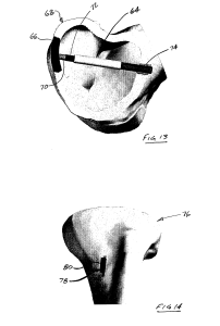
” For example FIG. 14 shows a tibia 76 having a system 78 in accordance with the present invention attached to it. The system 78 includes a linear actuator 80, which can be used to apply a static load, a cyclic load, or some combination thereof to the tibia 76. When a system, such as the system 78, includes two or more such actuators, one can be inserted in the cortical region and over time “grow” one side—e.g., the lateral side—and another can be inserted on the medial side to contract the bone. This effects an angular change at the joint line, and restores a more appropriate mechanical joint alignment.”<-For our purposes, we’d just set the two actuators to length bone.

ActuatorBone


FIG. 14 shows a system in accordance with another embodiment of the present invention, the system including a linear piezoelectric actuator to increase the length of a tibia;

actuators to increase tibia length

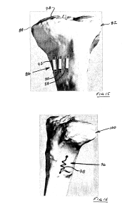
“FIG. 15 shows a system in accordance with another embodiment of the present invention, the system including a plurality of linear piezoelectric actuators to increase the length of a tibia as an alternative to an osteotomy”

Nowhere does it state that this would be limited to individuals with opten growth plates and in fact there are no visible growth plates on this bone.

This is an example of a linear actuator:

Note that I have no idea whether this actuator is sufficient in any way to provide a lengthening force on the bone.  It is just an example.

Note that the linear actuator is used in addition to the invention.  It seems that MIchael has considered using a linear actuator for a height increase device before.

” a swelling memory polymer can be used to provide expansion in a predetermined direction to a predetermined volume, thereby exerting pressure against the containing tissues. Shape memory alloys, such as Nitinol (Ni-TI) can also be used. Such alloys, commonly used in bone staples, can be formed as “muscle wires” and inserted into the cortical bone, where they lengthen in response to outside stimuli.”

“A shape memory alloy could also be formed as a spring, and configured to lengthen (or contract) upon application of an electrical current, for example, an 80 mA current at 20C”

This is how he describes inserting the device into the bone:

“creating an aperture[opening] in the bone proximate the articular surface, thereby making accessible an internal portion of the bone generally opposite the articular surface;
accessing the internal portion of the bone through the aperture in the bone; and
applying the at least one loading condition to the internal portion of the bone, thereby facilitating structural changes in the bone supporting the articular surface”

Then he describes inserting the device into the bone:

“wherein the at least one loading condition is applied to the bone with a joint restoration system including a housing having an aperture therethrough, and an elongate member configured for insertion into the aperture in the housing, the method further comprising attaching the housing to the bone such that the aperture in the housing is generally aligned with the aperture in the bone, and
wherein applying the at least one loading condition to the internal portion of the bone includes inserting the elongate member through the apertures such that the elongate member contacts the internal portion of the bone and applies a force thereto.”

“the joint restoration system further including a compression member configured to cooperate with the housing to apply a force to the elongate member, the method further comprising inserting the compression member into the housing such that it contacts the elongate member and imparts a force thereto, thereby facilitating the application of the force to the internal portion of the bone by the elongate member.”

Here he describes the bone lengthening method:

“The method of claim 1, wherein the at least one loading condition is applied to an external portion of the bone such that the certain structural change includes at least one of an increase in a length of the bone or a decrease in a length of the bone.”

” The method of claim 6, wherein the at least one loading condition is applied to the bone with a joint restoration system including an electromechanical actuator, the method further comprising:
attaching the actuator to the external portion of the bone; and operating the actuator to apply the at least one loading condition to the external portion of the bone.”

” piezoelectric devices will often have displacements in the 100’s of micrometers, which will not provide enough travel to effect desired bone growth in many patients. To overcome this limitation, the actuator is provided with a secondary movement mechanism. The secondary mechanism is configured to provide a ratcheting, positive lock that outwardly extends the extendable component by some discrete amount. This allows the application of a stepwise series of 100 micrometer piezoelectric adjustments, until a total bone displacement of 1-5 mm displacement is achieved.”

The paper A LINEAR ACTUATED TORSIONAL DEVICE TO REPLICATE CLINICALLY RELEVANT SPIRAL FRACTURES IN LONG BONES, describes one potential way a linear actuator can be applied to bone.  Although the device therein does not seem to be applied along the longitudinal axis as suggested by the patent but rather on the top and bottom of the bone.

This paper describes the use of a linear actuator to move a nail in distraction osteogenesis.  Note that in the patent above it specifically states there there is no osteomy required for this device to lengthen bone.

A paper from the LSJL scientists with a potential design of a knee loading device

I found another paper that studied the LSJL knee device here.

I happened upon this paper however which was published at a conference and has potential new insights.

FEA analysis of a portable knee rehabilitation device

“The knee loading device examined in this paper only remains effective for small levels of deformation. The intended displacement of the working device is very small, a maximum of only 6.35mm, and therefore the device cannot accommodate large deformations as such deformations will greatly decrease the effective range of motion of the device.”<-So the device mentioned in this paper can only deform the bone by a maximum 6.35mm and likely less than that as just because the device can be displaced by 6.35mm doesn’t mean it will displace the bone by 6.35mm.

According to mechanostat theory, you have to deform the bone by at least 1500microstrain or 0.15% of a bones original length to get into the plastic deformation range to actually stretch the bone out permanently.

The average adult femur is 2.34 cm in diamater and the maximum displacement is the same regardless of femur shape and size.  The diameter of the femur is what matters as the device loads laterally.  The maximum displacement force is well over what is needed to plastically deform the bone.  But remember that the maximum displacement force is not actually how much the bone is deformed and the bone is being loaded laterally and not being stretched.  By loading laterally a moderate stretching force should be applied but not by the entirety of the deformation.

So it is not that the device can induce an increase in deforming the bone but it is possible by changing the microenvironment via degradation of cortical bone, initiation of mesenchymal condensation, and an increase in chondrogenic signaling.

If you click on the link above to see the study and look at figures 2 and 3, you can see that the device looks remarkably like a clamp.

“The described joint loading modality applies lateral loads to synovial joints.”

“To apply such [the needed] load, a device would need to have a means of producing a transverse force directly to the end of a long bone, such as at the knee{My current working theory is the load needs to be applied at the the epiphysis with the intent to maximize bone on bone contact and not on the synovial joint} . A cyclic force applied in such an area would force a slight shift of the fluid within the bone towards the opposite end of the bone in a controlled fashion.  While no such device currently exists for use on humans{I think a clamp device can serve such a role}, a new joint study seeks to develop a portable device designed for human use to be used in future testing.”<-For our purposes we would likely want more fluid flow as to induce cortical bone degradation and mesenchymal condensation.

“a pressure of approximately 6.90KPawas used as the load for each vertical pad [in a potential joint loading device]. The pressure equates to approximately 40 N over the entire surface area which is the desired maximum load for the device.”

An Irwin Quick Grip 12-inch can generate 300lbs of force.

300lbs is 1334N which is well over 40N but it is likely that you are not going to be able to generate that force however it is also likely that you will generate over 40N of force.

“stress is equal to force divided by cross-sectional area and strain is equal to change in length divided by original length. Stress and strain are related by Hooke’s Law, which states that stress is directly related to strain by a factor known as the Modulus of Elasticity, which is unique to every material.”

To effectively measure strain we’d have to be able to measure microchanges in bone length which I do not see as being possible at this juncture.

“Given the duty cycle of 5 minutes of daily operation per patient with a 1
Hz frequency loading function during operation; or 300 cycles per operation, this device is designed to last for 2683 uses. The choice of 1 Hz as one example was linked to daily
human physical activities such as walking. The device is able to induce loads up to 20 Hz, and it is a future task to evaluate appropriate loading frequencies.”<-Given that walking does not traditionally increase bone length we would likely use a different frequency.  But using a clamp it’s very hard to get such frequencies.  20Hz is equal to 20times per second and it would be virtually impossible to rapidly unclamp and reclamp in that amount of time.

What my current LSJL method instead tries to progressively clamp harder and harder(while still being mindful not to clamp to the point of too much pain) to increase the number of “cycles”.

It should be noted that nothing in this study mentions using this device for longitudinal bone growth but the other studies that Yokota et al. have done on the joint loading modality suggests that it could.  If you look at figure 1B(in the study link above), you can see that joint loading puts pressure on the cortical bone from the medullary cavity.  At a sufficient enough pressure, there could be degradation of the cortical bone and there was evidence of this in a diagram in one LSJL study.  The degradation of cortical bone is highly significant as one of the key events of fusion is the joining of the cortical bone of the epiphysis to the diaphysis.  By degrading cortical bone, we can reverse some of the constraining effects of cortical bone on future longitudinal bone growth.  After all, a key event in distraction osteogenesis is the inducement of a cortical bone fracture.

New height increase supplement

Here’s a paper from Korea about a new longitudinal bone growth supplement that may be promising:

Effect of KH-BaRoKer-SeongJangTang based on traditional medicine theory on longitudinal bone growth

“KH-BaRoKer-SeongJangTang (KBS) is a recently developed formulation by using traditional drugs considering traditional medical theory of Oriental books such as ShinNongBonChoGyeong and JuRye, which has been used to improve the growth of child in Korea. Although KBS is usually prescribed to many children who are in retard for their age, its pharmacological effects have not been fully understood in experimental models. The aim of this study was to evaluate the effects of KBS on bone growth. Growth plate thickness and bone parameters such as bone volume/tissue volume (BV/TV), trabecular thickness (Tb.Th), trabecular number (Tb.N), connection density (Conn.D), and total porosity were analyzed by means of microcomputed tomography. Serum insulin-like growth factor-I (IGF-I) levels were measured by enzyme-linked immunosorbent assay. Hepatic IGF-I mRNA expression was analyzed by real-time polymerase chain reaction. Phosphorylation of signal transducer and activator of transcription5 (STAT5) was investigated using Western blot analysis and immunohistochemistry. The thickness of growth plate was increased by KBS. BV/TV, Tb.Th, TbN, Conn.D, and total porosity were improved by KBS. Hepatic IGF-I mRNA and serum IGF-I levels were elevated by KBS. Phosphorylation of STAT5 was increased with administration of KBS. These results suggest that KBS would be helpful to children who are in retard for their age through the elevation of IGF-I.”

Increase in Stat5 phosphorylation, increase in IGF-1, and increase in growth plate thickness could be indicative that the supplement could increase growth.

“Male ICR mice (4 weeks old) and diets were purchased from Dae-Han Experimental Animal Center (Eumsung, Republic of Korea), acclimated for 7 days, and then randomly assigned for 2 weeks to adequate protein (CON, 20% protein) or low protein diet (PEM, 4% protein) . The protein source used was casein. Except for the protein content, the two diets were identical and isocaloric. After 2 weeks, mice were divided into five groups, CON (adequate protein diet + distilled water (DW)-administered group); PEM (low protein diet + DW-administered group); KBS (low protein diet + KBSadministered group); Arg (low protein diet + Arg-administered group); Glu (low protein diet + Glu-administered group). The mice were fed indicated diet, administered each material three times a week for 12 weeks, housed four to six per cage in a laminar air-flow room, and maintained at a temperature of 22 ± 1℃, a relative humidity of 55 ± 1% throughout the study.”

So it seems that this supplement is an IGF-1 mimetic.  Whether this can increase height in children with normal IGF-1 levels depends on the bodies negative feedback mechanisms.

“Binding of GH to GHR activates receptor-associated intracellular tyrosine protein kinase Janus kinase 2 (JAK2), which phosphorylates signal transducer and activator of transcription 5 (STAT5). The phosphorylated STAT proteins translocate to the nucleus, where they bind to specific DNA sequences and regulate gene transcription. Among the signal cascades from the GHR, the JAK2-STAT5 pathway is regarded as a major pathway that mediates the action of GH on gene transcription in the liver. This pathway was shown to be responsible for the transcriptional action of GH on IGF-I. IGF-I is a mitogenic factor for various cells and plays an important role in cell growth and survival, and the majority of plasma IGF-I is biosynthesized in the liver”

Two of the ingredients of the supplement are listed to be arginine and glutamine which aren’t exactly game changing.  A detailed list of the ingredients are given in Table 2 of the paper(link provided).

KBS on bone thickness


The kbs growth plate and the general bone architecture does look a little bit better than the Arginine group.

“Effect of KBS on tibial growth plate thickness. (A) Representative 3D CT images of knee joint showing growth plate. (B) The thickness of excised bone growth plate was determined on five points. CON, adequate protein diet + DW-administered group; PEM, low protein diet + DW-administered group; KBS, low protein diet + KBS-administered group; Arg, low protein diet + Arg-administered group; Glu, low protein diet + Glu-administered group”

So the supplement didn’t have much more benefit over arginine.

“The lengths of proximal tibia growth plate in the CON and PEM groups were 112.82+/- 4.18 and 86.43+/-1.47, respectively. The growth plate lengths in the KBS, Arg, and Glu groups were 119.05+/- 6.48, 118.75+/- 4.81, and 87.82+/- 6.38, respectively. KBS and Arg significantly enhanced the longitudinal bone growth, whereas Glu did not”

Interestingly, the serum IGF-1 levels were lower than the contAnother major ingredient was a variation on the Hominis Placenta Extract.  Given that the source is the human placenta which is very powerful, it is possible that this extract has of yet undocumented effects.  And many species eat the placenta.rol group in the KBS group and lower than that in the Arginine group.

“Carthami Tinctorii Fructus increased the level of serum IGF-I and lengths of femur and tibia, however, its effect was very small and transient”

Something we can ascertain is that maybe excess IGF-1 levels don’t have that large of a benefit on height.  As the excess IGF-1 levels of KBS versus arginine groups did not result in increased height.

KBS and Arginine increased growth plate thickness by about the same amount so it’s hard tell whether KBS has additional effects beyond arginine.  The difference between KBS and Arginine growth plates are so small and the variance is so large that it’s hard to attribute the difference to anything other than normal variation.

It’s possible that KBS could increase longitudinal bone growth but whether that affect is greater than Arginine is unknown

Here’s the study on Carthami Tinctorii Fructus:

Determination of mineral content in methanolic safflower (Carthamus tinctorius L.) seed extract and its effect on osteoblast markers.

“Safflower (Carthamus tinctorius L.) seeds are used as a folk medicine to enhance bone formation or to prevent osteoporosis in Korea. Therefore, the methanolic extract of safflower seeds (MESS) containing high mineral content, such as calcium (Ca), potassium (K) and phosphorous (P), was evaluated for the role on osteoblast (Ob) markers of Sprague-Dawley rats. In serum of 3 to 11 weeks (wks) old rats, both osteocalcin (OC) content and bone-specific alkaline phosphatase (B-ALP) activity increased to their maximum levels in 4-7 wks. Hence, 3 wks old rats were selected for 8 wks oral treatment of MESS, resulted in the significant increase of Ob markers in serum such as OC content (4-8 wks), B-ALP activity (1-2 wks) and insulin-like growth factor I (IGF-I) level (1 wk), and the growth parameter such as the length of femur (2-8 wks) and tibia (4 wks). On the basis of Pearson’s correlation coefficient, there were a moderate correlation between OC and B-ALP at 8 wks, a low correlation between OC and IGF-I at 1, 4 and 8 wks, a moderate correlation between OC and femur length at 1, 2 and 8 wks, and a moderate correlations between OC and tibia length at 1 and 8 wks of MESS-treated groups. The result reveals that the changes of OC correlated at low to moderate level with the changes of B-ALP activity, IGF-I content and femur and tibia length in the MESS-treatment period. On the other hand, there were a strong correlation between IGF-I and femur length at 2 wks and moderate correlation between IGF-I and tibia length at 1, 2 and 8 wks of MESS-treated groups.

“A Korean herbal formulation, Gami-Honghwain, is comprised of crude ingredients from safflower seeds and hominis placenta.”

“There are reports about chemical components of the safflower. Its leaves contain eight flavonoids, some of which showed potent antioxidant activities. Its seeds also contain numerous polyphenolic compounds such as lignans, glucosides, flavonoids and serotonins”

The increase in tibia and femur length ranged from 3-5%.  Note though that a 3% increase of someone who’s 5’9″ brings them to 5’11”.  And at all data points the treated group had longer bones than the control group.

“. In the aqueous extract from safflower seeds, there are K (2.306 μg/g), P (1.043 μg/g), Mg (0.474 μg/g), Al (0.175 μg/g), Fe (0.100 μg/g), Ca (0.075 μg/g), Zn (0.070 μg/g), Na (0.066 μg/g), Cu (0.055 μg/g) and Sr (0.022 μg/g). In the MESS, there are Ca (3.752 μg/g), K (1.313 μg/g), P (1.161 μg/g), Na (0.177 μg/g), Fe (0.170 μg/g), Zn (0.042 μg/g), Mg (0.023 μg/g), Al (0.019 μg/g), Cu (0.015 μg/g) and Sr (0.002 μg/g).”<-None of these seem to be novel compounds except for Strontium which is found in small amounts in normal human food.  Since Strontium amounts are so small it would be possible to be deficient in it and extra dietary strontium may increase longitudinal bone growth.

How Does A Feminist Deal With Short Stature Height When Her Past Experience Is Only Body Acceptance Over Weight

An interesting article appeared in the online website The Daily UK today which made me think about the issue of height from a perspective which I never put any effort into before. I refer to the story of Lena Dunham standing next to Taylor Swift Here.

In certain parts of the internet, where men talk about the post-modern version of dating and relationships, a concept has come out called “The Red Pill”, which promotes this idea that what men today think women want and what women really want are very different. These men believe that at the most basic, instinctual level, each heterosexual female wants a dominant, strong, masculine men as based on our savage animal origins.

A central public celebrity which is brought up often to portray the stereotypical image of what is wrong with the American and/or Westernized women of today is Lena Dunham.

Lena Dunham Height

Lena Dunham has been vilified by many of these internet blogging men for being too self-accepting of the changes in the female figure. Weight has been traditional the issue/factor that the post-modern men and the post-modern female has the most contention over.

In this day and age where young girls are being constantly being exposed to images of strong powerful women who proclaim to be feminist, they are taught that it is okay to be overweight and not have the type of figure that men desire. Self-acceptance and being fully comfortable with one’s body is very important. In retaliation, the young heterosexual males find these type of women who prefer to stay “overweight” or “full-figured” difficult to find a relationship with stating that they want the old more traditional feminine type of women, which is obviously just a glorified fantasy of something which never really existed. .

Anger, Spite, and Venom has been spewed on both sides, sometimes going as far as threats of sexual assault and bodily harm. Of course, anyone can be sort of a problem as long as they hide behind a screen and stay in anonymity. A sense of self-entitled runs high on both sides.

However, now the issue of height has been brought up. This is where things get interesting. Lena Dunham has been branded a feminism of this modern age, which she seems to be comfortable with that label. When it comes to weight, she has almost no problems. She had taken pictures of herself naked squatting down holding a piece of cake before. She was being overly expressive in showing of the excess of her body to the world.

This recent story that has appeared suggests that she has to content with the other anthropometric measurement which is making her realize that maybe this whole movement of body acceptance is a little harder than believed.

Remember the basic tenet of trying to compare weight and height.

  • Weight is something you can change with a little bit of effort, work and discipline
  • Height is something that is almost impossible to change

What my point is that when people like Lena Dunham and other feminists are talking about weight issues, and body acceptance, they are focusing on the wrong thing.

There is no need to argue for or against any topic dealing with weight, because weight can be changed and molded to whatever level we desire. 

It is height that is what most important. Your level of stature is what is most important, which is what someone like Lena Dunham realizes she will always be inferior to. With her brand of feminism, she chooses to not wear high heeled shoes, which just makes her look even shorter to her female counterparts, who are trying to accentuate their “tallness”.

It is not the fat-shaming guy online that Lena has to content with (since she can easily dismiss those guy as losers) but another female, who happened to be taller than her, who chooses to dress in a classy way, with her tall female friends.

Suddenly you are put on the spot and realize just how “different” you look compared to the others, your own peers,, who you thought before was at the same level as you, only to see from a more objective point of view that they are much higher than you, literally. The other thing that you realize is that there is nothing that you can do about this factor, unlike weight which you can work on.

What do you do when you realize that you have been fighting the easy fight but have always shied away from the hard fight?

  • To gain acceptance of weight is easy.
  • To gain acceptance of height is near impossible.

If this girl was to really want body acceptance, she should have focused on height first and foremost and not even consider dealing with the issue of weight. I find it tragic that many modern feminists seem to be putting too much energy and focusing on the wrong thing in life. They should instead be working towards women and men finding body acceptance over their height, not weight.


The Questions that Must be Answered for a Height Increase Routine to Work

In evaluating potential height increase routines, there are questions that have to be answered true to potentially be effective.  Asking these questions before attempting a routine or supplement, will help to determine if that supplement or routine could possibly be effective although there are many supplements that can be synergestic.

1a.  Does the method increase longitudinal bone growth?

b. Or does the method stimulate an increase the thickness of the bone in a longitudinal direction(top of head or bottom of heel)?

c.  Or does the method target another tissue that is a determinant in height(for example: cartilage or skin at the top of the head)?

d. Does the method alter alignment(posture or loosening of ligaments via relaxin)?

The rest of these questions will involve assuming that 1a was the question that was answered yes to as b-d would have different follow up questions. Note that GH and IGF-1 are a maybe in terms of answering question 1a.  They tend to encourage tissue growth but do not necessarily encourage chondrogenic differentiation(although IGF-1 may).  Something like IGF-2 is something more likely to increase height as it is involved in an earlier development state.  And the growth plate is at an earlier development state than full bones.  GH and IGF-1 are present at all developmental states whereas something like IGF-2 is not.

2.  Does the method involve degradation of cortical bone?

b. Does the method stretch cortical bone(plastic deformation which requires extreme loads)?

This likely has to be an important criteria in the success of a height increase routine.  The end of growth involves the fusion of cortical bone between the diaphysis and the epiphysis.  Distraction osteogenesis involves the cracking of cortical bone.  Cortical bone is likely like a large constraint on longitudinal bone growth.  Note: LSJL likely degrades cortical bone via an increase in fluid flow.

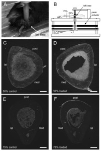
bone loading without drillingD is the LSJL loaded bone and C is not.  There’s definitely degration of the trabecular bone in D and F.  In D the LSJL cortical bone looks thicker and stronger which would make it harder to grow taller.  However in F there are some signs of degradation.  First, note that in E, that the inner bone is a fairly solid circular object but in F there is a part that juts outward on the upper lateral region of F this could be a sign of cortical bone degradation.    And note that 50% is the dead center of the bone which is not a likely target for increasing bone length.  75% is much closer to the epiphysis and since most longitudinal bone growth occurs at the epiphysis this is very promising that the necessary cortical bone degradation can occur to enable bone elongation to occur.

The bone loading only occurred for 3 days.  If bone loading had occurred longer it is possible that the protrusion of the inner bone would extend far enough to reach the outer point of bone enabling a neo-growth plate to form.  The mice were 14 weeks old which is fairly fair along the skeletal maturity process.  Mice growth plates don’t fuse but they do become dysfunctional and this is the time when growth cessation occurs.  So a potential protrusion caused by cortical bone degradation at a late skeletal maturity state is promising for the possible effectiveness of LSJL on adults.

3.  Does the method stimulate chondrogenic differentiation?

Bones have not yet been shown to be capable of interstitial growth and only through an intermediary tissue(cartilage) are they able to grow longer.  It would be possible to grow taller through the articular cartilage(which is capable of endochondral ossification) at the longitudinal ends of the bones but there does not as of yet seem to be an effective way of doing that.

The creating of an intermediary tissue like cartilage within the bone seems to be necessary to make the bones grow longer.  LSJL upregulates chondrogenic genes.

In addition, the chondrogenic intermediary must progress through the various stages and undergo hypertrophy to push the bone apart.  But it appears that endochondral ossification tends to be the standard procedure for chondrogenic tissue.

The Organ-On-A-Chip and 3D Bioprinting Conference Summary

I went to this conference for two main reasons. The first was to talk with a company that was selling a 3D Bioprinter. The 2nd was to listen to the lectures and presentations by the leading edge researchers in this field to see if anything that they are working on can be applied to what we are doing here.

Here is what I can say.

Every single industry, field, or niche has a few people who become very important in their chosen area of expertise. From this one conference, I did find out who are the main people we should be focusing on and following.

  • Dr. Lawrence (Larry) Bonassar
  • Dr. Anthony Atala
  • Dr. Warren Grayson

It turns out that a Dr. Jason Spector who was a speaker at the conference giving his talk  “Tissue Engineering, Bioprinting, and the “Reconstructive Ladder”” alluded to the fact that he was working with Dr. Bonassar on research with MSCs (Mesenchymal Stem Cells) to develop cartilage, whether it be fibrocartilage or hyaline cartilage. When I was listening to him, the name sounded very familiar until I realized that I saw the name before and actually wrote a big post on the work being done by Dr. Bonassar’s team in Cornell where they grew an implantable spinal disc. Refer to the post “This Researcher Succeeded In 3D-Printing Spinal Discs Allowing Adults With Closed Growth Plates To Grow Taller If They Desired – Big Breakthrough“. Dr Bonassar’s work and his Lab should be one of the primary focuses for us.

The other speaker that spoke about relevant information was a Dr. Paul Gatenholm (talk was “3D Bioprinting of Human Cartilage and Skin with Novel Bioink”, who apparently was a protege/student of Dr. Atala. He has a company CellLink where his son works at which has produced a type of bioink that is used in 3D Bioprinters. This bioink acts as the medium that stem cells would go into which is something similar to Hydrogel/Alginate/Extracellular Matrix/Scaffold. It is made from a derivative of cellulose.

Dr. Atala is sort of a super-star in the field of tissue engineering, regenerative medicine, and cartilage generation. He has been alluded to multiple times in the conference and I had referenced his research before as well on this website. Refer to the post ” Increase Height And Grow Taller Through Bioprinting And Electrospinning“. If you read this previous post, you would see that there was a Youtube video of Gabor Forgacs, who explained the revolution in regenerative medicine. He was the speaker at this recent conference which started everything off.

There was a third speaker Rahul Tare (talk was  “Application of Custom-Built Acousto-fluidic Perfusion Bioreactor for Cartilage Tissue Engineering” who revealed that he was also working with MSCs to engineer cartilage tissue.

It turns out that this conference I went to has people associated with the big players in this field. Most people there already knew each other.

In terms of the technical, I realized that bioprinting cartilage tissue that can be implanted back into the body is an endeavor which is probably impossible. In a discussion with Dr. Michael Gelinsky, he did not think that bioprinting a hyaline cartilage was viable at least for a long time. However, the idea of slowly developing chondrogenic tissue from implanted autologous chondrocytes and/or MSCs into a scaffold is probable, if not very doable. If Dr. Teplyashin’s group’s results are any indication, this step has already been done.

The problem which the researchers were trying to solve was over vascularization. Vascularization was the main problem that people who want to bioprint organs need to figure out. It was the bottleneck.

The other problem is over regulations. It turns out that many of the most scientifically advanced countries who are working in tissue engineering and regenerative medicine will not allow organs be implanted into the patients body.

Here are the list of 3D Bioprinters that I found out about.

  1. BioBots (3d Bioprinter)
  2. RegenHu 3d Bioprinter
  3. EnvisionTec Bioprinter
  4. Organovo’s 3D Printer
  5. Regenovo (China based)
  6. Qingdao Unique Products 3D Bioprinter
  7. Izumi International Inc (Deposition Machine turned into 3D Printer)

Some of the models were cheap but some were extraordinarily expensive.

Some others things I realized that I made a mistake on was to forget about the step of bioreactor. You need a bioreactor for the implanted cells into a culture or scaffold, with the growth factors, to proliferate in numbers. Dr. Spector revealed that to get the MSCs to differentiated into a tissue that is actually worth something, you need at least 200 million cells to work with. Anything less and the tissue that has been formed is not really enough.

So to make a correction on the steps, it would be

  • 1. Biopsy of bone marrow to get MSCs/ Extract part of the iliac crest for chondrocytes
  • 2. Use collagenase to dissolve the ECM around your desired cells
  • 3. Spin the solution to separate the cells from the other compounds
  • 4. Get the right type of cell medium (alginate, hydrogel, etc.) to put the cells in.
  • 5. Instead of medium, it could be a scaffold shaped in whatever way you want.
  • 6. Add some type of growth factor into the scaffold to help the cells differentiate or proliferate.
  • 7. Put it into a bioreactor to make the cells proliferate.
  • 8. Induce vascularization in the tissue (this is the tough part)
  • 9. Take the scaffold/medium out and implant the grown  tissue back into the person’s body. Wait until vascularization occurs to connect the new implant with the rest of the patient’s body.

Notice how I did not say bioprinting anywhere. Remember that there is more than 1 way to get the stem cells/chondrocyte implantation to work.

There is actually 2 ways to do this.

  1. You can use a 3D Bioprinter to bioprint a fully functional growth plate, with the chondrocytes inside in columnar structure form
  2. You can put extracted MSCs or chondrocytes into a scaffold (hard structure) and grow it into a bioreactor. You eventually implant the scaffold between the bones and wait for the scaffold to slowly turn into a hyaline cartilage layer of tissue ala pseudo-epiphyseal plate cartilage.

The 1st way is going to be very difficult, but not impossible. The 2nd way is not as “clean” but it should work.

There will be more conferences in the coming months worth looking into. They are….

  • 2015 4th Termis Tissue Engineering World Conference – Dr. Atala will be a keynote speaker there.
  • Biomaterials & Tissue Engineering Gordon Research Conference – Dr. Warren Grayson will be a speaker there.
  • Tissue Engineering, Synthetic Biology & Bioprinting (2016) – by SelectBio
  • Innovations in Cell-Based Regenerative Therapies Conference (MSC 2015)

What you sort of realize as you go down the list of speakers and attendees is that the same people was at this conference too. Eventually after so many conferences you meet the same 300 people over and over again.