Category Archives: Uncategorized

Articular Cartilage does contribute to bone growth in juvenile mice

If we can figure out how to grow via articular cartilage that will be a lot easier than to try to create neo-growth plates.

Spatial Regulation of Gene Expression During Growth of Articular Cartilage in Juvenile Mice.

“In juvenile mammals, the epiphyses of long bones grow by chondrogenesis within the articular cartilage. A bWe used laser-capture microdissection to isolate chondrocytes from the superficial, middle, and deep zones of growing tibial articular cartilage in the 1-wk old mouse and then investigated expression patterns by microarray. To identify molecular markers for each zone of the growing articular cartilage, we found genes showing zone-specific expression and confirmed by real-time PCR and in situ hybridization.Bioinformatic analyses implicated ephrin receptor signaling, Wnt signaling, and BMP signaling in the spatial regulation of chondrocyte differentiation during growth. Molecular markers were identified for superficial (e.g. Cilp, Prg4), middle (Cxcl14, Tnn), and deep zone (Sfrp5, Frzb). Comparison between juvenile articular and growth plate cartilage revealed that the superficial-to-deep zone transition showed similarity with the hypertrophic-to-resting zone transition{This is interesting, the study speculates that both the resting zone and deep zone may come from the same pool of epiphyseal chondrocytes}.ConclusionsLaser capture microdissection combined with microarray analysis identified novel signaling pathways that are spatially regulated in growing mouse articular cartilage and revealed similarities between the molecular architecture of the growing articular cartilage and that of growth plate cartilage.”

“While cell proliferation occurs sparingly in adult articular cartilage, it is much more common in juvenile articular cartilage. Growth of the articular cartilage reportedly occurs appositionally from the articular surface, suggesting that the SZ may contain actively dividing chondrocytes, or a progenitor cell population that is capable of generating new chondrocytes”<-these properties could be exploited to induce new longitudinal bone growth

“DZ[Deep Zone Articular Cartilage] expressed a high level of Wnt5b, which promotes chondrogenesis and inhibits hypertrophy ”

The development of articular cartilage: evidence for an appositional growth mechanism

“It is well-established that cartilage grows by a combination of matrix secretion, cell hypertrophy and cell proliferation. The extent to which this growth is by appositional, as opposed to interstitial mechanisms, however, remains unclear. Using the knee joints of the marsupial Monodelphis domestica to study cartilage growth, we have combined an immunohistochemical study of the TGF-β family of cartilage growth and differentiation factors between 30 days postpartum to 8 months, together with a stereological analysis of cartilage morphology during growth. Furthermore, to gain an insight into the generation of the characteristic zones within cartilage, we have examined the effects of intra-articular administration of bromodeoxyuridine, an agent that is incorporated into DNA during cell division and blocks further cell cycling. During early growth, TGF-β2 and -β3 were widely expressed but TGF-β1 was less so. After the formation of the secondary centre of ossification, all isoforms became more restricted to the upper half of the tissue depth and their distribution was similar to that previously described for IGFs, and PCNA-positive cells. Stereological analysis of tissue sections from the femoral condylar cartilage at 3 and 6 months showed that there was a 17% increase in total cartilage volume but a 31% decrease in cell density on a unit volume basis. Finally, cell-cycle perturbation with BrDU, which was injected into the knee joints of 3-month-old animals and analysed 1 and 4 months post-injection, revealed that the chondrocytes occupying the transitional zone were depleted 1 month post-injection, resulting in thinning of the articular cartilage. This effect was reversed 4 months post-injection. Immunohistochemical analysis revealed that BrDU-treatment altered the expression patterns of all TGF-β isoforms, with a marked reduction in labelling of TGF-β1 and -β3 isoforms in the upper half of the cartilage depth. Overall, the data lends further support to the notion of articular cartilage growing by apposition from the articular surface rather than by interstitial mechanisms.”

Articular cartilage and joint development from embryogenesis to adulthood

“Within each synovial joint, the articular cartilage is uniquely adapted to bear dynamic compressive loads and shear forces throughout the joint’s range of motion.”

“Early studies suggested that a region of proliferating cells “subjacent to the gliding surface of the joint” was responsible for interstitial growth of articular cartilage and increasing thickness of the articular surface. In this same study, Mankin and collaborators found that proliferation continued within deeper regions of the tissue and adjacent to the calcified cartilage, but ceased within the sub-superficial zone at later stages of postnatal growth. The presence of these two proliferative cell regions was confirmed by tritiated thymidine incorporation in the articular cartilage of immature rabbits. Later, Archer and collaborators confirmed the presence of a proliferative cell region in the superficial zone, suggesting that these cells were primarily responsible for the appositional growth and thickening of the articular cartilage postnatally

lateral expansion of the articular surface could be attributed to proliferation of cells within the superficial zone that would also give rise to daughter cells in a more rapidly proliferating cell population in the deeper zones leading presumably to vertical tissue growth.”

“In the growth plate, tremendous increases in chondrocyte volume contribute greatly to lengthening of long bones”

“chondrocyte volume in the middle and deep layers increased by over 8 fold from birth to 2 months of age, while overall decreases in cell density reflecting an increase in extracellular matrix production occurred during this same period.”

Hydrostatic Pressure and Encouraging Chondrogenic Differentiation

Hydrostatic pressure is a force we can manipulate via mechanical stimulation.

Hydrostatic pressure promotes the proliferation and osteogenic/chondrogenic differentiation of mesenchymal stem cells: The roles of RhoA and Rac1.

“Hydrostatic pressure can serve as an active regulator for bone marrow mesenchymal stem cells (BMSCs). [We investigate the roles] of cytoskeletal regulatory proteins Ras homolog gene family member A (RhoA) and Ras-related C3 botulinum toxin substrate 1 (Rac1) in hydrostatic pressure-related effects on BMSCs. Hydrostatic pressure promoted cell cycle initiation in a RhoA- and Rac1-dependent manner. RhoA played a positive and Rac1 displayed a negative role in the hydrostatic pressure-induced F-actin stress fiber assembly. RhoA and Rac1 play central roles in the pressure-inhibited ERK phosphorylation, and Rac1 but not RhoA was involved in the pressure-promoted JNK phosphorylation. Pressure promoted the expression of osteogenic marker genes in BMSCs at an early stage of osteogenic differentiation through the up-regulation of RhoA activity. Pressure enhanced the expression of chondrogenic marker genes in BMSCs during chondrogenic differentiation via the up-regulation of Rac1 activity. RhoA and Rac1 are critical to the pressure-induced proliferation and differentiation, the stress fiber assembly, and MAPK activation in BMSCs.”

Hydrostatic pressure applications are methods of applying mechanical loading that mimics the compressive forces borne by cartilage in a joint cavity“<-So we should be able to apply such mechanical load ourselves but in the bone rather than the cartilage to encourage a neo-ectopic growth plate.

“the activities of Rho GTPase signaling molecules are closely related to the differentiation and the fate of BMSCs.”

“the activation of the RhoA pathway and the inhibition of the Rac1 pathway under hydrostatic pressure promoted the assembly of the F-actin cytoskeleton, whereas the inhibition of the RhoA pathway and the activation of the Rac1 pathway blocked the F-actin cytoskeleton assembly.”

“the down-regulation of RhoA activity and/or pressure significantly blocked the phosphorylation of ERK1/2″<-LSJL increases ERK1/2 phosphorylation.

“RhoA activation inhibited the pressure-induced down-regulation of P-ERK1/2 expression and that RhoA played an important role in the regulation of ERK1/2 phosphorylation upon pressure stimulation.”

A combination of decreased RhoA activity and pressure stimulation (P/RhoA − group) achieved the maximum expression of the chondrogenic marker genes in BMSCs{So we have to find a way to decrease RhoA levels in the bone}. After two weeks of chondrogenic induction, the expression levels of the chondrogenic genes in the P/RhoA + group were significantly reduced compared with those of the P group. The up-regulation of RhoA antagonized the promoting effect of pressure on the chondrogenic differentiation of the BMSCs{So how do we downregulate RhoA?}. After 4 weeks of chondrogenic induction, the expression levels of Sox-9, Aggrecan and Col II in the RhoA −, P and P/RhoA − groups were significantly higher than those of the control group ”

” pressure inhibited ERK phosphorylation, suggesting that the induction of cell cycle initiation by pressure may not require a modulation of the cyclin D concentration, which involved the regulation of ERK activity through the LIMK protein.”

“pressure promoted the phosphorylation of JNKs but not ERKs in the BMSCs.”

the damage to the cytoskeletal structure and the inhibition of ROCK activity induced rounded cell morphologies and promoted the expression of chondrogenic marker genes in the BMSCs“<-Maybe hydrostatic pressure will induce more cytoskeletal damage over time and more chondrogenic differentiation will be induced naturally.

“Hydrostatic pressure regulates cell cycle initiation through both the RhoA/Rock and the Rac1 signaling pathways. At the same time, the mechanical stimulation promoted cytoskeletal assembly in BMSCs through the up-regulation of RhoA/ROCK activities, and activation of the JNK1/2 pathway by down-regulation of Rac1 activity. Hydrostatic pressure could also enhance expression of marker genes for early osteogenic differentiation through the up-regulation of RhoA activation or enhance the expression of chondrogenic marker genes in BMSCs during chondrogenic differentiation via the up-regulation of Rac1 activity ”
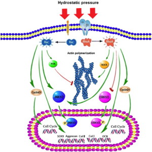
hydrostatic pressure effect

According to Flavoprotions: Advances in Research and Application: 2011, egf is a stimulator of Rac1.

There are several skin applicators of EGF but I couldn’t find any oral.
I’m not sure if applying it to the skin near the bone/joint region would work in stimulating Rac1 and I’m not sure if ingesting a product meant for the skin is safe nor if it will stimulate any desired region.  But the study suggests Rac1 versus RhoA is a key cellular distinction for osteoblasts versus chondrocytes.  And since there is as yet no RhoA inhibitor(but it’s being investigated due to cancer applications), stimulation of Rac1 via EGF is a worthwhile path to go down.  By encouraging chondrogenesis versus osteogenesis in the bone, it would be easier to great a neo-growth plate.

Rac1 promotes chondrogenesis by regulating STAT3 signaling pathway. has a possible suggestion of how STAT3 could also promote chondrogenesis,

“The small GTPase protein Rac1 is involved in a wide range of biological processes including cell differentiation. Previously, Rac1 was shown to promote chondrogenesis in micromass cultures of limb mesenchyme. However, the pathways mediating Rac1’s role in chondrogenesis are not fully understood. This study aimed to explore the molecular mechanisms by which Rac1 regulates chondrogenic differentiation. Phosphorylation of signal transducer and activator of transcription 3 (STAT3) was increased as chondrogenesis proceeded in micromass cultures of chick wing bud mesenchyme. Inhibition of Rac1 with NSC23766, janus kinase 2 (JAK2) with AG490, or STAT3 with stattic inhibited chondrogenesis and reduced phosphorylation of STAT3. Conversely, overexpression of constitutively active Rac1 (Rac L61) increased phosphorylation of STAT3. Rac L61 expression resulted in increased expression of interleukin 6 (IL-6), and treatment with IL-6 increased phosphorylation of STAT3. NSC23766, AG490, and stattic prohibited cell aggregation, whereas expression of Rac L61 increased cell aggregation, which was reduced by stattic treatment. Rac1 induces STAT3 activation through expression and action of IL-6. Overexpression of Rac L61 increased expression of bone morphogenic protein 4 (BMP4). BMP4 promoted chondrogenesis, which was inhibited by K02288, an activin receptor-like kinase-2 inhibitor, and increased phosphorylation of p38 MAP kinase. Overexpression of Rac L61 also increased phosphorylation of p38 MAPK, which was reduced by K02288. These results suggest that Rac1 activates STAT3 by expression of IL-6, which in turn increases expression and activity of BMP4, leading to the promotion of chondrogenesis.”

So BMP4, STAT3, and IL6 are all potential targets to induce chondrogenesis.

Contribution of the Interleukin-6/STAT-3 Signaling Pathway to Chondrogenic Differentiation of Human Mesenchymal Stem Cells.

“Mesenchymal stem cells (MSCs) are multipotent cells that can differentiate into chondrocytes. Articular cartilage contains MSC-like chondroprogenitor cells, which suggests their involvement in the maintenance of cartilage homeostasis by a self-repair mechanism. Interleukin-6 (IL-6) is a cytokine [which is] produced by MSCs in a steady manner and in large quantities. The purpose of this study was to investigate the involvement of IL-6 signaling in MSC differentiation into chondrocytes.
Human bone marrow-derived MSCs were cultured using a pellet culture system in medium containing transforming growth factor β3. Chondrogenic differentiation was detected by cartilage matrix accumulation and chondrogenic marker gene expression.
IL-6 was detected at a high concentration in culture supernatants during chondrogenic differentiation. The expression of the IL-6 receptor (IL-6R) was significantly increased, accompanied by markedly increased phosphorylation and expression of STAT-3. Addition of IL-6 and soluble IL-6R (sIL-6R) to the chondrogenic culture resulted in concentration-dependent increases in cartilage matrix accumulation and cartilage marker gene expression (type II collagen/aggrecan/type X collagen). Phosphorylation of the master transcription factor SOX9 was enhanced upon addition of IL-6 and sIL-6R. STAT-3 knockdown suppressed chondrogenic differentiation. IL-6 and the MSC markers CD166 and nestin were colocalized in macroscopically normal human cartilage taken from the lateral femoral compartment of knees with medial tibiofemoral osteoarthritis.
During differentiation of human MSCs into chondrocytes, the activation of IL-6/STAT-3 signaling positively regulated chondrogenic differentiation. The presence of IL-6 around MSC-like cells in the cartilage tissue was identified, suggesting that IL-6 contributes to homeostasis and cartilage self-repair by promoting chondrogenic differentiation.”

“cartilage contains chondrogenic progenitor cells with mesenchymal stem cell (MSC)–like characteristics”<-possibly actually more like epithelial cell characteristics.

“MSCs exhibit immunosuppressive activity and inhibitory effects on osteoclast differentiation via trophic effects, by releasing various humoral factors”

“MSCs produced high levels of IL-6 during chondrogenic differentiation”

“Chondrocytes are also capable of producing IL-6 upon stimulation, although under physiologic conditions (i.e., embedded in the cartilage matrix), chondrocytes are reported to have little ability to produce IL-6”

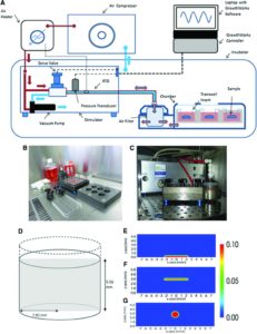
Evaluation of the Growth Environment of a Hydrostatic Force Bioreactor for Preconditioning of Tissue-Engineered Constructs

“To determine the effect of hydrostatic pressure on bone formation, chick femur skeletal cell-seeded hydrogels were subjected to cyclic hydrostatic pressure at 0-270 kPa and 1 Hz for 1 h daily (5 days per week) over a period of 14 days. At the start of mechanical stimulation, dissolved O2 and CO2 in the medium increased and the pH of the medium decreased, but remained within human physiological ranges.”

“Hydrostatic pressure has been shown to be an important mechanical stimulus for the direction of cell fate in various tissues, including articular cartilage, the intervertebral disc, bone, and the vascular system”<-We want to induce the cell fate of chondrogenesis.

“Osteocytes in the canalicula-lacuna network of load-bearing bones are subjected to physiological pressures of approximately 270 kPa”

“he heartbeat of chick embryos delivers a dynamic pressure of 4 kPa, the blood pressure is usually between 8–24 kPa, the hydrostatic pressure in the cerebrospinal fluid is around 1.2 kPa and the interstital fluid pressure is around 0.27 kPa”

“The application of hydrostatic pressure during tissue formation could result in enhanced transfer of small molecules, such as oxygen and CO2, into the tissue matrix and provide physical forces to cells and tissues

hydrostatic pressure machine

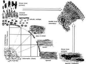
Biomechanics-driven chondrogenesis: from embryo to adult.

“Cartilage is relatively acellular, with chondrocytes only comprising 1–5% of the tissue by volume”

“Joint loading results in direct compression of chondrocytes inside a relatively impermeable matrix. Following tissue loading, hydrostatic pressure initially develops in the interstitial fluid, which is followed by fluid flow-induced shear. However, in time scales > 10 μs, the solid matrix begins to bear the applied load, resulting in deformation. Consequently, the cells residing in the matrix experience hydrostatic pressure, shear, compression, and, to a lesser extent, tension. This mechanical stimulation produces a signaling cascade, resulting in increased gene expression, matrix protein production, and intracellular ion influx”<-Our goal though is to induce hydrostatic pressure within the bone to induce chondrogeneic differentiation.

Spatiotemporal changes in progenitor cell adhesion molecule expression cause similar cells to transiently associate during chondrogenesis. However, cell-cell adhesion strength correlates linearly with cellular surface tension, irrespective of a homogeneous or heterogeneous interaction, suggesting surface tension as the primary driver of differential adhesion. Therefore, precartilaginous condensation may be the result of mesenchymal progenitor cells exhibiting similar surface tensions rather than similar biomarkers. Furthermore, disruption of surface tension inhibits differential adhesion“<-so altering surface tension may be key to inducing chondrogenesis and cellular biomarkers may not necessarily be a limiting factor on chondrogenic induction.

“the absence of gravitational force reduces precartilaginous condensations in mesenchymal limb bud cells”

“Biomechanics-driven development of cartilage from embryo to fetal stages and beyond. A, B) Progenitor cells migrate from the early mesoderm to sites of skeletogenesis (A), where they undergo precartilaginous condensations (B). C) Chondrocyte progenitors secrete cartilage-specific matrix and decrease expression of cell-cell interaction proteins. D) Proliferation continues at the subchondral growth front, while endochondral ossification occurs throughout the juvenile stages to transform cartilage into bone. E) Ends of long bones remain capped with a layer of articular cartilage throughout adulthood.”

” Intermittent and cyclic hydrostatic pressure and strain both help regulate matrix protein synthesis to affect macromolecular organization of collagen fibers, which, in turn, leads to changes in the mechanical properties of the tissue ”

“models predict that intermittent hydrostatic pressure inhibits degeneration and ossification of cartilage, while intermittent strain or shear stresses accelerate ossification and degeneration”

“HP does not result in deformation of incompressible media, so it is not expected to deform cells. Direct compression results in deformation of matrix and cells, which will also create fluid flow that is not observed with HP.”

Response to mechanical loading may be differentiation dependent.
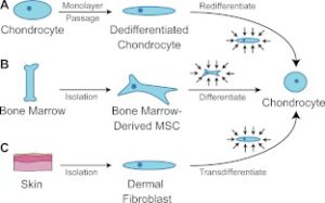
bonemarrowchondrodifferentiation

The above image is our intent where we intent to get bone marrow MSC’s to differentiate into chondrocytes.  However, we cannot isolate them in culture.

“Harnessing biomechanics to drive adult cell chondrogenesis. A) Following monolayer culture, chondrocytes rapidly dedifferentiate. Biomechanical stimuli, such as HP, can promote redifferentiation. B) Under mechanical stimulation, mesenchymal stem cells migrate and chondrodifferentiate. C) Mechanical stimulation can be used to induce transdifferentiation into chondrocytes.”

 Human mesenchymal stem cell responses to hydrostatic pressure and shear stress.

“the present study investigated the early responses of human mesenchymal stem cells (hMSCs) to intermittent shear stress (ISS) and to cyclic hydrostatic pressure (CHP) simulating some aspects of the biological milieu in which these cells exist in vivo. Production of nitric oxide (NO) and mRNA expression of several known mechanosensitive genes as well as ERK1/2 activation in the hMSC response to the two mechanical stimuli tested were monitored and compared. NO production depended on the type of the mechanical stimulus to which the hMSCs were exposed and was significantly higher after exposure to ISS than to CHP. At the conditions of NO peak release (i.e., at 0.7 Pa for ISS and 50,000 Pa for CHP), ISS was more effective than CHP in up-regulating mechanosensitive genes. ERK1/2 was activated by ISS but not by CHP. The present study is the first to report that PGTS2, IER3, EGR1, IGF1, IGFBP1, ITGB1, VEGFA and FGF2 are involved in the response of hMSCs to ISS. These findings establish that, of the two mechanical stimuli tested, ISS is more effective than CHP in triggering expression of genes from hMSCs which are bioactive and pertinent to several cell functions (such as cell differentiation and release of specific growth factors and cytokines) and also to tissue-related processes such as wound healing.”

“Exposure of the cell distal membranes to shear stress was achieved by flowing the cell-culture liquid medium through low-wall microchannels (μ-Slide) on whose surfaces (2.5 cm2) the cells were cultured; during these experiments, the medium flow was in the axial direction of the microchannels (μ-Slide).”

“PTGS2, IER3, EGR1, IGF1, IGFBP1, ITGB1, VEGFA and FGF2 genes are expressed in the response of human MSCs exposed to ISS, and that (ii) the FGF2 gene (but neither PTGES, EGR1 or VEGFA genes) are involved in the response of hMSCs to CHP. Last, but not least, the observed expression of mechano-sensitive genes (specifically, PTGS2, IER3, EGR1, IGF1, IGFBP1, ITGB1, FGF2 and VEGFA) returned to basal levels within 6 to 24 h after exposure of the hMSCs to the two mechanical stimuli tested”

DHT can increase IGF2 levels?

Dihydrotestosterone is a determinant of calcaneal bone mineral density in men.

Full study link->DHT and IGF2

“Hundred osteoporotic men with age matched normal were studied for serum levels of sex steroids, PTH, IGF system components, cytokines and bone turnover markers. Our findings show that serum DHT, IGF-I, IGF-II and IGFBP-3 levels were significantly decreased while IL-1beta and bone turnover markers were significantly increased in osteoporotic men compared to normal. Pearson correlation analysis revealed that serum DHT, IGF-I, IGF-II and IGFBP-3 levels were positively and strongly correlated with BMD, while serum IL-1beta levels were negatively correlated with BMD. Serum PTH, testosterone, estradiol, IGFBP-4, TNF-alpha, IL-4 and IFN-gamma levels were similar between the two groups. We observed that DHT levels significantly declined with age. However, the significant difference in DHT between the osteoporotic and normal groups is the same regardless of age. A multiple regression model adjusted for age demonstrated that DHT/BMD association is fairly stronger among those with osteoporosis than the normal. Our findings for the first time point out that DHT is an important determinant of BMD in men. Most importantly, the strong positive correlation of serum DHT with BMD offers new perspectives in understanding the role of non-aromatizable androgen in regulating bone metabolism in men, and might serve as a potential clinical marker in the diagnosis of male osteoporosis.”

Serum levels of estrogen and testosterone are slightly lower in osteoperotic men than normal.

IGF-II 955 ± 45(Normal) 722 ± 35(Osteoperotic)

“DHT increased the levels of IGF-I and IGFBP-3 in human osteoblastic cell line (hFOB/AR-6). DHT enhances the mitogenic effect of IGF-II in bone cells.”

This is the study that mentions the link between IGF-II and DHT:
Studies of the Mechanism by which Androgens Enhance Mitogenesis and Differentiation in Bone Cells

DHT did not increase the quantity of IGF-2.

“Because FGF and IGF-II are known to synergize with TGFB, the enhanced response to FGF and IGF-II due to DHT treatment may be dependent upon the increased production of TGFB.”

DHT pre-treatment approximately doubled the effect of IGF2.

Is The Real Height Requirement Of Men For Dutch Women In The Netherlands Now 6′ 3″ or 1.90 Meters Tall?

New evidence has come out suggesting that the real average height of ethnically pure Dutch men in the Netherlands may be as high as 1.92 Meters tall or 6′ 3.3″. This claim was something that I read from the book “The Tall Book” by Arriane Cohen. A claim like this is something that most people would write off as too incredible to believe, but I wanted to not just write off this claim completely. Anyone who has ever done a quick stroll through the streets of Holland or Friesland would reveal that the claim that 6′ 4″ is the average is not that farfetched.

First, we know that the Netherlands has a population of around 17 Million people, and the Ethnic makeup of the country shows that it is now a little less than 80% ethnically Dutch. In the Netherlands, there are large populations of Turkish, Albania, Arab, and North African communities. These immigrants usually come from poorer countries so the influx of immigrants has probably reduced the average height of the country to some level. How much? We take an educated guess

The last we checked, the average height of the Dutch, including the immigrant population was at 1.84 meters tall. So what would happen if the immigrant height factor was removed? We could make the arguement that the average height would get increased by 2-3 cms. A 1 cm increase would be too little since the non-dutch community makes up a very large part of the country, pushing the average height down. Anything over 4 cm would seem a little too large. It would be anything from 2-3 cms.

That means that if we were only looking at the pure ethnically Dutch males in the country, the real average height of the 18-30 year old group is more likely 1.87 meters tall.

Now, we have to remember that the Netherlands, like most other countries in the world is based on the Metric System. They use meters and centimeters, while the old British System which is used still be the UK, USA, and Australia uses feet and inches.

This means that sociologically speaking, if a native Dutch girl was to want a “tall” (relatively speaking) guy she would not say that the cut-off point is 1.80 meters tall, which is what the cutoff point is for most countries of the world, but say that the cutoff point is 1.90 meters tall.

In comparison, we can look at a country like South Korea, which a population of around 50 Million people. While the Wikipedia article says that the average height of men in South Korea is 1.74 m, a new study I found shows that the real average height is actually 1.75 meters tall (or 5′ 9″). Within this culture, the majority of girls prefer to date taller men, and since they are based on the metric system, there is a social rule that a man needs to be 1.80 meters tall (or 5′ 10.5″) to be considered dateable in certain girls’ eyes. And that is just the socially accepted cut-off point for the more superficial girls. To be considered “tall” in the Korean society, you are supposed to be at least 1.82 meters (5′ 11.60″). (Similarly, in the USA where the average height is believed to be 5′ 9.5″, the social rule of what is considered a “good” height for guys is 6′ 0″. The difference is 6. 35 cms.)

Notice that the difference in average height of men and what is considered tall is around 6-7 cms.

If we use this 7 cm rule and translate it to the dutch population, to be considered “tall” in this culture you would need to be 1.87 + 7 = 1.94 meters tall or 6′ 4.4″. Of course, there is no set requirement that a male needs to hit that “tall” category to be considered socially acceptable in height to be dateable for the majority of women in his group.

If South Korea could be used as an example, a 5 cm difference is what is required from average height. So for the Netherlands, 187+5 = 1.92 meters tall is the real cutoff point for young men these days.

Of course then you have to take consideration the non-ethnic dutch people/immigrants, so factoring that in, the height requirement gets reduced by 2 cm to just 1.90 meters tall. They would round down and say that they want a boyfriend to be at least 1.90 meters. To actually hit the right mark within only the dutch ethnic community, it would be probably closer to 1.93 meters or 6’4″.

To validate this idea even more, it was discovered recently that the real height of ethnically Dutch females is around 5′ 8.5″. On average we have found that the difference in height of men and women in a society is actually 5.5 Inches. This would suggest that the real average height of Dutch men is around 6′ 2″ or 1.88 meters tall.

So it would not seem that big of a stretch to ask that one’s boyfriend be just 2-3 cm taller than the average, at 1.90 meters.

For any people from the Netherlands, is this claim I made validate? Do your own life experiences, and casual observations agree with my claim?

What trait in women would be considered as universally unattractive as short stature in men?

For the longest time, men have tried to use the analog “Short is to Men” as “Overweight is to Women”. So Short:Men::Overweight:Women.

The usual rebuttal that men like to use when they meet a women who judges them based on height is to try to flip the situation back at the women and use the weight of the women as the response. However, I have found that the response is inaccurate, if not downright wrong. It is not weight for women. It is something else.

The real factor that men should be using as a response has been a factor of the human experience which is no longer allowed in American society (or most other developed Western nations today). If you use the real, most accurate response back at a women, people would be calling the guy a bigot.

When it comes to the issue of dating and relationship, men can complain that being short is bad towards their romantic life but what these men have been using as the female analog is incorrect. We’ve got it wrong. The analog for women is not weight (ala overweight means unattractive), but skin color. For most of the cultures and societies throughout history, the men have traditionally favored the lighter skin girls over the darker skin ones. Read Peter Frost’s book “Fair Women, Dark Men” and you would see that anthropologists have shown that nearly all (or maybe most) cultures prized light skin color, at least for the women (Refer to the claim by University of Washington sociologist Pierre L. van den Berghe). Remember that even in our language for old stories that are imprinted onto our culture’s psyche from passed down from history, we use terms like “fair princess” and “tall, dark, handsome”. These terms are used over and over again. But why?

Anthropologically speaking, the light skin color of men does not seem to have any positive correlation to how attractive they are. For men, our skin color does not determine how attractive we are, even though some men in some cultures (Indian for example) both the men and women want their skin to be lighter. Whereas Indian women use the cream Fair and Lovely, Indian men use the cream Fair and Handsome. The opposite is true too for men at least. In some cultures, it is more attractive if the men have darker skin (ie. Krishna from Indian mythology). Yes, the

For more examples, let’s go to South America and look at the natives before the Europeans ever set foot onto that continent. Look up the Chachapoyas people, who were overtaken by the Incans right before Pizzaro and the other Spaniards arrived. Before the Europeans ever came along, the native Incans still prized the women of Chachapoyas (cloud people) as being fair skinned and extraordinarily beautiful. Examples like these go against the old claim made by so many people in the modern era who think that European domination, imperialism and cultural influence for the last half 1000 years is why non-western/european nations and societies seem to want to have lighter skin, because they want to look more like their masters, the Europeans. This old theory has been disproved.

Before the Chinese ever met the Europeans who used force to destroy the old way of life, the Chinese culture prized light skin. The rulers of the land, like the Sultans and Shahs of Ancient Persia (Sassanid Empire) had harems, so they were able to choose the most beautiful young females in the land to have as concubines. These rulers would choose sisters who had slightly lighter skin than the dark skinned ones. These concubines would stay in the castles and never need to be in the fields growing and planting. They in turn became more and more white. The ones who did not get chosen to be in the castle had no choice but be a commoner, working the fields, and getting their skin turned darker from the sun over the years.

Side note: The old idea that East Asian societies (Japanese, Korean, Chinese, Vietnamese) developed this preference for lighter skin girls because in the old days, traditionally, the light skin was associated with upper class people not in the fields, and the darker skin was associated with lower class people who did work in the fields, is actually wrong. People have got it backwards. The society did not develop the preference for light skin as a result of the social association of light skin with high class. The truth is revealed to be the reverse. The social association of light skin with high class is from the result of the preference for light skin.

Just like the South Asians and East Asian cultures, The Arabs and Iranians are also not trying to emulate the physical features of the Europeans because of their influence. It seems that for the longest time, they have always prized women to have fair skin, blonde hair, and green/blue eyes. The area that the Arab empire tried to slaves from the most was always from the Northern lands. Look up the Arab slave trade which has been going on since the time before the beginning of Islam, and you would find examples of how female slaves from Europe who had fair skin, green eyes, and blonde hair would always fetch the highest price in the markets.

The Arab culture, the Indian culture, Iranian Culture, African America culture, Chinese, Korean, Japanese, Thai, Mexican, Hispanic, Brazilian, Chilean – In every single one of these cultures, people of lighter skin are on average considered more attractive than people with darker skin. India has Fair and Lovely. Korea has BB Cream. It is a global phenomena. If anything, if the American/Western/Australian culture had any type of real influence in changing the way the local Indians/Chinese/Iranians/Mexican/Filipino culture really thought about what would be considered attractive, these non-western nations would have gone with the standard, which is that darker tan-colored skin would be more attractive. However, that hasn’t happened. The people in the USA thinks darker skin is more beautiful. The people in India think lighter skin is more beautiful.

It seems to be only the USA (and maybe the UK, Canada, and the Australia) who have gone against the grain on this cultural preference for lighter skin. The change in thinking started back in the 1920s when Coco Chanel got sun burned while being in the sun in the French Riviera. After she came back, her fans loved her new skin color and that was how the Tan Craze started. Before the 1920s, American and British women wanted to be paler and whiter, like all the other cultures in the world. If you have watched any movies or tv about the british (or american) aristocracy from the 18th century, you would remember that they would put white powder on their face to whiten their faces, men and women. This is exactly the same thing that women in every other major culture in the modern age does.

Scientifically It turns out that the females on average have less melanin on the surface of their skin so they are usually lighter than their male counterparts.

At this point, I think we need to just admit to ourselves that for most cultures, being fair skinned colored as a women and being tall for a guy are what is considered attractive. From this point on, it is probably better to use the skin color factor for women when we are going to use the height factor for men as a way of comparing.

Of course, we know that in the modern american society, it is not politically correct to show discrimination towards people based on their skin color, even if it is a few shades too light or dark. Then again, we can look at the ethnic groups in our own society. Ask a good percentage of African American men and they would say that they prefer girls who have lighter skin than darker skin. Certain African American music performers have even gotten in trouble for stating their preference for women of lighter skin blatantly on radio, tv, or in interviews. (Neyo). The angry darker skinned african american girl might take extreme offense to this claim, and her self worth will come into question. How dare a fellow brother say that she is not beautiful, based on something that she can not control no less. Of course, we can show that this slip of the tongue by one young black guy is not a one time thing.

Doesn’t it seem interesting that the two “black” actresses that American pop culture consider to be “beautiful”, Halle Berry, and Beyonce Knowles, both have extremely light skin color{Tyler-Confounding variables: Both are bustier than normal and it’s very rare to find a completely black individual.  There’s a smaller pool to get attractive people from.  You’re more likely to find an attractive lighter black person than a pure dark black person due to numbers}? Ask most indian/chinese/russian immigrants who hasn’t adjusted to the racial sensitivity of America and don’t know how to be politically correct and they would claim that these “black” girls are mixed blood, explaining why they have such light skin color, and why they are more attractive than what they imagined in their own head of “american black girls” before they came to the US.

After centuries of battling the stigma associated with dark skin color, the American culture has managed to make progress on the issue of skin color. As for physical stature, there has not been as much. You are no longer allowed to make jokes of another person based on their race or skin color, but you can skin tease, harass, and joke another person based on their physical stature.

The question would be, can we change over time the cultural belief in a society’s belief system that tall men are more attractive then short men? I think we can implement rules to repress that subconscious biases from being so overt, but in the end, our real biases will come out.

To go against these biological ingrained biases is to fight against what is based on the rules of nature.

Evidence of LSJL arm length increase

Previously, I stated that my wingspan has increased from 72.5″ to 74.5″ but I didn’t have any proof because I didn’t take any before pictures.  I’ve been trying to get my wingspan increase but haven’t had significant enough measurements for undeniable proof.  I’m keep trying to increase height and wingspan but what I can do is try to create new photos to compare to the old ones

.arm length beforeHere’s an image from about 2012.  I tried to recreate something like this picture as best as I could.

arm length afterThis was from today.  The dumbell acts as sort of a constant.

The problem is the image is not 3D so it can’t account things like the dumbell tilt.  Right now my forearm measures 11 inches elbow to wrist “bump”.  Using the dumbell handle as a constant, I compared the forearm length elbow to wrist(although it was harder to identify wrist in the before picture.  I was pretty generous to the before picture.

Before forearm length as dumbell handle lengths: 2.4 dumbell handles

After: 2.48 dumbell handles

Which is about 3%.  My wingspan increased by 2 inches.  Assume a 0.5″ of that is in my left forearm.  .5″ is about 4.76% of 10.5″.

So given the inaccuracies of the photo I don’t think it’ll be easy to have definitive proof using the two unless someone has ideas of creating a new photo standardized against the first.