Category Archives: Uncategorized

Height Increase Techniques that are unlikely to work

1) Stretching(on the spine)

Read this summation of the theory of spinal stretching.

Although one study indicates that a twisting motion can increase height in some cases, other human studies have suggested that twisting can only reduce height in human models.  This is due to the nucleus pulposus not being mechanically stable in contrast to the annulus fibrosus which can grow.  In response to mechanically strain, there was degeneration in the nucleus pulposus and there is a medical term for this caused degenerative disc disease.

Now, other joints do not have nucleus pulposus like the joints of the ankle and knee so those can potentially grow in size.  Interesting though is that one study found that wingspan decreased more with age than overall height.  Which is contrary to what we would believe with discs being prone to degeneration.  There is an increase in the hand phalanxes continuously throughout lifeNow I received an increase of 0.8%(at least) in my right index finger metacarpal.  So the normal hand phalanx growth does not explain my growth.  That study suggests that articular cartilage can undergo endochondral ossification which can add appositional bone growth to the longitudinal ends of the bone resulting in longitudinal bone growth.

Now one thing is that nucleus pulposus is so prone to degeneration and can reduce height so much is that we can reduce degeneration.  For example by living in zero gravity, by strengthening the muscles surrounding the spine to reduce spinal load, or via spinal tractors/inversion to reduce load allowing water to return to the nucleus pulposus.

This height increase method listed here(Aquatic Vertical Suspension) resulted in an immediate height increase of about 4mm.

2) HGH

People have supplemented with HGH at levels at or greater than those who have “suffered” from gigantism reported no height gain.  Gigantism does not merely involve elevated HGH levels but also resistance to HGH suppression.  Gigantism also involves a tumor.  Two factors that are not involved in HGH supplementation.

There has been no evidence as of yet that HGH can induce exogenous growth plates to induce new longitudinal bone growth.  Although some anecdotal evidence of HGH induced growth has been reported.

3) Microcracks

There hasn’t been any evidence to suggest that microcracks can lengthen bone.  And mineralized bone seems to be incapable of interstitial growth(growth from within ala growth plates) due to high ECM stiffness.  Thus there needs to be an intermediary tissue involved like cartilage in order to lengthen bone in a roundabout way.  So to lengthen bone via microfractures you’d have to reduce ECM stiffness to allow for interstitial growth.  Here’s one possible method to reducing ECM stiffness using acid that has been suggested.

4)Bone Stretching

For example via the Rack.  The amount of load required to induce a bone fracture is  usually a colossal figure like 25000-lbs.  And the amount of load to induce plastic deformation(bone stretching or negatively compression) is usually very close to the fracture point.  This makes the generation of such loads problematic.  That’s why the design of LSJL is to try to induce mesenchymal condensation and neo-growth plate formation.

Creep Strain can induce plastic deformation at much lower loads.  However, a considerable continuous amount of load is required to induce 1-2 weeks.  I could not find any evidence of creep strain inducing plastic deformation in a positive way(stretching), only a negative way(compression).  I couldn’t find any other studies where length was even considered as only damage to the bone was measured.

Video Of Alexander Teplyashin’s Laboratory Lengthening Legs Of Sheep Using Stem Cell Implants

This is the 2nd video

There is some medical terms that can’t be translated like “stem cells” and “implant” so the video is quite accurate. Dr. Teplyashin is actually sort of a celebrity surgeon in the Moscow area. The rams/sheeps in the video are adult, so they are not going to get any taller. The implant goes into the bone after a small resection.

If you guys are interested in watching more videos about Teplyashin’s many video appearances on Russian Media TV , click here for the Youtube username Alexander Gaponov – https://www.youtube.com/user/elansky/videos

If any of you actually understands Russian, can you translate what they say in the short video?

Notice also in the video below how his lab follows general GLP (Good Laboratory Practices). This is not like what Gavrill Ilizarov did back in the 60s-80s with his version of leg lengthening.

I haven’t updated this website in a long time because I have recently started a new business venture. However, I promise that I will come back to this venture. There are other medical entrepreneurial ideas that I would like to pursue as well.

Aneurysmal Bone Cyst to increase height?

http://www.pedorthpath.com/about.html

Aneurysmal Bone Cyst<-may cause enlargement of affected bone.  Sometimes contain cartilage like-matrix.

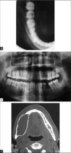
Aneurysmal bone cyst of the mandible: A case report and review of literature

“Aneurysmal bone cyst (ABC) is rare benign lesions of bone which are infrequent in craniofacial skeleton. ABC’s are characterized by rapid growth pattern with resultant bony expansion and facial asymmetry. We describe a case of ABC in a 25 year old male patient affecting the body of the mandible with expansion and thinning of the buccal and lingual cortical plates. Treatment consisted of surgical curettage of the lesion. A one year follow- up showed restoration of facial symmetry and complete healing of the involved site.”

“ABC is a benign cystic lesion of bone, composed of blood-filled spaces separated by connective tissue septa containing fibroblasts, osteoclast-type giant cells and reactive woven bone”

 Spontaneous healing of aneurysmal bone cysts. A report of three cases.

” We report three cases of spontaneous healing of aneurysmal bone cysts (ABC). In one case histological material was obtained after resection of the already ossified expansile mass discovered as a lytic lesion seven months previously. In the two other patients, spontaneous ossification of a radiologically presumed ABC in the lytic and expansile phase was observed after nine and seven months respectively. The healed lesions have remained stable at 12, 32, and 36 months respectively. These findings suggest that when the diagnosis can be made with confidence, and the lesion is in a location and at a stage that does not entail any risk of fracture or compression, expectant management should be considered. Our three patients were aged 22, 19 and 18 years, older than usual for developing ABC. This is also true for many of the few other reported cases of spontaneous or almost spontaneous healing and suggests that ABC has a greater tendency to stabilise in older patients.”

If you look at the results the bone expansion seems to be permanent.


Cortical Bone erosion

Cortical bone blocks height growth so it’s erosion is beneficial but only if coupled with replacement tissue(ideally cartilage).

Bone erosion in rheumatoid arthritis: mechanisms, diagnosis and treatment

“Bone erosion is a central feature of rheumatoid arthritis and is associated with disease severity and poor functional outcome. Erosion of periarticular cortical bone, the typical feature observed on plain radiographs in patients with rheumatoid arthritis, results from excessive local bone resorption and inadequate bone formation[so bone erosion occurs in everyone the balance is just switched in RA]. The main triggers of articular bone erosion are synovitis, including the production of proinflammatory cytokines and receptor activator of nuclear factor κB ligand (RANKL), as well as antibodies directed against citrullinated proteins. Indeed, both cytokines and autoantibodies stimulate the differentiation of bone-resorbing osteoclasts, thereby stimulating local bone resorption. Although current antirheumatic therapy inhibits both bone erosion and inflammation, repair of existing bone lesions, albeit physiologically feasible, occurs rarely. Lack of repair is due, at least in part, to active suppression of bone formation by proinflammatory cytokines. This Review summarizes the substantial progress that has been made in understanding the pathophysiology of bone erosions and discusses the improvements in the diagnosis, monitoring and treatment of such lesions.”

“erosions can also be observed in forms of arthritis other than RA, such as gout, psoriatic arthritis, spondyloarthritis and even osteoarthritis,”

“bone erosions typically emerge at the site at which the synovium comes into direct contact with bone (known as bare areas), suggesting that anatomical factors render these areas of juxta-articular bone susceptible to erosion”

“Anatomical factors that predispose skeletal sites for erosions include: the presence of mineralized cartilage, a tissue particularly prone to destruction by bone-resorbing cells; the insertion sites of ligaments to the bone surface, which transduce mechanical forces to the bone and could induce microdamage; and inflamed tendon sheaths (termed tenosynovitis), which pass by the bone surface, and enable the spread of inflammation from the tendon to the articular synovium“<-maybe the transmission of mechanical forces via ligaments could be exploited to induce height gain.

“The small bone channels that penetrate cortical bone carry microvessels and bridge the outer synovial membrane and the inner bone marrow space; these channels are also prone to erosive change early in the course of RA. The microvessels located within these channels facilitate homing of osteoclast precursor cells to the bone, which, upon contact with bone and receipt of the appropriate molecular signals, differentiate into osteoclasts. Widening of cortical bone channels, as a result of osteoclast-mediated bone resorption, is a typical early change in animal models of arthritis”

“Osteoclasts, giant multinucleated cells derived from the monocyte lineage, are the only cells capable of resorbing bone in the body.2,3 Osteoclasts are designed to resorb bone by adhering tightly to the bone surface through interactions with both integrins and extracellular matrix proteins, as well as by assembling junctions, which seal the bone surface and the osteoclast and thereby separate bone from the surrounding extracellular space. Proton pumps along the osteoclasts’ ruffled border then create an acidic milieu, enabling solubilization of calcium from bone. Matrix enzymes synthesized by the osteoclast, including cathepsin K, matrix metalloproteinase 9 and tartrate-resistant acid phosphatase type 5 (TRAP), degrade the bone matrix”

“Development of osteoclasts occurs locally in the synovial tissue as a result of expression of the two essential osteoclastogenic mediators, macrophage colony-stimulating factor 1 (M-CSF)and receptor activator of nuclear factor κB ligand (RANKL; also known as TNF ligand superfamily member 11). This process involves the migration of monocyte-lineage cells from the bone marrow into the secondary lymphatic organs and finally into the joints. The differentiation step at which monocytes enter the joint is unclear. Indeed, monocytes could be already committed to a certain monocyte lineage, such as M1 or M2 macrophages, dendritic cells or osteoclast precursor cells when they enter the joint. Some data suggest that TNF, a key proinflammatory cytokine expressed in RA synovial tissue, stimulates the migration of osteoclast precursor cells from the bone marrow into the periphery. In addition, TNF stimulates expression of surface receptors such as osteoclast-associated immunoglobulin-like receptor before the precursor cells enter the joint, and these receptors facilitate differentiation.  Once within the microenvironment of the joint, these cells are exposed to M-CSF and RANKL, and differentiate toward osteoclasts. Final differentiation into bone-resorbing osteoclasts is then achieved following contact with the bone surface.”

Inflammation plays a role in osteoclast formation.

Another potential LSJL design mentioned in a scientist paper

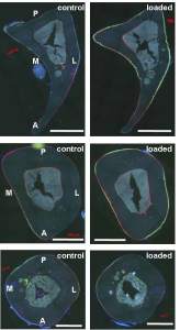
Todd Dodge is a scientist who works at Hiroki Yokota’s lab which developed the beginnings of LSJL.  Most of the paper is not relevant to LSJL except for his data that compares the inside of the control bone and an axial loaded bone.  The difference in the cracks is not as dramatic as the control and LSJL loaded bone.    This indicates that LSJL may alter bone structure more dramatically than typical loading and hopefully in such a way that facilitates neo-growth plate formation.  The key takeaway is the sample LSJL device provided in the picture at the end of this article and in the appendix of Todd Dodge’s paper.

Experimental and Computational Analysis of Dynamic Loading for Bone Formation<-Link to thesis at end of article

This paper is written by Todd Dodge who works closely with Hiroki Yokota who was involved in the initial ideas behind LSJL.

“Curvature in the structure of bone was hypothesized to enhance its damping ability and lead to increased bone formation through bending. In addition, loading at frequencies near the resonant frequencies of bone was predicted to cause increased bone formation, specifically in areas that experienced high principal strains due to localized displacements during resonant vibration“<-Is there a specific frequency for cartilage formation?

“Many types of applied mechanical loading of the skeleton have been proposed as potential treatments for osteoporotic conditions, including whole body vibration, axial loading or bending of long bones, and lateral joint loading. Each mechanical loading modality is thought to strengthen bone by causing dynamic fluctuations in intramedullary fluid pressure in areas that experience enhanced stresses and strains due to the applied load. This dynamic pressure gradient{This dynamic pressure gradient may also lead to stem cells differentiating into chondrocytes to form neo-growth plates} may cause fluid flow through the lacunocanalicular network of pores in the bone, causing a shear stress to be applied to osteocytes that inhabit those pores and channels. This shear stress may excite the osteocytes, causing activation of osteoblast activity and initiation of the bone remodeling process. Mechanical loading may also lead to application of force directly to osteocytes as strain in the bone matrix changes the shape of the lacunae and canaliculae{plastic changes in the bone matrix could lead to permanent lengthening? But it is unlikely given that bone is known for not being capable of interestitial growth}, imposing deformations on the osteocyte and its processes”

Todd Dodge used lateral knee loading on OVX rats.  1N of load was used at 5Hz for 5 min for a group of rats that was tail suspended.  Loading was done once a day for five straight days.  For a group of rats that were OVX 15Hz for 3 min for 10 days(5 days on, then one off)  12 week-old female sprague dawley rats.  So sort of medium along in the growing process with it starting to taper off.

“The slight increase in BMD in hindlimb-suspended mice may be attributed to additional mechanical stress being applied to the lumbar section of the body, possibly increasing bone growth in that area.”

Unfortunately longitudinal growth was not measured.

Here’s the tibia response to axial loading:

tibia load

Compare this to the LSJL holes which are much bigger between control and loaded and are larger.

“Future studies may incorporate not only cortical bone but also trabecular bone and growth plates, as well as surrounding tissues such as muscle, skin, and joints.”<-We may need to wait for further studies to get the results we need for LSJL.

“the e ffects of loading may not only be localized to nearby bones, but may have remote impacts in the spine.”

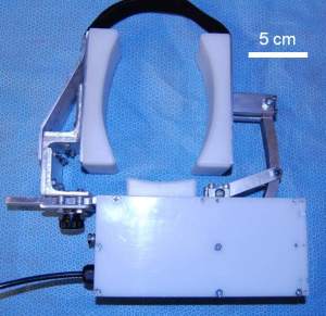
Here’s the knee loading device that he recommends, note though it is designed for BMD not for lengthening:

Knee Load device

Grow taller with prostacyclin?

Prostacyclin is available by prescription in brands such as Flolan.

Prostacyclin regulates bone growth via the Epac/Rap1 pathway.

“Growth plate cartilage is distinct from articular cartilage with respect to COX-2 mRNA expression: whereas articular chondrocytes express very little COX-2, COX-2 expression is high in growth plate chondrocytes, and is increased by Insulin-like Growth Factor-I (IGF-I). In bovine primary growth plate chondrocytes, ATDC5 cells and human metatarsal explants, inhibition of COX activity with non-steroidal anti-inflammatory drugs (NSAIDs) inhibits chondrocyte proliferation and ERK activation by IGF-I. This inhibition is reversed by PGE2 and PGI2, but not by PGD2 or TXB2. Inhibition of COX activity in young mice by intra-peritoneal injections of NSAIDs causes dwarfism. In growth plate chondrocytes, inhibition of proliferation and ERK activation by NSAIDs is reversed by forskolin, 8-Br-cAMP and a prostacyclin analog, iloprost. The inhibition of proliferation and ERK activation by celecoxib is also reversed by 8CPT-2Me-cAMP, an activator of Epac, implicating the small G protein Rap1 in the pathway activated by iloprost. Prostacyclin is required for proper growth plate development and bone growth.”

“Within the growth plate, chondrocytes resting within the reserve zone are recruited to the proliferative zone, wherein IGF-I stimulates cell division, which occurs along the long axis of the bone.”<-Can this recruitment be mimiced in adults to cause new growth plate formation?

“Terminally differentiated cells are found in the hypertrophic zone, wherein glycogen accumulation leads to dramatic cell hypertrophy.”

“IGF-I changes the ratio of activities of the mitogen activated protein (MAP) kinases, ERK1/2 and
p38; relatively high ERK1/2 activity drives proliferation, whereas relatively high p38 activity favors differentiation. The kinase cascades in which ERK1/2 and p38 participate presumably have indirect transcriptional effects, but little is known about the downstream transcriptional
targets of IGF-I in these cells.”

“chondrocytes treated with IGF-I that the growth factor significantly increases the expression of COX-2 in primary bovine growth plate chondrocytes. COX-1 and COX-2 catalyze the same rate-limiting step in the synthesis of prostaglandins, the conversion of arachidonic acid to prostaglandin H2 (PGH2). PGH2 is further metabolized by specific isomerases to produce a
variety of prostanoids, such as prostaglandins, prostacyclins and thromboxanes. COX-1 is constitutively expressed in almost all tissues. COX-2 is normally undetectable in most tissues, but its expression is rapidly induced by injury and proinflammatory factors, and in some tissues mitogens increase COX-2 expression”

“Growth plate chondrocytes release PGE2, which enhances their proliferation”

“whereas COX-2 expression is low in articular chondrocytes, the growth plate expresses
high levels of COX-2 mRNA, and IGF-I increases COX-2 expression and activity in these cells; the ability of IGF-I to stimulate chondrocyte proliferation andERKactivation is dependent on the presence of PGE2 or PGI2; PGI2 is more potent than PGE2 in supporting proliferation and ERK
activation; and PGI2 signals via cAMP activation of the Epac/Rap1 pathway.”

“ibuprofen and celecoxib blocked IGF-I-stimulated proliferation in both primary chondrocytes and in ATDC5 cells at concentrations near or above the IC50 for COX-2 (eg, 50 M ibuprofen, or 2 M celecoxib for primary chondrocytes).”<-Does ibuprofen stunt growth?

“Both the ibuprofen and celecoxib-treated mice were significantly smaller than the mice that received vehicle only”<-and they specifically used doses comprable to those used in children.

“the effects of the protein kinase A inhibitor H89 in chondrocytes is dose-dependent;
low doses tended to increase IGF-I stimulated proliferation slightly, whereas higher doses (such as 10 M), blocked chondrocyte proliferation”

“COX-2 inhibition negatively affects growth plate development in mice by blocking the ability
of IGF-I to activate ERK1/2 and stimulate chondrocyte proliferation; that PGI2 (and to a lesser extent PGE2) and their analogs are able to reverse this blockade suggests that the ability of NSAIDs to suppress chondrocyte proliferation is due to direct inhibition of COX activity. At
least in vitro, we found that the ERK inhibiting effects of celecoxib on chondrocytes are rapidly reversed by a PGI2 analog, suggesting that the dwarfism in mice treated with NSAIDs is due to direct inhibition of COX activity.”