Category Archives: Uncategorized

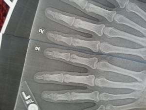
HUGE LSJL update-x-ray proof of LSJL

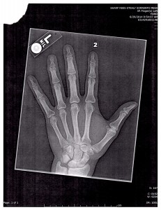
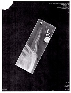
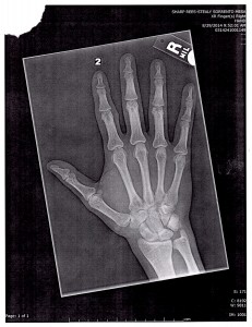
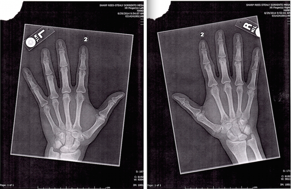
I loaded my right index finger and my left thumb.  This post isn’t finished because I still have to break things down via gimp and it’s a lot harder to measure the bones than I thought.   But I wanted it up right away to get feedback right away. Maybe someone could help measure the bones of the right and left index finger and the right and left thumb.  I need someone to measure the bones of the right and left index finger to confirm that I’ve grown.  The x-ray’s are standardized as you can see by the centimeter mark on the bottom of the page.  Help me with this and we can prove LSJL.

One thing to note about the x-rays.  Is that the whiter something is on an x-ray the denser it is while the blacker it is the less dense it is.  So darker areas of the bone could be some indication of the possibility of a micro growth plate in that region.

X-Ray left hand:

img007

img009

X-Ray right hand:

img006

img008

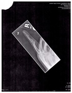
No visible growth plates but that does not rule out microgrowth plates.  You’d need a lot more than a an x-ray to detect micro growth plates.  The difference in finger length does not seem to be due to swelling nor are there signs of osteoarthritic degeneration.  The bumps seem to be due ganglion cysts(see below).  The epiphysis on the right clamped finger is noticeably thicker.  If you look at the lateral view of the right versus the left finger then the difference is extreme and noticeable.

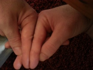
Here’s a side to side comparison between the two fingers:

20140829_143800The right index finger is longer than the left although I will have to do further analysis with GIMP or have Michael or someone do independent analysis.  Same with the left thumb(clamped thumb being longer than the right).

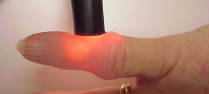
Turns out that I don’t really have osteophytes on my finger but really it’s closer to a ganglion cyst.  See this image here of this photo with someone with ganglion cysts and it’s very similar to how my finger looks:

1547906sThe lump is completely in the soft tissue and cannot contribute to height (see the x-rays)

—————————

Michael: This is what I can say currently after studying the X-ray of the left & right index fingers placed right next to each other. I stared for a full 10 minutes at the picture that Tyler uploaded but could not tell any differences in in the distance between the interphylangeal joints of the right hand compared to the left hand, at least for the index fingers. This is from his own comparison pictures.

However, when I took his pictures and put the pictures together (like below), after carefully looking at the X-ray, there does seem to be a noticeable long bone length difference, where the most proximal bone of the right index finger was just SLIGHTLY longer than the one on the left hand. You have to really look at the specific bone very closely but the difference is noticeable, when viewing the left-right hand X-Ray pictures below from a higher up perspective. When they are put to side to side, there is a difference. Of course, then the question would be, was the right index finger always longer than the leftindex  finger to begin with, from natural bone growth, before ever starting LSJL clamping?

When I looked at the left and right thumb bone X-Rays of the lateral perspective, the difference in thickness of the X-Rays is very noticeable. However, to me, it sort of looks like that the RIGHT thumb is thicker laterally than the LEFT one.

The Problem: I am now in a hotel for a conference in San Francisco and I don’t have an exact ruler on hand. All the measurements are done by eye. To be absolutely sure, I would need to find a ruler though, and maybe even go to a Kinkos or FedEx to blow up the X-ray pictures so that a ruler can be used to really do the most exact measurement possible.

What is obvious though is that the overall right index finger is longer. When you compare the skin edge of the right hand index finger to the one on the left hand, it is very noticeably longer.

X Rays of Metacarpal

LSJL may inhibit Src activation in chondrocytes

This isn’t a breakthrough study but it’s always nice to see a new LSJL study coming out.

Distinctive Subcellular Inhibition of Cytokine-Induced Src by Salubrinal and Fluid Flow.

“A non-receptor protein kinase Src plays a crucial role in fundamental cell functions such as proliferation, migration, and differentiation. Inhibition of Src is reported to contribute to chondrocyte homeostasis. In response to inflammatory cytokines and stress to the endoplasmic reticulum (ER) that increase proteolytic activities in chondrocytes, we addressed two questions: Do cytokines such as interleukin 1 beta (IL1β) and tumor necrosis factor alpha (TNFα) induce location-dependent Src activation? Can cytokine-induced Src activation be suppressed by chemically alleviating ER stress or by applying fluid flow? Using live cell imaging with two Src biosensors (i.e., cytosolic, and plasma membrane-bound biosensors) for a fluorescence resonance energy transfer (FRET) technique, we determined cytosolic Src activity as well as membrane-bound Src activity in C28/I2 human chondrocytes. In response to TNFα and IL1β, both cytosolic and plasma membrane-bound Src proteins were activated, but activation in the cytosol occurred earlier than that in the plasma membrane. Treatment with salubrinal or guanabenz, two chemical agents that attenuate ER stress, significantly decreased cytokine-induced Src activities in the cytosol, but not in the plasma membrane. fluid flow reduced Src activities in the plasma membrane, but not in the cytosol. Src activity is differentially regulated by salubrinal/guanabenz and fluid flow in the cytosol and plasma membrane.”

Interesting that fluid flow reduced Src activation in the plasma membrane but the Cytosol and for Salubrinal it was vice versa.  The plasma membrane is the outside covering of the cell so it makes sense to be more affect by fluid flow as it would be in direct contact with it.  Whereas cytosol is the fluid on the inside of the cell. It would be highly significant if we could identify that LSJL mainly affected proteins in the plasma membrane but not so much the cytosol.

“Src is one of the integrin-dependent signaling proteins involved in mechanotransduction, and it plays critical roles in various cellular processes including proliferation, apoptosis, migration, adhesion, and differentiation. To mediate such a variety of cellular processes, Src’s distinct subcellular activation pattern is required. Src is mainly stationed in the cytosol near the endosomes, and activation of Src requires its translocation to the plasma membrane through the cytoskeleton”

“the shear stress of 2–10 dynes/cm2 has been shown to affect chondrocyte signaling and metabolism either positively or negatively”

“Cells were pretreated with Cytochalasin D (CytoD) for 1 h to disrupt the actin cytoskeleton or with MβCD for 1 h to extract cholesterol from the plasma membrane. CytoD partially blocked Cyto-Src activation, and it completely inhibited Lyn-Src activation. MβCD reduced both Cyto-Src and Lyn-Src activations, although to a lesser degree to Cyto-Src. Collectively, these data suggest that the actin cytoskeleton and lipid rafts are essential components for cytokine-induced Lyn-Src activation”

“Lyn-Src was responsive to fluid flow in a magnitude-dependent manner. In response to shear stress at 5 dynes/cm2, a rapid inhibition of Lyn-Src activity was observed (9.7% decrease). In contrast, shear stress at 10 dynes/cm2 led to its activation (14.9% increase). However, Cyto-Src activity was not altered at any magnitude of shear stress”

According to one article, inhibition of Src kinase activates the chondrocyte phenotype.  The article however did not distinguish between Src activities in the cytoplasma or plasma membrane.  It’s possible for instance that only Src activities in the cytoplasm discourage the chondrogenic phenotype and fluid flow would therefore have no effect.

LSJL Progress Update 8-26-14

In my previous progress post I continued to establish that I had in fact grown due to finger clamping.  I have been using the new LSJL method which emphasizes clamping force rather than duration.  I’ve been clamping right index finger, left thumb, elbows, wrists, knee, ankle, calcaneous, and experimentally toes. Michael suggested using two clamps at the same time and I’ll have to try that.  I’ve been trying to find alternative ways to get X-rays but looks like Michael was right and the best bet is Urgent Care.  It’s going to be a pain in the butt but I don’t think I have a choice.

I’ve been gradually increasing the clamping force.  I think it’s key to be clamping on the synovial joint.  Since the bone is so strong, it’s likely that a lot of the LSJL affects are due to clamping the synovial joint.  If a clamp only the epiphysis of one bone than I can seemingly clamp forever but if I clamp more at the synovial joint I can tolerate much less.  It’s interesting to note that I did grow in the arms from a wingspan of 72.5″ to 74.4″ and the way I’m been clamping the elbow is more on the humerus side because the bone structure is so awkward.  So I can’t clamp exactly in the the middle of the humerus and ulna.  However, I have still gotten results there.   So I think it’s more important to clamp in an area where you get some sort of feedback from your body rather than to clamp in some specific spot.  If you can clamp an area forever, you’re doing it wrong.   Right now I’m clamping areas between 100 to 140 seconds and that’s after working up to it.

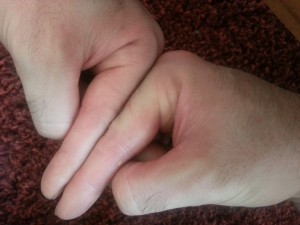
20140826_123611Here’s another finger angle with the tips aligned.  You can see that the knuckle is higher on the right finger.  But yeah, yeah, yeah,  you have to accept X-rays.  And I have to make sure I get both hands in the x-ray.  Although if there are neo-growth plates in the right finger that would be enough regardless of a comparison.  Although it’s possible that the growth was due to another mechanism say fibrocartilage, etc.

As for my legs and arms, there are signs that I am growing.  Last measured wingspan was 74.5″ but since arms can be stretched a little bit it’s not enough.

As for legs, they seem longer and my jeans and shorts seem to be a little higher but nothing definitive yet.  Now when I have a chance I’m clamping twice a day.  At least once a day seven days a week.

What I Learned From Attending The Annual 3D Printer World Expo
What I Learned From Attending The Annual 3D Printer World Expo

3D Printer World ExpoSince the annual 3D Printer World Expo was being held this year at Seattle, (more specifically Bellevue, WA) I bought tickets to attend the expo and listen to the speakers talk about the revolution that is taking place right at this time, which will revolutionize the world in the coming decades to come.

The event was being held from August 22-23, 2014 which concluded tonight. I decided only to attend the conference the first day, and talk with all of the people at the exhibits to see how far they have come along in being able to develop products for medical application.

There was three main reasons why I decided to attend.

First, I realize that the revolution of 3D Printers is going to bring about a paradigm shift in the form of a disruptive technology, similar to how the company Uber is trying to transform the taxi service industry. It is absolutely critical to keep up with the real edge of what is going on in the world today. At the expo, I was able to try out the Oculus Rift that people in the tech world have all been going crazy for, with the people from Prizmiq.

Second, I wanted to see who is already trying to create biocompatible implantable hyaline cartilage, by combining the fields of tissue engineering and stem cells, for what I believe will be the real revolution for our endeavor.

Third, I have started to go into research on the developing technology of turning humans into androids/cyborgs. I have already jumped on the BioHacking bandwagon, promoting the idea set forth by David Asprey, but I feel like there is much further we can go to integrate the electronic world with the biological world.

I took pictures, got free stuff, and talked to people who are in the industry who are promoting their businesses and products.

For our purposes though, I asked each of companies that was selling or promoting 3D Printers the question, “Are you guys selling 3D Printers that has the ability to print bio-compatible, in vivo implantable functional human cartilage?

Almost all of the companies I talked to said no, except maybe 1 company (Nytec). When I asked just how fine the filaments threads can get to in terms of accuracy in making these 3D Models, the smallest I found was around 7-8 microns, with the upper end being around 200 microns.

Here is some things which they did tell me which is being created.

PEEK BIo-Material (poly-ether-ether-ketone) – Unlike the PLA and the ABS type plastic extruded filament material that is traditionally used to make 3D models, there is a another material that is coming out known as PEEK Material. It has been used apparently for the last decade and has slowly had multiple variations/derivatives of the PEEK material made. Its various materials characteristics makes it the most attractive material to be used for in vivo implants. (Refer to study “PEEK Biomaterials in Trauma, Orthopedic, and Spinal Implants” or “In vivo biocompatibility testing of peek polymer for a spinal implant system: A study in rabbits“)

Organovo – This company I have done research before, and it seems like they are the main company to focus on. The people who organized the 3D World Expo when I asked them specifically named this company for 3D Printing Implantable Tissues. I originally referred to Organovo when I wrote about the possibility of using the biomedical practice of Bioprinting aka Electrospinning in the post “Increase Height And Grow Taller Through Bioprinting And Electrospinning“. On the company website, they state the following…

“…Our 3D bioprinted human tissues are constructed with precision from tiny building blocks made of living human cells, using a process that translates tissue-specific geometries and cellular components into 3D designs that can be executed by an Organovo NovoGen Bioprinter. Once built, the bioprinted tissues share many key features with native tissue, including tissue-like cellular density, presence of multiple cell types, and the development of key architectural and functional features associated with the target native tissue.”
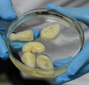
RegenovoRegenovo – This seems to to be China’s answer to Organovo. From the 3DPrintersIndustry.com website, they say that the China based Regenovo is still about 15 years away from being available to the average patient. It is a type of”…medical grade 3D bioprinters that is still under development and showing promising results“. The researchers at Regenovo have already been able to bioprint out a fibrocartilage based ear type tissue, as well as potentially also noses.

Since Regenovo is already able to print out nose and ear structures in about 1 hour!!, they should be able to re-create the chondrocyte column like structure that is found in the epiphyseal hyaline cartilage which can be a mass-produced synthetic growth plate which can be popped into a bone resection to lengthen the bone. The researchers are from some university in Hangzhou and are working with Independent Intellectual Property Rights (IIPRs). The developer named Xu Ming-en is able to create a 4-5 inch wide ear cartilage part within 1 hour already using the regenovo 3d bioprinter.

From the website 3DPrinterWorld.com website, what we are seeing is that along with Organovo & Regenovo, there are labs in Universities around the world like Wake Forest, Cornell, and the University of Iowa who are all working on very similar areas of focus, aka cartilage and spinal disc regeneration.

Taulman3D.com – This company supposedly got a FDA approval also a day ago for using their implantable nylon based material for medical applications.

Selective Laser Sintering (aka SLS) – This is a type of way of 3D Printing method based on using a laser to add more material on the object you are trying to shape and form. Currently, I know very little about this type of manufacturing technique, although it seems to be one of the manufacturing industries most common approaches to making parts.

One company I talked to say that SLS might be able to be used to make implantable parts. I forgot which one though by now.

However, what I have noticed is that due to the nature of the filament (aka raw materials) you are heating up and extruding through the extruder head, these materials like the most two types of filament material, PLA and ABS just can’t be used for medical application, since they would poison the person who tried to put the thermoplastic either in their body or on their skin. Now, there are many, MANY, companies right now who are using 3D Printers to build and make prosthetics, (like for instance Coyote Design & Mfg based in Idaho) by making bone like-tissue which will be placed along the skin but very few companies have been able to succeed in printing things that would work okay inside the human body.

Here is something I didn’t realize maybe months ago when I was writing about the possibility of using 3D Printers to print out our own internal organs. Currently, there is maybe about just 2 dozen types of materials that you can use as the raw material in the filament to make the models and shapes that you want. Here are some of the most common types…

  • ABS
  • HIPS
  • PLA
  • NYLON
  • PVA
  • TPE
  • T-GLASE

What to take away from it all… 

I have been trying to combine all of the areas/fields that I am interested in and doing research on into one. So far, that has led me into the field of 3D Printers and finding the right type of collagen/scaffold configuration to create new cartilage tissue.

It has already been shown by at least 2 sources that making a 10 cm wide piece of fibrocartilage layer is already possible, taking just around 1 hour. I am wondering just how hard would it be to turn that ear/nose piece already made in multiple university labs around the world into hyaline cartilage and make the type of functional implantable cartilage that can expand and make our bones grow volumetrically. Not too hard I suppose.

If the researchers really wanted to, they would be able to get the new growth implant implantations available for the general public’s use within maybe just 3-5 years if they actually put all their effort and focus into it.

To end this post, I give you a vision of what will almost definitely come about in the future, with the forward being presented by former Lucasian Professor of Cambridge, Dr. Stephen Hawking, on how medical breakthroughs have allowed him to live and function beyond the years expected of him when he was first diagnosed with Lou Gehrig’s Disease decades ago.
Sclerostin Inhibitors Promotes Bone Growth and Protects Against Cartilage Degradation

Sclerostin Inhibitors Promotes Bone Growth and Protects Against Cartilage Degradation

Sclerostin InhibitorDue to the nature and subject which we do research on, we often find that our work/research is referenced by certain websites and forums which focuses on talking about very controversial and questionable subjects. The tracking software for the website revealed that the PUA Hate forum was at one time linking to the website, before it was taken off line from the amount of negative media attention it got after what one of its former members Rodger Elliot did.

It took maybe 1 month before some other places similar to PUA Hate started to link to us, with almost the exact same type of social mentality. We are referring to the forum SlutHate.com. I guess for a certain percentage of the male population in any society, there is a lot of anger towards women. I don’t promote these philosophies and don’t agree with what they think. However, sometimes the members do link to something which reveals a very interesting find. That is what happened this time.

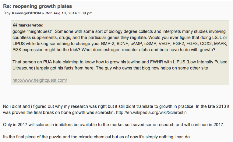
One thread on the sluthate forum talked about the possibility of “reopening the growth plates” and a poster going by the name RevengeOfDOM mentioned a chemical which I have not heard about before. Refer to the clipped picture of his post below…

Sclerostin InhibitorsIt seems that this guy has also been doing his own research on growing taller like us in parallel. It is just that he does not publically publish what he finds. His mention of the class of chemicals which can inhibit this compound known as Sclerostin shows that he understands the stuff very well.
After looking at his other posts, he gave a rather good explanation on how to increase the width of one’s shoulder, talking about the costal cartilage in the sternum area, which we just did, so he knows what he is talking about. Technically, young guys can increase their shoulder width by making the costal cartilage in the ventral sternal area expand through weight lifting and taking HGH in as an adult.

I just did a quick search through Google and PubMed to see what is the potential of this compound class. Here is what I found…

I personally have not had the time to look over those studies above which I just linked to. However, there was one study which made me realize that this chemical compound has huge potential.

The reason why this study was the one that was most interesting was a recent find which showed that the reason why Osteoblasts can’t transdifferentiate into Chondrocytes is due to the conanical Beta-Catenin/Wnt Pathway. A quick guess would be that any chemical that can inhibit the conanical Beta-Catenin/Wnt pathway would mean that maybe Osteoblasts could possibly turn into chondrocytes, and thus regular bone tissue would turn into cartilage tissue.

(Note: Interestingly, Tyler talked about the possibility of inhibiting Sclerostin in a post which I did not read yet “New LSJL study(but bone effects only)

Of course the entire process to reopen growth plates obviously wouldn’t be that easy and simple. This type of research is amazingly hard to get right, since there are probably thousands of young men in the world currently looking into trying to do what we are trying to do, scouring through the PubMed database for interesting studies.

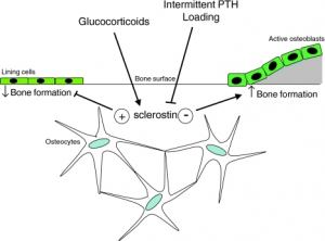
In addition to these PubMed published papers, other sources show that it has bone growth potential. In an article entitled “Novel Osteoporosis Drug Could Change Treatment: Study – Early results indicate romosozumab can rebuild bone” by the Colonial Heights Surgery Center, the chemical compound which is called romosozumab was shown to be able to increase the BMD (Bone Mineral Density) in Lumbar vertebrate bone much more significantly than any of the commercial types of BMD stimulants that is commonly used. (Quote:The new drug uses an antibody to block the function of sclerostin, a protein the body produces to naturally inhibit bone growth”) When the romosozumab was compared to other compounds like teriparatide (Forteo) and alendronate (Fosamax), which is supposed to be some type of biphosphonase. If I remember correctly, I think I wrote a piece on Fosamax about a year ago looking at its potential for bone growth but ultimately concluded that these types of chemicals would never increase the bone’s volume, but the bones’s BMD.

These new class of sclerostin inhibitors are much better than those other compounds at increasing the BMD. For a better understanding on the effects of decreasing the effects of Sclerostin, refer to the study “Targeted Deletion of the Sclerostin Gene in Mice Results in Increased Bone Formation and Bone Strength published back in 2008 and written by Xiaodong Li, Michael S Ominsky, et. al.

So how effective is this compound for height increase or bone growth?

When you are looking for a type of chemical compound group which has promise in promoting bone mineral density and reversing osteoporosis in old age, these sclerostin inhibitors seem to do an amazing job. The compound known as romosozumab is probably just one in that class maybe.

However, we can already seen that at least for one person, they did not see any practical results. As they said on the forum, “…it did not translate into practice“. It would sound like that this person got their hands on sclerostin somehow and injected it or orally took it and did not see any height gain.

However, they said that these inhibitors wouldn’t be available until 2017 to the general public. It seems that he is going to wait until 2017 when these “breakthrough” chemical compounds finally reach the general public and the general market before starting his research again.

Based on my personal opinion though, I don’t think this chemical would be able to do what we are hoping it would do.

Increasing bone growth through BMD is a useless way to do it. It may in fact make the job even worst. However, I would still need to look further at its anabolic effects on cartilage tissue, and what happens when it inhibits the Wnt pathway.

I am not saying that this compound has no potential. It actually has a lot of potential, if the research on its cartilage tissue regenerative abilities turn out to be really high.

That is why I conclude this post by saying that I will at some point go back to this class of chemicals to see just how well can it repair the damage from articular cartilage degeneration.

LSJL Update-8-18-2014

So I’m still working on getting a hand x-ray.  I’m contacting facilities to see if they take walk-ins.  No replies so far. I also read you may be able to get an x-ray at the airport(but that was in context of a full body scan)?  I’m looking for alternative medicine facilities that do x-rays but I’ve read they need a doctors script too.  I don’t really want to go do the Urgent Care but it looks like I may not have a choice.  If anyone has any experience let me know. Here’s the last update.

20140817_150801The right finger could be longer since last update but not sure.

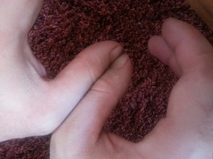
Here’s a thumb pic:

20140817_150737I’ve been clamping my left thumb but not my right and from this picture you can see the left thumb is slightly but significantly longer.  The osteophytes are much less noticeable in my left thumb than my right index finger but they are there if I feel around the clamping region.

Interestingly, I clamp each of the three joints of the finger.

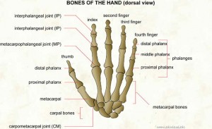
Here’s an image of the finger bones:

027 Bones of the hand (dorsal view)I clamp the two interphalangeal joints from side to side  but at the metacarpophalangeal joint I clamp from top to bottom.   The epiphysis near the MP on the index proximal phalanx of my right (clamped) finger is thicker than my left finger but not as enlarged as the epiphysis near the IPs of the distal and middle phalanx.  The IPs of the clamped joints are also rougher than the MP which is clamped overhead rather than side to side.  So some of the enhanced epiphyseal thickness may be due to LSJL growth stimulation and other effects may be due to irritation from the clamping surface on the joint.  If I can get an x-ray it will be interesting to compare the two IPs to the MPs.  For a period of time, I tried clamping the two IPs from top and bottom but didn’t get noticeable results from that.  Top to bottom clamping may be more effective in the MP than the IPs due to differences in joint structure.  You can feel much more pressure generated by top to bottom clamping of the MP than you can the IPs.

As for leg clamping, I am increasing the duration and intensity of the clamping.  I may be getting taller but I cannot yet rule out placebo(like I have with my thumb and index finger which are too noticeable and significant to be placebo) or measurement error.  I grew about 1 1/2 inches in wingspan for sure but I don’t have photographic evidence.  I’ll have to keep monitoring there.  In a week if I don’t have noticeable leg progress, I’ll bite the bullet and go to urgent care to get the x-rays.