Category Archives: Uncategorized

Chondrodiatasis Is Tensile Loading The Epiphyseal Growth Plate Cartilage Longitudinally Without Fractures

Chondrodiatasis Is Tensile Loading The Epiphyseal Growth Plate Cartilage Longitudinally Without Fractures

ChondrodiatasisFor a long time I’ve wondered whether it was possible to do more than just stretch out the bone tissue to make the overall bones longer, and it seems that there have been a few recent articles which came out showing that it is indeed possible to stretch out cartilage (epiphyseal growth plate) tissue. More than just stretching the cartilage, it seems that it would not even result in any type of fractures.

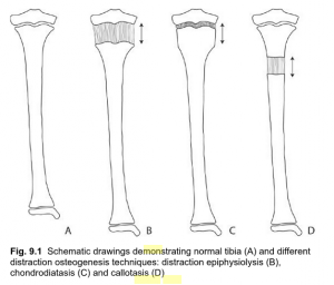
This medical technique is known as chondrodiatasis. It seems that it is so rare and so uncommon that there is almost no information on it except in a few sources from Google. Even orthopedic surgeons are not that too familiar with this idea. (There is a 2nd type of physeal distraction called distraction epiphysiolysis which is much faster but does result in fractures and the cartilage ossifying by the trabecular bone tissue).

In the medical reference book Musculoskeletal Tissue Regeneration: Biological Materials and Methods by Vacanti and Pietrzak there is a small section dedicated to alternative ways to distract bones which is much less step intensive.

There are two types. You have…

1. Distraction Epiphysiolysis – This method is the distraction of the growth plate at a fast rate of 1-1.5 mm per day. The fast rate of separation between the epiphysis and metaphysis results in the cartilage developing fractures, which also leads to the physeal cartilage to turn into trabecular bone. The experiment done by Zavialov and Plaskin back in 1967 showed the change of the cartilage into bone. We won’t focus on this type of physeal distraction technique too much because of the fact that the cartilage is almost immediately converted into bone through advanced levels of osteogenesis.

2. Chondrodiatasis – This is the type of physeal distraction that would be worth our time to look at. It is very slow in fact, usually less than 0.5 mm a day. It seems that tension stress causes the chondrocytes to increase in activity. This technique also increases the level of osteogenesis but there is no evidence (yet) that the cartilage will convert into bone tissue.

Sliedge and Noble back in 1978 showed that they could increase the thickness of lab rabbit’s growth plates by 150% without the presence of fractures. De Bastiani distracted it at just 0.25 mm a day and the chondrocytes cells went through hyperplasia but the overall form of the cells did not seem to change. When the rate of longitudinal growth after physeal distraction was checked, there was no decrease in bone longitudinal growth seen.

Remember however that for any type of bone or cartilage distraction, the surgeons would still drill two pin holes into the bone to pull. It seems that there is no way to get away from having an external fixator used.

However, there will always be complications associated…

Surgical Complication #1: Because we are talking about the growth plates, which are often already so thin, to distract them by only 0.25 mm a day would be very hard to perform.

Surgical Complication #2: The physis would actually get damaged.

Surgical Complication #3: This is the major issue, which seems to be many incidences where growth of the bones seemed to completely stop, which is slightly concerning.

Out of the three ways that you can distract a bone, both of the ways to distract cartilage has proven to be not that effective. That is why the surgeons have almost always just focused on callotasis, instead of these two other types of surgical techniques.

Surgical Complications

Looking through the PubMed archives, there were probably only a dozen studies which ever mention this term “Chondrodiatasis“. We picked the ones which are worth looking over and linked them below…

  1. Chondrodiatasis-controlled symmetrical distraction of the epiphyseal plate. Limb lengthening in children.
  2. Early physeal closure after femoral chondrodiatasis
  3. A large-deformation, finite-element study of chondrodiatasis in the canine distal femoral epiphyseal plate

We will also make a medical reference to the text “Physeal Injury Other Than Fracture” By Hamlet A. Peterson.

While the initial studies showed us that maybe it is possible to lengthen a bone by distracting the epiphyseal cartilage, which would mean no osteotomy necessary, these later studies showed that maybe it is not such a good idea.

The first study gave us hopes and showed that the surgical technique might be viable. There was no visible lesions, infections, or loss in vascularization.

It is the 2nd study which showed that this technique did have its own share of problems. It seemed that when the bone lengthening was done, almost immediately after the surgery was over, the cartilage ossified over. The end result was that the lengthen bone actually shrank. The end result was that this type of surgery was not recommended for children who are too young or have just a small limb length discrepancy.

The last study done on the femoral growth plates of lab dogs was not as informative as I hoped. All it showed that was if you are going to be pulling the bone, the cortical bone and the area where the cartilage and the bone touch will experience a high level of stress.

Conclusion

These procedures known as Chondrodiatasis and Distraction Epiphysiolysis are not well known because they are probably almost never done anymore. It seems that the amount of complications where the cartilage will ossify to prematurely was the reason most orthopedic surgeons will not try to length bones by doing distraction on the cartilage. Distracting the bone tissue seems to be much less complicated and more straight forward, with much more surgical evidence and examples.

Triptorelin Can Increase Adult Height For People Developing Precocious Puberty

Triptorelin Can Increase Adult Height For People Developing Precocious Puberty

Someone recently messaged me information about the existence of a type of drug called Triptorelin which is used by endocrinologists to help young children develop taller adult height. It did trigger my interest so I did only a little bit of research on this drug and these are 5 sources which had the most relevance to the effects of triptorelin on growth.

  1. Final height after long-term treatment with triptorelin slow release for central precocious puberty: importance of statural growth after interruption of treatment. French study group of Decapeptyl in Precocious Puberty
  2. Final height and timing of menarche after treatment for idiopathic central precocious puberty (CPP)
  3. My daughter & Triptorelin (Decapeptyl 3.75mg)?
  4. Three-month sustained-release triptorelin (11.25 mg) in the treatment of central precocious puberty
  5. Effect of GnRH Analogs Leuprolide-Acetate and Triptorelin on Bone Mineral Density in Girls with Central Precocious Puberty

The basic idea is that for a minority of females, they will experience something known as central precocious puberty aka CPP. The gonads will start releasing their steroids about 2-3 years earlier than was they are supposed to. The ultimate result is that the girls will end up with shorter than average final adult height. To inhibit the gonads of the female, the egg, from releasing too much of the steroid, which I believe is just estrogen, the medical researchers used this compound called Triptorelin.

We learn from the 2nd PubMed study that two groups were made, the control group and the group with the girls that were given the treatment. The dosage and timing was every 4 weeks at a dose of 3.75 mg. It was shown that the triptorelin could hold back the initiation of menarche by almost 1.4 years, when the experimental and control groups were compared. What is surprising with the 2nd study is that the average heights of the two groups tested had only about a 2.3 cm difference, which the researchers felt was statistically insignificant.

As for the 1st study, it was probably more insightful. The study was tested on both girls and boys. The Final Height aka FH was compared to control groups and what the predicted final adult height of the control groups would be. For the girls, the average difference was around 4.8 cm but the range of the differences was extremely large (even over 10 cm for certain subjects in the group). For the boys, the increase in height was even more noticeable. Of course, they were compared to the group of kids who did not get any type of GnRH Agonist treatment. Compared to the the predicted height before treatment, the actual increase in height was on average exactly where the predict height was calculated to be.

What is probably most interesting is that apparently triptorelin can actually have a detrimental effect if the treatment is given after a certain age, specifically about at age 11 for the girls. 

Beyond these few studies, there are commercial viability in the drug for children who are noticeably shorter but have not gone through puberty yet. The company called Debiopharm Group has been working on the drug. The website for Debiopharm Group which showed that…

January 13, 2014 – Debiopharm Group™ (Debiopharm), a Swiss-based global biopharmaceutical group of companies with a focus on the development of prescription drugs that target unmet medical needs and companion diagnostics, announces that it has completed the recruitment of patients for its Phase III clinical study in Central Precocious Puberty (CPP) with triptorelin 22.5 mg.

My personal opinion is that while triptorelin may be useful, we maybe should look into the dozen other compounds we have already found which is probably more effective in inhibiting the stunted growth in children. Triptorelin is not the best compound to use since its effects is very depend on the age of the kid for when they start getting the treatment.

For us, as adults, this type of synthetic compound, which is a type of GnHR agonist, is not useful. If it is going to be successful in some type of effect, you want to start on it as early as possible, and quit using it around the age of 12.0-12.5 years of bone age. The source of this info was from the study “Analysis of the factors affecting auxological response to GnRH agonist treatment and final height outcome in girls with idiopathic central precocious puberty

Restoring Lost Height From IVD Collapse Is Already Possible But What About Healthy Thick Discs?
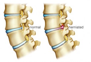
Restoring Lost Height From IVD Collapse Is Already Possible But What About Healthy Thick Discs?

Restoring Lost HeightEver since I put up the contact us button to the sidebar which is linked to our contact us web page the amount of messages and information that is reaching the website email has increased 5X. With most of the messages, the readers of the website have really found some amazing resources which prove some concepts which we suspected was being done already.

In the past, we’ve found multiple patent proposals on how it would be possible to restore the lost height people have after suffering something like a bulging disc or herniated or had their discs crushed by the amount of mass that on the upper torso of the body. We all know that over time, as people get older, they usually loss height. Having any type of medical condition which affects the back (or more specifically the lower back like the L4-L5 region) can make the height lost much greater.

I personally have read a few forum threads around the internet where people loss even upwards of 1.5-2 inches after developing a serious back problem like kyphosis, which is hunched back. Of course, it could be even worse than just 2 inches.

If their is a medical cause making the person loss height from disc degeneration, there are now multiple ways to treat that and restore height. I list the following articles and sources that the people who have messaged us have given us…

Source #1: New stem cell transplant holds promise for treatment of degenerative disc disease

Senior author, Wenchun Qu, MD, PhD, of the Mayo Clinic in Rochester, Minnesota – stem cell therapy for disc degenerative disease might be a potentially effective treatment…. not only did disc height increase, but stem cell transplant also increased disc water content and improved appropriate gene expression…. prepare for translation of stem cell therapy for degenerative disc disease into clinical trials….The increase in disc height was due to restoration in the transplant group of the nucleus pulposus structure, which refers to the jelly-like substance in the disc, and an increased amount of water content, which is critical for the appropriate function of the disc as a cushion for the spinal column, the researchers concluded.

Source #2: Intervertebral disc regeneration in an ex vivo culture system using mesenchymal stem cells and platelet-rich plasma

Here are excerpts of the abstract which we just copy and pasted below….

An ex vivo degenerative intervertebral disc (IVD) organ culture system… a stem cell and growth factor-based therapeutic approach for the amelioration of IVD.

…different therapeutic regimens including: (1) mesenchymal stem cell derived from eGFP-transgenic porcine (MSC-GFP), (2) platelet-rich plasma (PRP) and (3) MSC-GFP/PRP combined treatment, and confirmed in in vivo animal model…. Col II and aggrecan were found upregulated and chondrogenic matrix deposition increased…

PRP that has been shown to promote nucleus pulposus (NP) regeneration also resulted in significant increased levels of mRNA involved in chondrogenesis and matrices accumulation…. repeated and supported by in vivo porcine degenerative system.

Moreover, the disc height index (DHI) was significantly increased in both in vivo MSC-GFP and PRP regeneration groups

Source #3: Molecular and genetic advances in the regeneration of the intervertebral disc

Rapid advances… have resulted in several promising methods to facilitate the translation of laboratory-based techniques to clinical disc regeneration…. Tissue engineering strategies that employ (1) biomimetic scaffold materials, (2) differentiation-driving bioactive molecules, and (3) multipotent cells to enhance disc regeneration are being developed at a rapid pace.

The paper was not one of those papers written to say something new, but to organize all the possible ways that the current medical community knows how to restore the discs. Here was a list of the ideas that tissue engineering researchers around the world have tried…

1st Approach:  Disc Regeneration Via Morphogens and Mitogens

  1. Delivery of exogenous proteins: Growth factors and cytokines
  2. Factors with biologic activity: Mitogens, morphogens, and intracellular mediators (ex. TGF-Beta1, TGF-Beta3, IGF-1, etc)
  3. Transforming growth factor beta superfamily; morphogens and biologic mediators of intravertebral disc development
  4. In-vitro (and In-Vivo) study of BMP-2 for IVD regeneration
  5. BMP-7 (aka OP-1) induces similar in vitro effects as BMP-2 in IVD cells
  6. In vitro and in vivo studies of BMP-14 (GDF-5)
  7. More work is needed to ascertain effects of lesser known bone morphogenetic proteins family members (like BMP-4 and BMP-13)

2nd Approach: Cell-Based Regenerative Strategies

  1. MCSs
  2. MCSs from bone marrow which can differentiate into nucleus pulposus like phenotype
  3. In vivo experimentation demonstrates the promise of marrow-derived mesenchymal stem cells
  4. Multiple niches of mesenchymal stem cells display regenerative properties

3rd Approach: Intervertebral Disc-Derived Cells

  1. Cells derived from intervertebral disc… rejuvenate endogenous cells
  2. Notochordal cells

4th Approach: Advances in Gene Therapy and Delivery

  1. Altering genotype of cell population, transcription/translation sustains protein synthesis
  2. Direct in vivo gene therapy: Direct administration of infecting agent(s) to host
  3. Adenoviral vectors expressing numerous bone morphogenetic proteins
  4. Sox9 gene delivery in human IVD cells
  5. Primary human IVD cells respond favorably to adenoviral infection with growth factors
  6. Treatment of IVD cells with lim mineralization protein-1 causes increased production of proteoglycans both in vitro and in vivo

5th Approach: Multi-Faceted Approaches to Disc Regeneration

  1. In vivo performance of cell-seeded scaffolds

6th Approach: Hydrogel-Based Scaffolds: A Promising Method for Nucleus Regeneration

  1. In vivo growth factor delivery from scaffolds

7th Approach: Enhancing Disc Regeneration by Slowing The Degenerative Process

  1. Proteinase inhibitors utilized in gene therapy-based approaches
  2. Statins act as intracellular inhibitors of MMP production and enhance BMP-2 mRNA expression
  3. Lactoferricin causes increased PG accumulation and downregulated catabolic processes
  4. TNFα-stimulated gene product TSG-6 with inter-α-inhibitor downregulate MMP activation
  5. Etanercept is an approved TNF-α inhibitor
  6. The oral administration of CPA-926 an esculetin prodrug

Source #3: Duke Bioengineers Develop New Approach to Regenerate Back Discs

In a proof-of-concept study published online in the journal Biomaterials… a new biomaterial designed to deliver a booster shot of reparative cells to the nucleus pulposus, or NP — the jelly-like cushion naturally found between spinal discs. The NP tissue distributes pressure and provides spine mobility, helping to relieve back pain.

  • Primary Goal: “Our primary goal was to create a material that would be liquid at the start, gel after injection in the disc space and keep the cells in the location where they’re needed,”
  • Secondary Goal: “Our second goal was to create a material that would provide the delivered cells with the environmental cues to promote their persistence and biosynthesis.”

The most common types of problems: …re-implanting NP cells, or even stem cells, can delay disc degeneration. Several companies already offer cell delivery strategies, but the methods are poor and ineffective. (using the currently available cellular delivery strategies, 100 percent of the injected NP cells leak out of an IVD site within three to four days after injection. “They allow the cells to quickly migrate out of and away from the injection site,” Francisco said.

The Duke team’s delivery strategy keeps the cells in place and provides cues that mimic laminin, a protein in native nucleus pulposus tissue. Laminin is normally found in juvenile but not degenerated discs and allows injected cells to attach and remain in place with the delivered biomaterial. Laminin may also enable the cells to survive longer and produce more of the appropriate extracellular matrix or structural underpinning of the discs that help stop degeneration, Setton said.

The researchers… developed a gel mix designed to reintroduce NP cells to the intervertebral disc (IVD) site. The gel mixes together three components:

  1. the protein laminin-111 that has been chemically modified
  2. two polyethylene glycol (PEG) hydrogels that can attach to the modified laminin.

Separately, these substances remain in a liquid state. The gel, however, holds the cells in place upon injection.

What they did: ….injected the gel into rats’ tails… puncturing the tail’s thin outer layer, they held the needle in place for one minute, delivered the injection to the rat’s IVD site, and closed the injection spot. The solution began to solidify after five minutes and was completely set at 20 minutes.

The Result: …more than 14 days after injection significantly more cells remained in place when delivered within the biomaterial carrier compared to cells delivered in a liquid suspension.

“The concept is that these cells will be promoted to produce matrix that can support tissue regeneration or arrest degeneration,” Setton said….

The exact study that the article above refers to is Injectable laminin-functionalized hydrogel for nucleus pulposus regeneration.

Source #4: NOVOCART® DISC

is a high-quality autologous cell compound in an innovative hydrogel for the biological reconstruction of partially damaged discs following disc prolapse

Biological disc reconstruction

…biological reconstruction of partially damaged disc tissue before massive degeneration… transplantation of in vitro cultivated chondrocytes from autologous disc tissue,or ADCT (Autologous Disc Cell Transplantation).

ADCT is comparable to joint ACT (Autologous Chondrocyte Transplantation)… In both cases, isolated from the biopsy material, the cultivated cartilage cells induced the desired reconstruction.

One further special feature…is the hydrogel used, which fixes the cells in the damaged disc… also anti-inflammatory.

Description of ADCT with NOVOCART® DISC

Transplantation of the cultivated disc cells can take place three months after extraction by way of operation of the prolapsed disc. The relatively long period between tissue extraction and cell transplantation must be adhered to, to ensure that the protective fiber ring (annulus fibrosus) is fully healed following the intervention.

So what do these 4 sources which was sent to us suggest?

We know that there are at least 2 different research teams at universities which are doing research to figure out how to restore the function and shape of IVDs after they have suffered through discs degeneration, whether due to just normal old age or some type of degenerative disease.

Since these teams are able using whatever technique to restore the shape of the IVD, they have also increased the thickness, which translates to mean that the person regained lost height.

Whether it is a scaffold implant, or cells like MSCs, or growth factors, the discs can be saved and given new life.

The first point is this – We now have multiple options on how to restore lost height from the collapse of the IVDs.

However, can we do anything if the discs are healthy and we want to increase our height?

I suspect that for most people, we are not fully maximized on how tall we can be. The majority of people would be able to gain upwards of even 1.25 inches in extra height, if we can correct our posture, and then our IVD structural integrity. If we wanted to, we could use these developing technologies to make sure that our nucleus pulposus matrix is strong and rich in the right collagen material.

However, there are a small group of people who have the discs already full. Maybe we are 18-20, with a completely healthy body but our only problem is that we suffer from familial short stature. For these people, who have already done all the stretching, the disc decompressions, and maybe tried even certain traction machines, I would suggest that we would no longer focus on the discs, but to focus on the material next to the discs.

However, the problem is probably going to have more to do with medical ethics than a technical problem. Most doctors would never agree to sticky a needle into the spinal area of a healthy young adult. There is a great fear that a needle stuck in the wrong area would cause a disc to become punctured or even slight paralysis.

In a rather old post I did, it was shown that for young adults, around the 17-23 range, many of them have a very thin layer of articular cartilage that does run between the bone tissue of the vertebrate bone and the nucleus pulposus of the discs. That layer of cartilage however is so thin that it is very hard to see. It was guessed that the layer is most likely about 1-2 mm thick. However, we can work with that.

If might be possible to stimulate that layer of articular cartilage to go through hypertrophy using a small injection of IGF-1 directly into the cartilage tissue. We know that a local injection of IGF-1 into the bone layer under the periosteum would increase the rate of long bone longitudinal growth, as we had revealed in a previous post “Increase Height And Grow Taller Using Local Subperiosteal Injection Of Growth Factors IGF-1 And TGF-Beta Percutaneously” as long as the epiphyseal growth plate cartilage was intact.

Since the young adult who wants to grow taller, but has full discs, still has maybe 1-2 milimeter of cartilage in every connecting segment between the discs and the vertebrate bone, it should be reasonable to inject the IGF-1 (or TGF-Beta) locally next to the layer of thin articular cartilage. Since there is 33 vertebrate bones in the human skeletal system, we probably have 20 or so thin layer of cartilage we can try to stimulate using small needles.

If we can use an X-ray to double check the location of the needle injections, to make sure that we don’t puncture a disc or hit a nerve ending, it could work. The hypertrophic effect of injections of the growth factor into 20 of these thin layers can add up to maybe 2-3 extra inches, but that is a very wildly optimistic situation.
Why Chiropractor Jean Pierre Meersseman Of Milan Lab Could Help Their Clients Increase In Height

Why Chiropractor Jean Pierre Meersseman Of Milan Lab Could Help Their Clients Increase In Height

Jean Pierre MeerssemanI enjoy reading Mark Cuban’s posts on his thoughts at Blog Maverick and there was one post he wrote back in 2013 where he said that one of the best biological investments a person can give their kids and turn their kids into superhuman was to save the blood that is inside the umbilical cord, which is known as cord blood. One of the commenters noted that Mark should check out some of the really interesting physical therapy techniques and methods being practiced by this physical therapy consulting company called Milan Lab.

We looked on the Milan Lab website. They describe themselves as “AC Milan’s high tech interdisciplinary scientific research centre.” The goal of the research center has been to use whatever means necessary from whatever scientific field and branch to find ways to make soccer players healthier, stronger, and recover from injury faster.

The fact that in the first sentence on the description on the website the word “interdisciplinary” is used suggest that the people who run the consulting and/or research center does whatever is necessary to help out the players in AC Milan. The founder, Jean Pierre Meersseman seems to be someone who only focuses on results and the end result. His willingness to try out and combine any field of biomedical research together to find a way to help out the players shows that he probably has done extensive research, and searched through all the resources that are available to find esoteric ideas and fringe concepts to incorporate into his system.

We quote the following from the article “Milan lab man brings unorthodox science to Premier League” below…

“…doing the unusual with the unorthodox, combining his specialisms of kinesiology and chiropractic with traditional approaches. Still the question needs to be asked: what would the sceptics make of how he treated Seedorf?…    It’s not accepted in evidence-based medicine but I don’t give a damn about that,” he says, genially but firmly. “I’ve seen it work. We’ve done over one million tests at Milan. And our mathematicians and engineers have developed a formula which has a high success rate of predicting and managing injuries.”

This guy is bringing on people from fields that traditionally have no relationship with biology or medicine, engineering and mathematics to figure out how to measure and quantify sports injuries and how the body works.

Many people have been wondering this “What is this guy, Jean Pierre Meerseeman’s secret sauce?

Obviously Jean would not tell anyone what it is. My guess is that he spent maybe half a decade or more reading over as many scientific journals, sports medicine journals, academic journals, pubmed studies, and read old soviet olympic medalist trainer’s notes which the average physical therapist does not do. His willingness to look over the obscure and unique probably means that he has discovered, and made a derivative of, some some old, unknown method which was developed unknown researchers before him.

There was a rather well known YouTube video (available here) which shows the football player Terrell Owens ask Kobe Bryant about his recent visit to the Los Angeles physical therapist and his use of a technique known as Platelet-Rich Plasma Therapy (PRP Therapy) to repair the articular cartilage in his knees. (Kobe was being interviewed for almost a full hour by Jimmy Kimmel for Up Close) Bryant apparently flew to Germany and had the PRP Therapy done twice and his knees seemed to be much better. The name that Kobe mentioned was a neuromuscular physical therapist named Barrence Baytos and this guy was some type of genius in his profession.

There are people like Baytos and Meersseman who are supposed to be in the area of sports medicine or physical therapy and injury rehabilition treatment who probably know unique tricks and ways to get the human body to perform in optimum functino that even doctors trained in the best medical schools and orthopedic residency and fellowships are not aware of.

If I was a betting man, I would guess that Jean Pierre Meersseman and similar physical therapist know rolfing, feldenkrais method, the alexander method, and physical therapy well enough to actually help people who desired to become taller, in a more permanent way than any normal chiropractor or physical therapist would be able to. Their years of research into obscure studies and ideas probably lets them understand the mechanics and ability of the human body very well.

If there is anyone in the world who would be able to help a professional athlete increase their height, it would be most likely Jean Pierre from the AC Milan Lab since he has dozens of specialists which act as consultant. They do dozens of test on every single part of the body and try figure out how the individual parts of the body work together. If any guy can perform a miracle, this guy probably can.

Breakthrough: How Mechanical Loads incourage zone of ranvier and growth plate formation

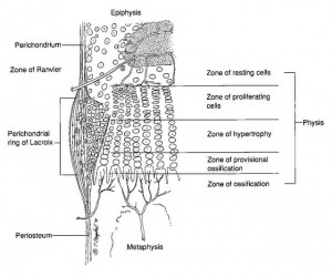
Here’s a diagram showing the zone of Ranvier in relation to the growth plate:

zone of ranvier

If we know what stresses are responsible for forming growth plates we can re-induce these mechanical stresses to form new growth plates.

This study would be more significant except it was published in 1988 before the scientific community began to focus more on genes and cellular epigenetics.  Just because the highly undifferentiated cells of a juvenile or infant may be induced to differentiate into the cells composing the Zone of Ranvier and in turn the growth plate does not mean that adult mesenchymal stem cells will do the same thing.

And I think going back to these old studies is a good thing as they focued more on mechanical factors rather than retrovirus genetic engineering.

One study found that interstitial fluid flow upregulates the mesenchymal condensation gene Msx2 but downregulates the chondrogenic gene Col2a1.  This is not necessarily a bad thing as mesenchymal condensation in a neo-Zone of Ranvier would be key to forming new growth plates and those genes would not be chondrogenic until they became a growth plate.  In fact in this study the Zone of Ranvier experienced more osteogenic than chondrogenic stimuli so it would be no surprise that a mesenchymal stem cell that may eventually become a growth plate chondrocyte may not express chondrogenic genes initially.

This study provides additional evidence for LSJL as LSJL compresses the bone and the joint thus altering the “Loading history” of the bone and joint.  Since the shear strain, caused by LSJL fluid flow, needs to be high this could explain why I’m getting more results for my fingers than my legs.

The role of mechanical loading histories in the development of diarthrodial joints.

“The role of mechanical loading history in chondroosseous development at the ends of long bones is explored using two-dimensional finite element models of chondroepiphyses. Loading histories are characterized in terms of discrete loading cases defined by joint contact pressure distributions and an associated number of loading cycles. An osteogenic stimulus throughout the chondroepiphyses is calculated following the theory that cyclic octahedral shear stresses promote endochondral ossification and cyclic compressive dilatational stresses inhibit ossification{Octahedral stresses are shear stresses that are acting on octahedral planes inside the bone.  The plane whose normal vector forms equal angles with the coordinate system is called octahedral plane.  But basically shear stress which is what LSJL can induce. A compressive dilational stress is the increase in volume per unit volume of a homogeneous substance.  So for example if you squeezed a stress ball and it got bigger}. The resulting distributions for the osteogenic stimulus predict the appearance of the secondary ossific nucleus and the shape of the developing bony epiphysis. The zone of Ranvier and the formation of articular cartilage and the growth plate are predicted by the models{Considering that the zone of Ranvier is the basis of the growth plate this is key}. Tissue stress histories constitute an important influence during skeletal morphogenesis {And we can expose the tissue to different stresses ala LSJL}.”

“Alterations in joint loading or motion can alter the pattern of ossification and growth in developing bone ends, and a reduction in joint forces can delay the appearance of the secondary ossific nuclei”

“The sequence of cartilage proliferation, maturation, degeneration, and ossification is the normal process for all cartilage in the appendicular skeleton. This process is (a) accelerated by intermittently applied deviatoric (shear) stresses (or strain energy) and (b) inhibited or prevented by intermittently applied compressive dilatational stresses (hydrostatic pressure).”

“The appearance of the secondary ossific nuclei in both chondroepiphyses was predicted. Increased osteogenic stimuli were also calculated at the edge of the advancing ossification front where the zone of Ranvier (ossification groove) forms. The areas where the osteogenic stimuli were low define those cartilaginous regions that become the growth plate and the articular cartilage.”

“the ossific nucleus appears in an area of high shear (deviatoric) stresses the edge of the advancing ossification front (zone of Ranvier or ossification groove) also experiences high shear stresses, and the joint surface, where articular cartilage forms, is exposed to high-magnitude hydrostatic compression”<-Since the zone of Ranvier is the key forming new growth plates, inducing high shear stress would be key to forming new growth plates and LSJL can induce shear stress via interstitial fluid flow.  According to the study Interstitial fluid flow: the mechanical environment of cells and foundation of meridians., interstitial fluid flow induces shear stresses.  Here’s Michael’s summary of LSJL.

In both the convex and the concave chondroepiphysis, a state of high hydrostatic pressure is created directly beneath the loaded contact surface. In the interior regions of both chondroepiphyses, areas of high-magnitude octahedral shear stress are created. The location of these areas within the central zone of the chondroepiphyses shifts with the direction of the applied loading”<-If we mimic this state we can achieve chondroinduction.

“the shape of the developing bony epiphysis will depend on the geometry of the bone end. In the model with a convex joint surface, the developing ossific nucleus is stimulated to produce a sphere-shaped bony epiphysis.  The model with a concave joint surface is stimulated to create a flatter, more disc-shaped bony epiphysis.”

“The stored energy in cartilage under mechanical loading is primarily in the form of deviatoric or shear energy owing to the nearly incompressible nature of this tissue. Some of this stored deviatoric energy is lost in hysteresis upon unloading. The energy dissipated during intermittent mechanical loading must be accounted for by a change in internal energy and/or a change in the temperature of the cartilage. It is possible that the direct mechanical alteration of cells or the increased temperature associated with energy dissipation caused by intermittent shear stresses could increase mitotic activity or activate specific biochemical pathways in the cells”

Here’s another study with some information on the Zone of Ranvier:

Stem Cells and Cartilage Development: Complexities of a Simple Tissue

“[The] biochemical composition [of cartilage] is uniquely suited to providing a combination of tensile strength with deformability, giving it mechanical properties that resemble those of a shock absorber, thereby dissipating forces across the bones, preventing them from fracturing during normal activity.”

“The balance between mechanical stiffness and flexibility is itself the result of interaction between the thin type II collagen fibrils, giving tensile strength, within which are trapped molecules of aggrecan, which are highly negatively charged and so bind water avidly”

“When unloaded, the water content of cartilage is about 70% of the wet weight. Under deforming load water flows out and when the load is reduced it flows back in, damping the effects of these forces. Damage to either the type II collagen or aggrecan may lead to loss of cartilage function”<-Maybe when LSJL loads the cartilage it results in water flowing out and possibly other nutrients and maybe even growth factors that can result in neo-growth plates.

“During cartilage development [cartilage transforms] from a relatively simple isotropic tissue with a high cell density and homogeneous distribution of collagen fibrils to an anisotropic tissue with a low density of chondrocytes growing in vertical columns and a unique arrangement of collagen fibrils.”

Articular cartilage chondroprogenitor cells are derived from migration of mesenchymal cells out of the zone of Ranvier niche.  In early development these cells may accumulate in the surface zone and drive the process of appositional growth of cartilage but with maturity they become dissipated throughout the cartilage.”<-So maybe LSJL can drive these cells back into the zone of ranvier?

Growth Of Ear Cartilage On Mice Using Biodegradable Porous Scaffolds By Dr. Charles Vacanti

Growth Of Ear Cartilage On Mice Using Biodegradable Porous Scaffolds By Dr. Charles Vacanti

Biodegradable Porous ScaffoldsSomething that I have been getting into in recent months have been to watch the new TV shows that have come out depicting the fictional literary figure Sherlock Holmes. It has been a great experience comparing the two shows, Elementary and Sherlock, to see how well Sherlock Holmes is portrayed on TV. On the recent episode of Elementary, (Season 2 Episode 17 “Ears to You”) the final conclusion made by Holmes was that the former wife who disappeared managed to grow her a new set of her own ears on her back and have the ears cut off by her new plastic surgeon husband to send to her old husband for ransom money (If you haven’t already seen the show, sorry about the spoiler).

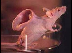
I didn’t focus too much on the show except the part in the end when Holmes mentions this most unique of research done by a certain Dr. Charles Vacanti where he managed to regrow almost a human sized ear on the backs of lab mice. That reminded me of this article I found which showed that a 3-dimensional growth plate like material was formed through. Based on what I’ve read about how the process is done, the steps are the following….

  1. Find the raw material to make the scaffold. Often the scaffold is made from alginates and/or hydrogel. The scaffold is known by other names as well, like synthetic biodegradable polymer substrates.
  2. Get the biodegradable porous scaffold molded/buffed/grinded into the shape and size that you want.
  3. Implant some chondrocytes and/or the MSCs into the scaffolds thoroully so that the entire scaffold is loaded in all the porous holes with the cells.
  4. Implant with the cells (whether chondrocyte, osteoblasts, or MSCs) some type of growth factor, like a peptide.amino acid/unique protein type, BMP (2, 6, 7, and/or 9), GDF (5 or 9), or TGF-Beta (1,2, and/or 3)
  5. Place the scaffold with the cells and growth factor into the space/position/cavity in the area of the body as you desire.
  6. Over time, while the scaffold dissolves and gets absorbed by the body, the cells multiply and take over, turning into the type of tissue that is usually made by the excretion/waste of the cells.

In the case of the lab rats, the cartilage formed from the chondrocytes embedded led to the formation of the ears, which are fibrocartilage in nature. For this particular experiment, the ear lobes were not functional, since there was no inner ear canal, there was no connection of the cartilage to the nerve cells in the periphery, and there was probably very little vascularization into the fibrocartilage. Those the the technical difficulties that Vacanti experienced. However, the idea is definitely interesting and I think those technical difficulties will be easily removed with some slight alterations in the overall tissue engineering process.

However the proof of concept and ideas is valid. That is what excited me. That was also why I started to google the term “biodegradable scaffold cartilage” into Google and Google Scholar and Google Patent to see what turns up. It is most likely that I will be more focused on disecting whatever unique patents and studies I find from this specific search term into Google.

I am almost positive that within 6 months to 1 year, at least I will no longer be writing non-scientific posts. I want to focus exclusively on looking at tissue engineering, tissue regeneration, and stem cell differentiation and stimulations exclusively. That is the area which I think has the most potential.