Category Archives: Uncategorized

The Connection Between Relaxin And Possible Height Increase

Me: Ever since we started to find stories and posts on the internet mothering and pregnancy boards where women have come forward to state that their height increase from their pregnancy, the topic of relaxin as a possible height increase hormone has been raised. Tyler has recently done a post on the subject (link HERE).

Tyler: Added a new study directly below.  The most likely cause of the pregnancy height gain some females experience is due to increase in tendon length as there doesn’t seem to be any logical connection between relaxin and longitudinal bone growth.  Although the cattle pelvic height growth study seems to indicate that relaxin could somehow induce articular cartilage endochondral ossification results in pelvic height growth.

The effect of relaxin on the musculoskeletal system.

“Relaxin circulates during pregnancy emanating from the corpus luteum and placenta”

“In rodents, circulating relaxin peak concentrations at the end of pregnancy reach 100 ng/mL, two times greater than in human”

“seven known relaxin family peptides (RXFP) are structurally related to insulin which include relaxin (RLN)1, RLN2, RLN3, and insulin-like peptide (INSL)3, INSL4, INSL5, and INSL6”

Relaxin alters cartilage and tendon stiffness by activating collagenase“<-collagenese are enzymes that break the peptide binds in cartilage.

The study notes that Relaxin increases length in cartilage and tendons(Fig 2).

Relaxin has a role in osteoclastgenesis.

Relaxin treatment in pregnant cattle increased pelvic width and height, but not in other joints such as wrist and knee“<-Maybe the reason that relaxin didn’t increase other joint height is due to receptors?  The Relaxin receptor is RXFP1.  According to the study Relaxin Receptors in the Human Female Anterior Cruciate Ligament, women but not men had relaxin receptors in the anterior cruciate ligament.

Relaxin appears to decrease knee articular cartilage stiffness through induction of collagenase-1, MMP-1, and MMP-3, which reduces collagen content and expression in fibrocartilaginous cells. Modulation of MMPs to loss of collagen by hormones may contribute selectively to degeneration of specific joints fibrocartilaginous explants facilitated by proteinases. The degradation of extracellular matrix in fibrocartilage is synergized by β-estradiol. Relaxin exerts its effect through binding to RXFP1 and RXFP2 receptors. The ratio of RXFP2 in knee meniscus of pregnant rabbits was shown to be more than RXFP1, which may indicate differential role of these receptors in the remodeling of fibrocartilage. Comparison of collagen content in articular cartilage of nonpregnant and pregnant rabbits showed that the total RNA levels and chondrocyte metabolism decreased during pregnancy.”

One study has noted that porcine relaxin is capable of modifying type II collagen expression in chondrocyte cells.  Another study found that relaxin increased tendon length so tendon length could be related to the height grain in pregnancy.

This is my attempt in trying to see whether any PubMd studies I have found would be worth mentioning in relevance to our research. I would cite and copy below 5 studies.

Analysis & Interpretation:

From the 1st study…

I would assume Tyler in his post used the study “Pelvic development as affected by relaxin in three genetically selected frame sizes of beef heifers”. The really interesting thing is that the study seems to show that the entire pelvic bone area increased in size. The term “primiparous beed heifers” refer to female cows which are going through their first birthing experience. The issue with the study is of course that cows were used than humans. Also it doesn’t really go and try to explain how it was possible that relaxin caused the increase in pelvic height increase or pelvic width increase. I am now cow expert so I don’t know whether the cows were still growing by the standards of humans. What I do know is that cows which are going through their first birthing experience can easily be young enough to still have growth plate cartilage so what we see in pelvic bone height can be just the excess release of whatever hormones female cows will go through when they are pregnant. From the controlled study we see that relaxin being introduced does cause clear increases in pelvic bone height and width.

For humans, we have to remember the way human female reproductive systems work. The indication that human females can grow is from the start of the menses cycle. The first menses is known as the menarche. Most girls would have their menarche during the puberty stage, but before the growth plates ossify completely. This means that before girls stop growing taller, they would develop the ability to have children and go through the human gestation process. In bovine, we would guess it is the same thing. In conclusion, from just this one study we can see that in cows, the hormone relaxin has the ability to increase pelvic height. Since the pelvic bone outline/structure does indeed contribute to the overall human height, the increase in pelvic height we see in young female bovine may be able to be translated to young human females. However the age values we find from the internet discussion boards and forum suggest that the relaxin is doing something more extraordinary since the women are already even in their late 20s or 30s when they notice the height increase. If relaxin is just only a muscle relaxant, how does it actually make pelvic bone taller? Just something to wonder about right now.

{Tyler: “Pelvic height was determined by measuring the linear distance from the approximate midpoint of the dorsal surface of the symphysis pubis to the ventral surface of the prominent junction of the third and fourth segments of the sacral vertebrae”<-Doesn’t seem to factor in tendon length.

“pelvic growth during the last 10 days before parturition[child birth] can be modified by the intracervical administration of relaxin.”

“The mechanism of pelvic canal expansion in cattle is unknown. The pelvic canal may increase in area as a result of relaxation of sacroiliac ligaments, formation of interpubic ligaments, and modification of the pubic symphysis by transforming symphyseal cartilage[a type of cartilage joint] and bone{if it does this then relaxin could have very strong height increase potential}“<-growing from the pubic symphysis would be like growing from the articular cartilage which would have very nice height increase implications.  The study that discsusses this is called Dystocia in Cattle by LE Rice unfortunately I could not find this study.

From the 2nd study…

If we however look at the 2nd study I have linked, we can see that Relaxin, specifically the Relaxin Family Peptide Receptor 1 (RXFP1) can be the precursor to a mechanism which triggers many of the other pathways and hormones we have looked at before. including NO and cGMP. Somehow it disrupts the TGF-β1/Smad2 axis through a signal process that involves the ERK/pERK/NO/cGMP pahway. One set of proteins I have been trying to get to researching more into are the MMPs. Somehow the RXFP1 increase matrix metalloproteinase (MMP) expression. My knowledge on MMPs are nonexistent at this point so I can’t really breakdown what the abstract is really talking about. However the researchers would conclude with…

“These findings demonstrated that H2 relaxin signals through a RXFP1-pERK-nNOS-NO-cGMP-dependent pathway to mediate its anti-fibrotic actions, and additionally signals through iNOS to up-regulate MMPs; the latter being suppressed by TGF-β1 in myofibroblasts, but released upon H2 relaxin-induced inhibition of the TGF-β1/Smad2 axis”

So I guess the two main things to take away from this study is that the H2 relaxin seems to inhibit/suppress the differentiation of myofibroblast and also increase the gene expression of the MMPs.

From the 3rd study…

Relaxin seems to regulate the expression of two types of MMPs, MMP-9 and MMP-13. Relaxin is a type of ligand that attaches to a certain type of substrate or receptor which will accept it. There is two types of relaxin receptors described in this study, Relaxin family peptide receptors 1 & 2, called RXFP1 & RXFP2 respectively. It would seem that the RXFP1 is the one that has the real regulating power on MMP9 & MMP-13 expression while the RXFP2 doesn’t seem to regulate the MMP expression. There are quite a few pathways involved in the relaxin’s regulating ability including the PI3K, AKT, ERK, and a few other pathways or compounds which I am not familiar with at this time. The main thing to take away from this study is that relaxin has some regulating ability over the extracellular matrix and MMP expression.

From the 4th study…

From the 3rd study we learn that there seems to be at least two main relaxin receptors, called the relaxin family peptide receptors, RXFP1 and RXFP2. It seems that while the first receptor RXFP1 is for the actual relaxin compound in a ligand-substrate match, the 2nd receptor is for matching with something called insulin like peptide (INSL)3. Both of the receptors and thus both of the compounds increase the cAMP level but through two different pathways, also both going through a compound called a G-Alpha. At this point I don’t understand or know what most of the abstract is talking about but the key to understand is that the relaxin and another compound very similar to it both increases cAMP expression. Like what we find in the 2nd and 3rd studies, relaxin can increase the up-regulation of a few key compounds which we have looked at before in our research.

From the 5th study…

It would seem that this compound we have been looking at through the last 5 studies is well known as a an anti-fibrotic element. It is a peptide hormone that inhibits fibrosis of different types, but for this study specifically the cardiac fibrosis type. When you take TGF-Beta or Angiotensin Type II (Ang II) it would cause accelerated fibroblast differentiation into myofibroblasts. Relaxin seems to inhibit the differentiation of fibroblasts which are treated with Ang II, IGF-1, or TGF-beta. This was found from detecting that the expression of alpha-smooth muscle actin and collagen decreased. MMP-2 expression was also noted to have increased from relaxin under the presence of the Angiotensin II and TGF-beta.

The researchers would conclude with…

“These coherent findings indicate that relaxin regulates fibroblast proliferation, differentiation, and collagen deposition and may have therapeutic potential in diseased states characterized by cardiac fibrosis”

Conclusion

At this point, I would say that relaxin is something that I would need to study further since it and it’s receptors the RXFP1 and RXFP2 have some regulating function towards many types of the MMPs, MMP2, 9, and 13. It also have control over the expression of cAMP and fibroblast differentiation ability.


Biol Reprod. 1986 Mar;34(2):363-9.

Pelvic development as affected by relaxin in three genetically selected frame sizes of beef heifers.

Musah AI, Schwabe C, Willham RL, Anderson LL.

Abstract

Purified porcine relaxin was administered into the cervical os on Day 278 of gestation to determine its effects on pelvic development in three genetically selected frame sizes of primiparous beef heifers. Heifers were categorized as small, medium and large frame based upon their genetic composition. Pelvic height, pelvic width and cervical dilatation were determined from Day 270 to 2 days postpartum. On Day 270, heifers were assigned at random to one of three treatments: vehicle control, n = 16; relaxin once (3,000 U), n = 14; and relaxin twice (2 times 3,000 U 12 h apart), n = 17. Each heifer-frame size was represented in each treatment. Relaxin caused marked increases in pelvic height and width, as well as in the rate of linear increase (cm/day) of these parameters (p less than 0.05). These linear increases in pelvic height were 510, 264 and 204%, and pelvic width, were 280, 213 and 204% of the respective pretreatment rates for small, medium and large heifers. The rate of linear increase in pelvic width was greater than pelvic height in all heifers, but maximal in small-frame heifers; relaxin attenuated these intrinsic differences. For small heifers, the rate of linear increase in pelvic width was 121 and 145% of increases for medium and large heifers, respectively, before treatment, and 160 and 200% after treatment. The rate of postpartum involution of pelvic width was -0.03, -0.36 and -0.50 cm/day and, for pelvic height, -0.02, -0.27 and -0.29 cm/day in small, medium and large heifers, respectively.(ABSTRACT TRUNCATED AT 250 WORDS)

PMID: 3955148     [PubMed – indexed for MEDLINE]    Free full text

From PubMed study 2 link HERE

PLoS One. 2012;7(8):e42714. doi: 10.1371/journal.pone.0042714. Epub 2012 Aug 22.
Relaxin signals through a RXFP1-pERK-nNOS-NO-cGMP-dependent pathway to up-regulate matrix metalloproteinases: the additional involvement of iNOS.
Chow BS, Chew EG, Zhao C, Bathgate RA, Hewitson TD, Samuel CS.
Source

Florey Neuroscience Institutes, University of Melbourne, Parkville, Victoria, Australia.

Abstract

The hormone, relaxin, inhibits aberrant myofibroblast differentiation and collagen deposition by disrupting the TGF-β1/Smad2 axis, via its cognate receptor, Relaxin Family Peptide Receptor 1 (RXFP1), extracellular signal-regulated kinase (ERK)1/2 phosphorylation (pERK) and a neuronal nitric oxide (NO) synthase (nNOS)-NO-cyclic guanosine monophosphate (cGMP)-dependent pathway. However, the signalling pathways involved in its additional ability to increase matrix metalloproteinase (MMP) expression and activity remain unknown. This study investigated the extent to which the NO pathway was involved in human gene-2 (H2) relaxin’s ability to positively regulate MMP-1 and its rodent orthologue, MMP-13, MMP-2 and MMP-9 (the main collagen-degrading MMPs) in TGF-β1-stimulated human dermal fibroblasts and primary renal myofibroblasts isolated from injured rats; by gelatin zymography (media) and Western blotting (cell layer). H2 relaxin (10-100 ng/ml) significantly increased MMP-1 (by ~50%), MMP-2 (by ~80%) and MMP-9 (by ~80%) in TGF-β1-stimulated human dermal fibroblasts; and MMP-13 (by ~90%), MMP-2 (by ~130%) and MMP-9 (by ~115%) in rat renal myofibroblasts (all p<0.01 vs untreated cells) over 72 hours. The relaxin-induced up-regulation of these MMPs, however, was significantly blocked by a non-selective NOS inhibitor (L-nitroarginine methyl ester (hydrochloride); L-NAME; 75-100 µM), and specific inhibitors to nNOS (N-propyl-L-arginine; NPLA; 0.2-2 µM), iNOS (1400W; 0.5-1 µM) and guanylyl cyclase (ODQ; 5 µM) (all p<0.05 vs H2 relaxin alone), but not eNOS (L-N-(1-iminoethyl)ornithine dihydrochloride; L-NIO; 0.5-5 µM). However, neither of these inhibitors affected basal MMP expression at the concentrations used. Furthermore, of the NOS isoforms expressed in renal myofibroblasts (nNOS and iNOS), H2 relaxin only stimulated nNOS expression, which in turn, was blocked by the ERK1/2 inhibitor (PD98059; 1 µM). These findings demonstrated that H2 relaxin signals through a RXFP1-pERK-nNOS-NO-cGMP-dependent pathway to mediate its anti-fibrotic actions, and additionally signals through iNOS to up-regulate MMPs; the latter being suppressed by TGF-β1 in myofibroblasts, but released upon H2 relaxin-induced inhibition of the TGF-β1/Smad2 axis.

PMID: 22936987 [PubMed – in process] PMCID: PMC3425563

From PubMed study 3 link HERE

Mol Cell Endocrinol. 2012 Nov 5;363(1-2):46-61. doi: 10.1016/j.mce.2012.07.006. Epub 2012 Jul 24.
Relaxin induces matrix-metalloproteinases-9 and -13 via RXFP1: Induction of MMP-9 involves the PI3K, ERK, Akt and PKC-ζ pathways.
Ahmad N, Wang W, Nair R, Kapila S.
Source

The University of Michigan, 1011 North University Avenue, Ann Arbor, MI 48109-1078, USA.

Abstract

We determined the precise role of relaxin family peptide (RXFP) receptors-1 and -2 in the regulation of MMP-9 and -13 by relaxin, and delineated the signaling cascade that contributes to relaxin’s modulation of MMP-9 in fibrocartilaginous cells. Relaxin treatment of cells in which RXFP1 was silenced resulted in diminished induction of MMP-9 and -13 by relaxin, whereas overexpression of RXFP1 potentiated the relaxin-induced expression of these proteinases. Suppression or overexpression of RXFP2 resulted in no changes in the relaxin-induced MMP-9 and -13. Studies using chemical inhibitors and siRNAs to signaling molecules showed that PI3K, Akt, ERK and PKC-ζ and the transcription factors Elk-1, c-fos and, to a lesser extent, NF-κB are involved in relaxin’s induction of MMP-9. Our findings provide the first characterization of signaling cascade involved in the regulation of any MMP by relaxin and offer mechanistic insights on how relaxin likely mediates extracellular matrix turnover.

Copyright © 2012 Elsevier Ireland Ltd. All rights reserved.

PMID:  22835547     [PubMed – in process]

From PubMed study 4 link HERE

Mol Pharmacol. 2006 Jul;70(1):214-26. Epub 2006 Mar 28.
Relaxin family peptide receptors RXFP1 and RXFP2 modulate cAMP signaling by distinct mechanisms.
Halls ML, Bathgate RA, Summers RJ.
Source

Department of Pharmacology, P.O. Box 13E, Monash University, Clayton, VIC 3800, Australia.

Abstract

Two orphan leucine-rich repeat-containing G protein-coupled receptors were recently identified as targets for the relaxin family peptides relaxin and insulin-like peptide (INSL) 3. Human gene 2 relaxin is the cognate ligand for relaxin family peptide receptor (RXFP) 1, whereas INSL3 is the ligand for RXFP2. Constitutively active mutants of both receptors when expressed in human embryonic kidney (HEK) 293T cells signal through Galphas to increase cAMP. However, recent studies using cells that endogenously express the receptors revealed greater complexity: cAMP accumulation after activation of RXFP1 involves a time-dependent biphasic pathway with a delayed phase involving phosphoinositide 3-kinase (PI3K) and protein kinase C (PKC) zeta, whereas the RXFP2 response involves inhibition of adenylate cyclase via pertussis toxin-sensitive G proteins. The aim of this study was to compare and contrast the cAMP signaling pathways used by these two related receptors. In HEK293T cells stably transfected with RXFP1, preliminary studies confirmed the biphasic cAMP response, with an initial Galphas component and a delayed response involving PI3K and PKCzeta. This delayed pathway was dependent upon G-betagamma subunits derived from Galphai3. An additional inhibitory pathway involving GalphaoB affecting cAMP accumulation was also identified. In HEK293T cells stably transfected with RXFP2, the cAMP response involved Galphas and was modulated by inhibition mediated by GalphaoB and release of inhibitory G-betagamma subunits. Thus, initially both RXFP1 and RXFP2 couple to Galphas and an inhibitory GalphaoB pathway. Differences in cAMP accumulation stem from the ability of RXFP1 to recruit coupling to Galphai3, release G-betagamma subunits and thus activate a delayed PI3K-PKCzeta pathway to further increase cAMP accumulation.

PMID:  16569707   [PubMed – indexed for MEDLINE]      Free full text
From PubMed study 5 link HERE

Endocrinology. 2004 Sep;145(9):4125-33. Epub 2004 May 20.
Relaxin modulates cardiac fibroblast proliferation, differentiation, and collagen production and reverses cardiac fibrosis in vivo.
Samuel CS, Unemori EN, Mookerjee I, Bathgate RA, Layfield SL, Mak J, Tregear GW, Du XJ.
Source

Howard Florey Institute, Gate 11, University of Melbourne, Parkville, Victoria 3010, Australia. c.samuel@hfi.unimelb. edu.au.

Abstract

Cardiac fibrosis is a key component of heart disease and involves the proliferation and differentiation of matrix-producing fibroblasts. The effects of an antifibrotic peptide hormone, relaxin, in inhibiting this process were investigated. We used rat atrial and ventricular fibroblasts, which respond to profibrotic stimuli and express the relaxin receptor (LGR7), in addition to two in vivo models of cardiac fibrosis. Cardiac fibroblasts, when plated at low density or stimulated with TGF-beta or angiotensin II (Ang II), accelerated fibroblast differentiation into myofibroblasts, as demonstrated by significantly increased alpha-smooth muscle actin expression, collagen synthesis, and collagen deposition (by up to 95% with TGF-beta and 40% with Ang II; all P < 0.05). Fibroblast proliferation was significantly increased by 10(-8) m and 10(-7) m Ang II (63-75%; P < 0.01) or 0.1-1 microg/ml IGF-I (27-40%; P < 0.05). Relaxin alone had no marked effect on these parameters, but it significantly inhibited Ang II- and IGF-I-mediated fibroblast proliferation (by 15-50%) and Ang II- and TGF-beta-mediated fibroblast differentiation, as detected by decreased expression of alpha-smooth muscle actin (by 65-88%) and collagen (by 60-80%). Relaxin also increased matrix metalloproteinase-2 expression in the presence of TGF-beta (P < 0.01) and Ang II (P < 0.05). Furthermore, relaxin decreased collagen overexpression when administered to two models of established fibrotic cardiomyopathy, one due to relaxin deficiency (by 40%; P < 0.05) and the other to cardiac-restricted overexpression of beta2-adrenergic receptors (by 58%; P < 0.01). These coherent findings indicate that relaxin regulates fibroblast proliferation, differentiation, and collagen deposition and may have therapeutic potential in diseased states characterized by cardiac fibrosis.

PMID: 15155573    [PubMed – indexed for MEDLINE]       Free full text
Update #9 – Research On Pulsing Electrical Devices – December 1, 2013

Update #9 – Research On Pulsing Electrical Devices – December 1, 2013

Pulsing Electrical DevicesWhile my research might have slowed down since last year I have not stopped looking deeper into the research. I post less but the stuff that I do post are going to be more subtle and revolutionary to the overall research than before. I have just recently gotten back doing some serious research on the field. What I have found may have some types of revolutionary consequences.

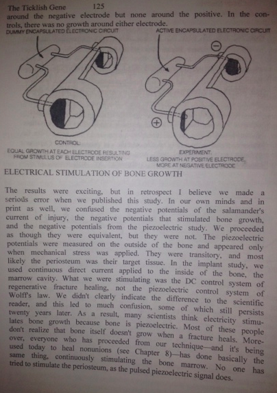
Currently I am reading up on the effects of emitting electrical current to bones to see how they react. I am following VERY closely to the research the late Dr. Robert O Becker did back in the 80s and 90s.

His research on the piezoelectric properties of bone has a connection to the research that Dr. Brighton did. They even collaborated together on a few studies or patents together. The Book that made him famous is The Body Electric.

For further reading on his research you can read the study Electrical Stimulation of Partial Limb Regeneration in Mammals.

This area of research is where I think a lot of my time will be spent on for at least the next 3-4 months, while still editing the main article for the guide on Limb Lengthening Surgery.

I will be writing a post on a patent called “Methods for modulating chondrocyte proliferation using pulsing electric fields (Patent # – US 20080039901 A1)” which was created by James Kronberg. Kronberg is famous for writing the seminal scientific paper “A pulsing electric field (PEF) increases human chondrocyte proliferation through a transduction pathway involving nitric oxide signaling.“. This is a very clear evidence that using a type of PEMF Device would indeed cause the longitudinal growth rate of long bones to be increased, at least if the electrical stimuli is used in either a pulsing fashion or alternating current (AC) fashion in an intermittent way. I had just shown in a recent post that using a function generator or pulse generator would indeed work in stimulating bone for increased longitudinal growth. “AC Electrical Signals Differentiates MSCS Into Bone and Cartilage Tissue Versus Adipose (Breakthrough)

He had also created the patents “Compact biomedical pulsed signal generator for bone tissue stimulation” – James W. Kronberg (5217009)

We did find more evidence of his patent and proposals in the patent Digital electronic bone growth stimulator (Patent #: US05413596)” . This one was filed in 1993 and issued in 1995. 

From the Patent: A device for stimulating bone tissue by applying a low level alternating current signal directly to the patient’s skin. A crystal oscillator, a binary divider chain and digital logic gates are used to generate the desired waveforms that reproduce the natural electrical characteristics found in bone tissue needed for stimulating bone growth and treating osteoporosis. The device, powered by a battery, contains a switch allowing selection of the correct waveform for bone growth stimulation or osteoporosis treatment so that, when attached to the skin of the patient using standard skin contact electrodes, the correct signal is communicated to the underlying bone structures.

Note: There is a researcher from Harvard Medical School with a similar last name, Kronenberg, (Henry M Kronenberg) . who wrote the article “Developmental regulation of the growth plate.” and “How PTHrP controls growth plate chondrocytes” but they are two different people.

Overall Summary: This is a sort of wrap-up on the different areas of research I am currently up to. The focus is on seeing just how would sending a certain type of AC or Pulsing Electrical Current of a certain frequency, pulsing rate, and amplitude would have the most benefit.

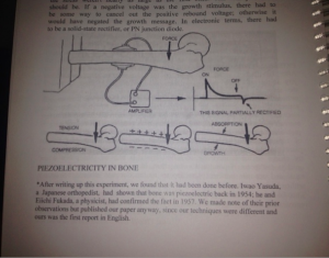
Electrical Stimulation of Bone GrowthYou can see to the picture to the right that there is actually a huge part of the book The Body Electric dedicated to figuring out what are the effects on bone remodeling due to electrical stimuli. Becker along with Charlie Bachman had postulated that bone matrix was a biphasic semiconductor. They were not sure whether the Collagen or the Apatite was the N-part of a semiconductor. The connection between the apatite and the collagen would be where the P and the N join together to create a P-N Junction.

It seems that back then, no medical researchers were researching what were the effects of electrical stimuli on the periosteum. Becker was one of the few who did and his way was to stimulate the periosteum, which is the only part of the bone which still had progenitor cells before they differentiated into the osteogenic lineage. Those cells would be the cambium cells in the inner layer of the periosteum.

What about my own height increase?

As for my own personal height increase journey, I have increased my dosage of Glucosamine Sulfate to around 2000 mg per day.

I am also trying to add the supplements of Vitamin K2 (aka menaquinone) and Vitamin K3 to the formulation. We did find evidence that consumption of Vitamin K2 seemed to help older women increase height and did a post about it.

While I have not gotten a chance to measure myself and see if my height has increased, something that has happened is that I also don’t feel any type of lower back pain anymore. Just a month ago before I started to take the Glucosamine Sulfate consistently my lower back was in a sort of chronic pain.

AC Electrical Signals Differentiates MSCS Into Bone and Cartilage Tissue Versus Adipose (Breakthrough)

AC Electrical Signals Differentiates MSCS Into Bone and Cartilage Tissue Versus Adipose (Breakthrough)

These days I don’t get to do too much research on this website anymore. My duties are now more towards making money and proving a stable living for my future wife (yes, I am supposed to be getting married in the Summer of next year if everything goes to plan). I had once said that the main reason people give up on this endeavor is if they get have children. Life takes over and this dream of figuring out how to become taller takes a sort of backseat. I am not sure how this website and the research will continue once I do get married. I don’t plan to have kids anytime soon so maybe I still can push through a little more research.

I did want to give you guys a little bit of good news though, which is from a patent I found. It is entitled “Alternating electric current directs, enhances, and accelerates mesenchymal stem cell differentiation into osteoblasts and chondrocytes but not adipocytes – Patent #: WO 2013049598 A1″. I actually found the patent when I was trying to find which patents from Google referenced Dr. Robert O. Becker’s research that was done 20-30 years ago.

The 2nd reference: HRONIK-TUPAJ MARIE ET AL: “Osteoblastic differentiation and stress response of human mesenchymal stem cells exposed to alternating current electric fields“, BIOMEDICAL ENGINEERING ONLINE, BIOMED CENTRAL LTD, LONDON, GB, vol. 10, no. 1, 26 January 2011 (2011-01-26), page 9, XP021088361, ISSN: 1475-925X, DOI: 10.1186/1475-925X-10-9

The patent just got published in the last few months from what I would guess are a bunch of professors, post-docs, and graduate students from the University of Texas System.

The quick abstract – A method for directing, enhancing, and accelerating mesenchymal stem cell functions using alternating electric current. Mesenchymal stem cells are preferentially directed to either osteoblast or chondrocyte lineages, but not to the adipocyte lineage, when exposed to alternating electric current.

So why is this patent such a breakthrough?

I haven’t taken the time to dissect the patent but this is what I do know. The bone marrow of humans change as they get older. When the human is young, their bone marrow is more of a red color, which signifies healthy hematopeietic cells. I think I will let Wikipedia explain it a little better than me…

From the Wikipedia article on Bone Marrow – The two types of bone marrow are medulla ossium rubra (red marrow), which consists mainly of hematopoietic tissue, and medulla ossium flava (yellow marrow), which is mainly made up of fat cells. Red blood cells, platelets, and most white blood cells arise in red marrow…. At birth, all bone marrow is red. With age, more and more of it is converted to the yellow type; only around half of adult bone marrow is red. Red marrow is found mainly in the flat bones… and in the cancellous (“spongy”) material at the epiphyseal ends of long bones such as the femur and humerus. Yellow marrow is found in the medullary cavity, the hollow interior of the middle portion of long bones.

Anyone who has ever done research extensively on adult height increase would know that it is critical that one can get the MSCS that are still inside the epiphysis outer cortical bone tissue to turn into the right type of tissue. Most of us are already adults, so the MSCS that are left in the long bone ends/epiphysis, in the marrow, already are surrounded by yellow type bone marrow , which is just fat tissue or adipocyte.

I had reference Dr. Carl Brighton multiple times in this website, and his patent about using Capacitative Direct Current Electrical Signals to stimulate Epiphyseal Plate Increase growth was the find that sparked this idea inside of my head that maybe electrical stimuli is one of the major keys.

However, my engineering background from taking multiple Electrical Engineering courses and the Electrodynamics course from Junior level undergraduate physics programs made me always felt that Alternating Current (AC) was probably much better than DC (Direct Current) Emission.

I had found from other patents that Dr. Brighton had to his name published in the late 1980s showed that he kept on talking about these potential ways to up-regulate the expression of a lot of genes who go on to make proteins that are found in the extracellular matrix of hyaline cartilage. All of these involved setting some type of electrical emitting device to a specific frequency,

Examples:

  • Up-regulation of bone morphogenetic protein (bmp) gene expression in bone cells by electromagnetic signals – WO 2005070136 A3
  • Regulation of matrix metalloproteinase gene expression using specific and selective electrical and electromagnetic signals – WO 2004112708 A3
  • Resolution of aggrecan gene expression using specific and selective electrical and electromagnetic signals – WO 2004029210 A3
  • Regulation of type ii collagen gene expression using specific and selective electrical and electromagnetic signals – WO 2004033644 A3

From our 4th example, these are the exact figures on what he claims are the set points for the numerical values…

  • …the generating step comprises the step of generating a specific and selective electric field having an amplitude of approximately 20 mV/cm, a sine wave configuration, a duty cycle of approximately 1/12, and a frequency of approximately 60 kHz.
  • …the step of applying the specific and selective electric field to the cartilage tissue for a duration of approximately 30 minutes every 24 hours.
  • …the generating step comprises the steps of selectively varying the amplitude, duration, duty cycle, frequency, and waveform of the specific and selective signal until the up-regulation of the gene expression of type II collagen mRNA in the cartilage tissue by the generated field is substantially optimized.

This shows that Brighton went away from the idea of applying DC Currents, and moved on to AC Currents. If he was not using AC Current, then he was using pulsing electrical currents. This was the other proposed idea which I had predicted in one of my last breakthrough posts “Using An Electrical Pulse Massager Physiotherapy Device To Increase Longitudinal Growth In Vivo For Open Growth Plates (Big Breakthrough)

So either one of two things to control gene expression of specific cartilage protein stimulation…

  1. He is using AC Current Signals
  2. He is using pulsing electrical signals.

There is a difference between the function shape of the two types of electrical signals.

The Implication

I think we have found very good evidence that instead of using a DC signal to stimulate cartilage regrowth, we should be trying either Pulsing Electrical Signals or AC Electrical Signals. That is what is going to keep the few Mesenchymal stems cells still left in the bone marrow of the medullary cavity which already consists of half of adipocyte/fat tissue cells to differentiate specifically into cartilage and bone tissue.

Of course now there are two questions that develops.

  • Is it better to be emitting AC Electrical signals or Pulsing Electrical signals?
  • Which values for the following parameters (1. amplitude, 2. duration, 3. duty cycle, 4. frequency, and 5. waveform of the specific and selective signal) should we have to set the device (whether function generator, pulse generator, TENS Unit, Pulse Massager, etc. ) to to get the MSCS to differentiate specifically towards the chondrogenic line?

ERR-lambda

Cartilage-Specific Overexpression of ERRγ Results in Chondrodysplasia and Reduced Chondrocyte Proliferation.

“To assess the effect of increased ERRγ activity on cartilage development in vivo, we generated two transgenic (Tg) lines overexpressing ERRγ2 via a chondrocyte-specific promoter; the two lines exhibited ∼3 and ∼5 fold increased ERRγ2 protein expression respectively in E14.5 Tg versus wild type (WT) limbs. On postnatal day seven (P7), we observed a 4-10% reduction in the size of the craniofacial, axial and appendicular skeletons in Tg versus WT mice. The reduction in bone length was already present at birth and did not appear to involve bones that are derived via intramembranous bone formation as the bones of the calvaria, clavicle, and the mandible developed normally. Histological analysis of P7 growth plates revealed a reduction in the length of the Tg versus WT growth plate, the majority of which was attributable to a reduced proliferative zone. The reduced proliferative zone paralleled a decrease in the number of Ki67-positive proliferating cells, with no significant change in apoptosis, and was accompanied by large cell-free swaths of cartilage matrix, which extended through multiple zones of the growth plate. We identified known chondrogenesis-associated genes with at least one predicted ERR binding site in their proximal promoters, as well as cell cycle regulators known to be regulated by ERRγ. Of the genes identified, Col2al, Agg, Pth1r, and Cdkn1b (p27) were significantly upregulated, suggesting that ERRγ2 negatively regulates chondrocyte proliferation and positively regulates matrix synthesis to coordinate growth plate height and organization. ”

” in a cartilage-specific ERα-deleted mouse, appendicular bones developed normally, but exposure to high levels of estrogen failed to reduce bone length as it did in wild type (WT) mice, indicating that ERα was required for the natural deceleration of bone growth that occurs in mice upon sexual maturity”

Ironically, ERR-lambda decreased FGFR3 expression despite reducing height.

“Overexpression of ERRγ2 in a cartilage-specific manner leads to dose-dependent abnormalities in the axial and appendicular skeletons due to alterations in Cdkn1b expression and chondrocyte proliferation as well as differentiation-maturation- matrix synthesis”<-This is despite that ERRy2 results in overexpression of many anabolic chondrogenic genes.  Thus, it only takes one misexpressed gene to ruin longitudinal bone growth.

thumbnail

IGF2 is very likely able to form new growth plates

height gene proteins

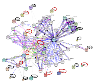
Click on the image for a better view.  IGF2 is a strong candidate for increasing height including adults giving it’s role for inspiring progenitor cells that form new growth plates.  The problem is how to increase it.

Target genes for height increase are as follows, for KO genes supplements or activities that inhibit these genes will increase height.  For OE genes supplements or activities that upregulate these genes will increase height.  For biphasic genes both overexpression and knockout decrease height.  There are many biphasic genes but they are not listed.

KO(inhibit these)(Black):
FANCC
NOG
NPR3
RNF135
SOCS2
STC2
GPC3
IGF2R
GPR30
POMC

OE(stimulate these)(Red):
NPPC
PLAG1
SHOX2
Twist1
CNP
IGF2
IGF1
Akt1
CTGF(CCN2)

Notice how close four genes are in proximity to IGF2: IGF2R, GPC3, POMC, GPER.  NPPC has a direct relationship with NPR3 with NPPC increasing height and NPR3 decreasing height.  IGF2 is a strong central locus for increasing height also being connected to IGF1 and Akt1.  LSJL increases Akt-phosphorylation by the way.

It turns out that the growth plate will regrow as long as their are progenitor cells, this area is called the zone of RanvierIGF2 could play a role in making these growth plate progenitor cells.

Since IGF2 is such a strong target for height growth how can we increase IGF2 levels for height?

Loss of imprinting of IGF2 and the epigenetic progenitor model of cancer.

“While IGF2 usually supports normal cellular growth, LOI of IGF2 may lead to overexpression of the gene and moreover global chromatin instability.”

“differentiated cells present in adult tissue that can acquire the ability to become undifferentiated and behave like stem cells, or progenitor cells”

” In contrast to mutations in the genetic sequence of DNA, epigenetic changes occur beyond the level of DNA and alter the protein-DNA complex that forms chromosomes. One such epigenetic factor is parental imprinting”

“Insulin-like growth factor 2 (IGF2), a gene whose end action is to stimulate general growth, is usually imprinted such that only the paternal allele is expressed. When LOI occurs, the maternal allele may also be expressed and some studies have, indeed, correlated LOI of IGF2 with increases in expression“<-So we can cause Loss of imprinting in adults to increase IGF2 expression in adults.

“The ICR for the IGF2/H19 locus is located in the 5’ flanking region of the H19 gene and 90kb downstream of IGF2. The ICR on the maternal allele is unmethylated, while the ICR on the paternal allele is methylated. This methylation of the paternal allele ICR blocks the transcription factor CCCTC-binding factor (CTCF) from binding and creating a physical barrier that stops downstream enhancers from augmenting IGF2 promoters. This effectively silences the maternal allele”

“Mouse models have shown that CTCF binds to regions near the IGF2 promoter, as well as the ICR, and subsequently forms CTCF-CTCF dimers, creating an intrachromosomal loop. The CTCF dimer then interacts with the SUZ12 (suppressor of zeste 12 homolog) domain of polycomb repressive complex 2 (PRC2) which methylates histone H3 lysine residue 27 (H3K27) causing silencing of the maternal allele.  CTCF [synthesizes] decoy CTCF proteins. When introduced to cells, the decoys bind to the unmethylated ICR and IGF2 promoter but do not interact with SUZ12, thereby rendering Enhancer of zeste homolog 2 (EZH2), another part of PRC2, unable to methylate histone H3K27, resulting in reactivation of the imprinted allele{so the decoy CTCF are key to causing loss of imprinting of IGF2}. Neither intact CTCF sites nor hypermethylation at the ICR is sufficient for maintaining paternal allele silencing, and sequences outside of the CTCF binding sites at the ICR are needed for silencing”

“Thus, LOI of IGF2 may result from a variety of causes including: aberrant ICR methylation, a decreased expression of PRC2, a mutation of the ICR, or altered PRC2 H3K27 methylation. In contrast to the maternal allele, the paternal ICR is methylated, thereby blocking CTCF and PRC2 binding”

“A two-fold increase in IGF2 expression results in a 131% increase in offspring growth. Circulating IGF2 ligand has been shown to regulate crosstalk between the WNT and IGF1R pathways, which can lead to activation of either the phosphoinositide 3-kinase (PI3K)-AKT or the Ras-MAPK (mitogen-activated protein kinase) pathways that control metabolism, growth, differentiation, and apoptosis ”

“IGF2 expression levels thirty fold higher than wild type were not sufficient to develop tumors until senescence.”<-Elevated IGF2 levels do not directly cause cancer.  Just more cells = more opportunities for malfunction=increased likelihood for cancer.

“LOI of IGF2 causes a bi-allelic expression of the gene, resulting in the overexpression of the IGF2 protein”

” a diet lacking synthetic methyl donors, including folic acid, vitamin B12, choline, and methionine, could cause LOI of IGF2 in murine models”<-Not really something you want to do however.

IGF2 injections were shown to induce height growth in an LSJL related study.

There are no known supplements that increase IGF2 in humans and I don’t know of any therapies involving injections.

Using CTCF decoy proteins may be a way though:

Interruption of intrachromosomal looping by CCCTC binding factor decoy proteins abrogates genomic imprinting of human insulin-like growth factor II.

“Monoallelic expression of IGF2 is regulated by CCCTC binding factor (CTCF) binding to the imprinting control region (ICR) on the maternal allele, with subsequent formation of an intrachromosomal loop to the promoter region. The N-terminal domain of CTCF interacts with SUZ12, part of the polycomb repressive complex-2 (PRC2), to silence the maternal allele. We synthesized decoy CTCF proteins, fusing the CTCF deoxyribonucleic acid-binding zinc finger domain to CpG methyltransferase Sss1{not an easy thing to do homemade} or to enhanced green fluorescent protein. In normal human fibroblasts and breast cancer MCF7 cell lines, the CTCF decoy proteins bound to the unmethylated ICR and to the IGF2 promoter region but did not interact with SUZ12. EZH2, another part of PRC2, was unable to methylate histone H3-K27 in the IGF2 promoter region, resulting in reactivation of the imprinted allele. The intrachromosomal loop between the maternal ICR and the IGF2 promoters was not observed when IGF2 imprinting was lost. CTCF epigenetically governs allelic gene expression of IGF2 by orchestrating chromatin loop structures involving PRC2.”

” A maternally transmitted microdeletion of two CTCF binding sites in the ICR results in biallelic IGF2 expression and H19 silencing in Beckwith-Wiedemann syndrome”<-A syndrome that results in increased height.

Here’s a paper that states that Paxillin could be involved in IGF2 related growth:

Paxillin-dependent regulation of IGF2 and H19 gene cluster expression.

“Paxillin (PXN) is a focal adhesion protein that has been implicated in signal transduction from the extracellular matrix. Recently, it has been shown to shuttle between the cytoplasm and the nucleus. When inside the nucleus, paxillin promotes cell proliferation. Here, we introduce paxillin as a transcriptional regulator of IGF2 and H19 genes. It does not affect the allelic expression of the two genes; rather, it regulates long-range chromosomal interactions between the IGF2 or H19 promoter and a shared distal enhancer on an active allele. Specifically, paxillin stimulates the interaction between the enhancer and the IGF2 promoter, thus activating IGF2 gene transcription, whereas it restrains the interaction between the enhancer and the H19 promoter, downregulating the H19 gene. We found that paxillin interacts with cohesin and the mediator complex, which have been shown to mediate long-range chromosomal looping. We propose that these interactions occur at the IGF2 and H19 gene cluster and are involved in the formation of loops between the IGF2 and H19 promoters and the enhancer, and thus the expression of the corresponding genes. These observations contribute to a mechanistic explanation of the role of paxillin in proliferation and fetal development.”

“focal adhesion proteins can be found not only at focal adhesion contacts but also inside the nucleus and they can shuttle out of it and back in”

” The block of CRM1-dependent export pathway causes accumulation of paxillin inside the nucleus ”

“reactivation of IGF2 expression on the maternal allele has been previously reported in a number of human tumors and tumor cell lines”<-controlled reactivation could help us grow taller.

“SMC1A and MED23 play a role in paxillin-dependent regulation of the H19–IGF2 gene cluster. ”

 An essential role for IGF2 in cartilage development and glucose metabolism during postnatal long bone growth.

“Postnatal bone growth involves a dramatic increase in length and girth. Intriguingly, this period of growth is independent of growth hormone and the underlying mechanism is poorly understood. Recently, an IGF2 mutation was identified in humans with early postnatal growth restriction. Here, we show that IGF2 is essential for longitudinal and appositional murine postnatal bone development, which involves proper timing of chondrocyte maturation and perichondrial cell differentiation and survival. Importantly, the Igf2 null mouse model does not represent a simple delay of growth but instead uncoordinated growth plate development. Furthermore, biochemical and two-photon imaging analyses identified elevated and imbalanced glucose metabolism in the Igf2 null mouse. Attenuation of glycolysis rescued the mutant phenotype of premature cartilage maturation, thereby indicating that IGF2 controls bone growth by regulating glucose metabolism in chondrocytes. This work links glucose metabolism with cartilage development and provides insight into the fundamental understanding of human growth abnormalities.”

“Newly formed chondrocytes are proliferative and morphologically round, but eventually become flat chondrocytes to form the ‘columnar zone’”

“the Igf2 null growth plate cartilage was shorter and disproportionally thinner than the WT.  The mutant growth plate was generally well formed, but its hypertrophic zone was disproportionally larger and the epiphyseal zone shorter. In addition, there was a clear delay in SOC formation in the mutant, which contributed to the shortened cartilage template ”

“IGF2 deficiency caused a shortening of the prehypertrophic zone ”

” Our prior study on human adult articular chondrocytes also failed to detect Akt activation by IGF2, suggesting that IGF2 may act differently than IGF1 in chondrocytes ”

“the regulatory role of IGF2 on chondrocyte maturation and matrix production in endochondral ossification is mediated by its activity on glucose metabolism in chondrocytes.”

“TheIgf1r null mouse has shorter bones, but the IR null mouse has a normal bone length.  However, both knockouts had a reduced hypertrophic zone which is consistent with the phenotype of the Igf1 knockout.  Igf2 null bones, on the other hand, exhibit a disproportionally larger hypertrophic zone. These data indicate different roles of IGF1 and IGF2 on cartilage development. Consistent with this notion, although IGF2 overexpression promoted postnatal growth, it failed to compensate the phenotype caused by the loss of IGF1 ”

“IGF2 is unique in its ability to bind to IGF2R, and it has been shown that the direct binding of IGF2 to IGF2R stimulated proteoglycan synthesis and induced calcium influx in chondrocytes, as it occurs even in the presence of an antiIGF-IR antibody.On the other hand, knockout of Igf2r exhibits increased skeletal growth{Maybe because this encourages IGF2 to bind to different receptors? Thus,none of the single knockout of the potential receptors exhibits the same phenotype as that of the Igf2 null mouse. However, it is possible that the phenotype of the Igf2 null mouse is a result of its binding to multiple receptors, together with interaction of multiple IGF-binding proteins and subsequent complex downstream signaling”

Phd2

Inhibiting Phd2 may be a potential drug target to help people grow taller.

Conditional Deletion of Prolyl Hydroxylase Domain-containing Protein 2 (Phd2) Gene Reveals its Essential Role in Chondrocyte Function and Endochondral Bone Formation.

“The hypoxic growth plate cartilage requires hypoxia-inducible factors (HIFs)-mediated pathways to maintain chondrocyte survival and differentiation. HIF proteins are tightly regulated by prolyl hydroxylase domain-containing protein 2 (Phd2) mediated proteosomal degradation. We conditionally disrupted the Phd2 gene in chondrocytes by crossing Phd2 floxed mice with Col2α1-Cre transgenic mice, and found massive increases (>50%) in the trabecular bone mass of long bones and lumbar vertebra of the Phd2 conditional knockout (cKO) mice caused by significant increases in trabecular number and thickness and reductions in trabecular separation. Cortical thickness and tissue mineral density at the femoral mid-diaphysis of the cKO mice were also significantly increased. Dynamic histomorphometric analyses revealed increased longitudinal length and osteoid surface per bone surface (OS/BS) in the primary spongiosa of the cKO mice, suggesting elevated conversion rate from hypertrophic chondrocytes to mineralized bone matrix as well as increased bone formation in the primary spongiosa. In the secondary spongiosa, bone formation measured by MS/BS and MAR were not changed but resorption was slightly reduced. Increases in the mRNA levels of Sox9, Osterix (Osx), Col2, Aggrecan, ALP, Bsp, VEGF, Epo, and glycolytic enzymes in the growth plate of cKO mice were detected by quantitative RT-PCR. Immunohistochemistry revealed an increased HIF-1α protein level in the hypertrophic chondrocytes of cKO mice. Infection of chondrocytes isolated from Phd2 floxed mice with adenoviral Cre resulted in similar gene expression patterns as observed in the cKO growth plate chondrocytes. Our findings indicate that Phd2 suppresses endochondral bone formation, in part, via HIF-dependent mechanisms in mice.”

“hypertrophic chondrocytes can transdifferentiate into osteoblasts and contribute to trabecular, endosteal, and cortical bone formation”

“In chondrocytes, HIF-1 increases the expression of VEGF and promotes angiogenesis in the
surrounding perichondrium ”

Only deletion of Phd2 in chondrocytes seems to increase height.  Deletion of Phd2 in say osteoblasts seems to decrease height.  Also Phd2 cKo seems to result in shorter overall body length. It’s just the primary spongosia that’s increased in length.