Grant with potential implications on growth plate regeneration

Here’s the grant Cartilage progenitor cells for growth plate regeneration

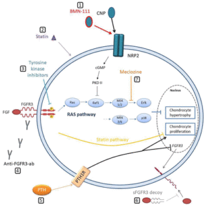
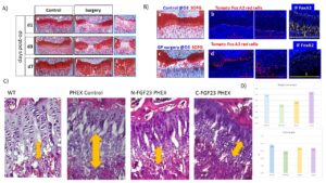
“Growth plate (GP) injuries result in growth arrest, formation of a “bony bar”{a technique that gets rid of the bony bar could potentially be used to make a new growth plate in adults} and angular limb deformities in children. Novel therapeutic approaches directed towards prevention of bone formation and growth arrest have to integrate cellular grafts, biomaterials and growth factors with the ultimate goal of recapitulating the complex zonal organization of the growth plate. One endogenous source of cartilage progenitor cells is thought to be the resting zone of the growth plate. Until now, the lack of specific marker(s) for the resting zone restricted the examination of this population. Currently, the evaluation of potential strategies for growth and cartilage disorders can mainly be achieved in vivo, therefore we proposed to use genetic modified mice to characterize the GP population and to characterize its development. Mice are the most appropriate model to use for several reasons: 1) GP is a tissue hardly approachable in vitro 2) we aim to characterize the GP population, and GP dynamics during the process of growing for which there is no optimal in vitro assays 3) we chose PHEX hemizygous mice for the study of GP dynamics because it is a well stablished model of X-linked hypophosphatemia (XLH){also called vitamin D resistant rickets}, which has being used for decades to study growth plate ossification. An other thing to keep in mind is that the standard treatments for XLH patients do not completely rescue the rickets and bone deformities. Serious side effects such as nephrocalcinosis and hyperparathyroidism have also been observed. Antagonizing FGF23 activity with antibodyes (Burosumab treatment){here’s some papers that indicate that FGF23 inhibition may potentially be part of a growth plate reactivation treatment.  FGF23 impacts bone mineralization so that may be why FGF23 inhibition could potentially help with growth plate restoration } is a recent and very promising therapy. However, this treatment requires at least a monthly infusion, is very costly and alleviates symptoms in many patients but not all. Hence, it is of the utmost necessity to identify more affordable strategies for therapy. Dr. Santos’ Laboratory demonstrated that inhibiting the MAPK pathway (FGF23 downstream pathway) in PHEX mice partially rescues growth impairment by normalizing the GP structure, specifically in the hypertropy zone. Nevertheless, it is not completely understood how FGF23 inhibition affects GP dynamics or how this is translated into a growth rescue and whether this treatment would be suitable for paediatric patients. Consequently, we will utilize the PHEX mice and FGF23 to 1) gain a better understanding of GP development and 2) look for alternative therapies to antagonize FGF23 activity.”

“FoxA2+ cells exhibit high clonogenicity and longevity. Moreover, FoxA2+ cell number expand in response to trauma and the data suggest that these cells participate in the production of hyaline cartilage, allowing for successful cartilage regeneration”

“Unlike other cartilage regions such as articular cartilage, GP has the ability to regenerate.”

Since Wild Type was the longest we don’t know it this treatment would have any impact in healthy developing individuals.

Some research with implications on longitudinal appositional growth(dinosaurs)

Endochondral ossification is not the only mechanism by which one can grow taller but also by appositional growth on the longitudinal ends whether by articular cartilage endochondral ossification or by direct bone deposition.  This study indicates that dinosaurs may have grown via non-endochondral ossification dependent mechanisms.  {Edit:  I emailed BM Rothschield and he said that all longitudinal bone growth identified occurred via longitudinal means but I think there’s a lot of interesting stuff here like that vascularization is key for growth plates.

An apparently phylogeny-independent method for identification of skeletal (longitudinal) growth cessation (skeletal maturity) in birds

“Identification of skeletal maturity is of interest as a measure of species longevity and for identifying its maximal achievable size/mass. Measurement of age on the basis of growth arrest/accentuation lines and external fundamental system evidences cessation or at least extreme slowing of circumferential bone growth{circumferential is appositional/periosteal growth which is mainly bone width but could potentially increase bone length albeit very slowly}. Such intramembranous (periosteal)-derived growth is distinct from the endochondral ossification responsible for longitudinal growth and therefore achievable organismal size/mass. As subchondral transcortical channels are required for nourishment, their loss should identify cessation of longitudinal growth{if we can restore subchondral transcortical channels then maybe we can restore longitudinal bone growth}. Predicated on phylogenetic bracketing/relationship and shared anatomical structures with and without growth plates, birds represent an appropriate model for the study of dinosaur ontogeny. Persistence of transcortical subchondral channels in the long bones of birds are examined at ×100–200 magnification and correlated with bone length. Transcortical channels are present in subchondral articular surfaces, but disappear when terminal longitudinal growth is achieved. Articular vascular channels perforating articular surfaces from within the bone are detected. Loss of penetrating channels is interpreted as evidence of skeletal growth cessation, identifying the longitudinal bone length at which skeletal growth cessation has been achieved. The current study provides evidence that maximal bone length does correlate with endochondral cessation growth. Failure of circumferential growth reduction/cessation to correlate with bone length may be related to lack of synchronicity of periosteal-based circumferential growth with the endochondral process responsible for bone lengthening{so the author states that circumferential growth can contribute bone length potential but it does not happen in practice}. Loss/closure of articular vascular channels may be the most reliable measure of a bird’s achievement of maximal growth (indicating cessation of appendicular element lengthening).”

“As trans-cortical channels through subchondral (that just below the articular cartilage) bone are the major source of nutrients for continued longitudinal growth, it is hypothesized that atrophy or loss of those articular vascular channels would preclude continued growth of bone – at least longitudinally.”<-this may be why mechanical loading could contribute to longitudinal bone growth by making this delivery more efficient by driving nutrients and potentially stimulating the channels to be open for longer

Even calcified cartilage (which persists in the long bones of some groups) retains trans-surface (articular) channels (Rothschild and Tanke, 2007) and represents the opportunity for continuing growth“<-BM Rothschild the author of this study has also studied dinosaur bones

” epiphyseal bone expands “into the cartilage anlage until the interface forms a calcific layer that arrests vascular invasion.” That shuts off the marrow, and the vascular supply thereby available, from the epiphyseal cartilage”<-if we can degrade that layer then we can restore longitudinal bone growth

““proximal end of humerus grows faster than distal, “because the internal pattern of spongiosum depends on the direction of bone pressure.””<-thus again that bone pressure can affect bone growth.

“Trans-articular surface canals (articular vascular channels perforating articular surfaces from within the bone marrow) are detected, independent of avian order or family, confirming loss of the blood supply necessary for longitudinal growth.”<-so we if can keep the blood supply than we can keep growing for longer.  Note distraction osteogenesis basically restores the blood supply via fracture

” “canal-like contacts between the articular cartilages and the medullary cavities of epiphyses” supports the interpretation that continued growth is dependent upon persistence of such channels. While the loss of articular vascular channels does not identify an individual’s actual age, it does allow identification of the longitudinal bone length at which skeletal growth cessation has been achieved.”

a linear relationship between LAG/EFS{growth arrest lines} and bone length is not established in dinosaurs. However, there is a confounding issue: Circumferential growth is the result of intramembraneous (periosteal) bone formation, while bone lengthening is the product of a very different process, endochondral bone formation”<-meaning dinosaurs were likely able to grow via mechanisms other than longitudinal bone growth.

” Longitudinal histologic section of bones provides evidence that growth had ceased, at least by documenting loss of trans-cortical channels. It is unclear that such a histological approach would provide significant additional information (on longitudinal growth cessation) to that observed by microscopic examination of joint surfaces. At this time, loss/closure of articular vascular channels may be the most reliable measure of a bird’s achievement of maximal growth (indicating cessation of appendicular element lengthening)”

Here’s more on dinosaurs also by BM Rothschild

Identification of growth cessation in dinosaurs based on microscopy of long bone articular surfaces: preliminary results

“As applied to bone, ‘determinate growth’ identifies an upper limit to size and the point when normal endochondral ossification ceases. This contrasts with ‘indeterminate growth’, which proceeds through the entire life of the animal. In this study, a non-destructive method, epi-illumination surface microscopy of the articular surfaces of long bones, is applied for the first time in 40 taxa of non-avian dinosaurs to determine cessation of endochondral growth. Thereby, the presence or absence of articular vascular channels between the endochondral bone and the cartilage is assessed. As articular vascular channels are the major source of nutrients for continued longitudinal growth, atrophy or loss of those channels would preclude continued growth of bone. We correlated our findings with published histological data and bone length measurements. We found articular vascular channels in all assessed dinosaur groups, but some individuals showed a loss of detectable articular vascular channels – what we interpret as evidence of longitudinal skeletal growth cessation. This observation contrasts with the hypothesis of continuous indeterminate growth in dinosaurs, at least for the taxa identified here, in which channels have been documented as closed or closing over{so dinosaurs must have grown via mechanisms other than endochondral ossification}. The new method introduced here provides a phylogenetic tool for definitively distinguishing new ‘dwarf’ species from juveniles of known species. Furthermore, this study confirms the rarity of skeletally mature dinosaurs discovered to date and indicates that we have only begun to witness the full extent of dinosaur growth.”

“Growth measured by LAGs and by EFS is intramembranous in derivation. It is a very different process from the endochondral ossification (in which bone replaces cartilage), which characterizes longitudinal bone growth”

“trans-cortical channels through subchondral (i.e., just below the articular cartilage) bone are the major source of nutrients for continued longitudinal growth, it is hypothesized that atrophy or loss of those articular vascular channels would preclude continued growth of bone—at least longitudinally. Even calcified cartilage (which persists in the long bones of some dinosaur groups) retains trans-surface (articular) channels and represents the opportunity for continuing growth{!}.

“epiphyseal bone expands ‘into the cartilage anlage until the interface forms a calcific layer that arrests vascular invasion’. This shuts off the marrow, and the vascular supply thereby available, from the epiphyseal cartilage”

” in [some] taxa (e.g., Plateosaurus) the bone length is not correlated with ontogeny as measured by slowing/cessation of intramembraneously based circumferential growth”

“failure to identify growth cessation in Tyrannosaurus rex.”

Does this study disprove LSJL and growth plate loading? No

Here’s a study that shows that growth plate loading can stunt growth but it also shows that it can enhance growth in one vertebrae which emphasizes the importance of developing the proper loading method.

In vivo dynamic loading reduces bone growth without histomorphometric changes of the growth plate

“This in vivo study aimed at investigating the effects of dynamic compression on the growth plate. Rats (28 days old) were divided into three dynamically loaded groups, compared with two groups (control, sham). A device was implanted on the 6th and 8th caudal vertebrae for 15 days{but note not on the 9th vertebrae which grows as we’ll see later}. Controls (n = 4) did not undergo surgery. Shams (n = 4) were operated but not loaded. Dynamic groups had sinusoidal compression with a mean value of 0.2 MPa: 1.0 Hz and ±0.06 MPa (group a, n = 4); 0.1 Hz and ±0.2 MPa (group b, n = 4); 1.0 Hz and ±0.14 MPa (group c, n = 3). Growth rates (µm/day) of dynamic groups (a) and (b) were lower than shams (p < 0.01). Growth plate heights, hypertrophic cell heights and proliferative cell counts per column did not change in dynamic (a) and (b) groups compared with shams (p > 0.01). Rats from dynamic group (c) had repeated inflammations damaging tissues{this group had the highest frequency of loading}; consequently, their analysis was unachievable. Increasing magnitude or frequency leads to growth reduction without histomorphometric changes. However, the combined augmentation of magnitude and frequency alter drastically growth plate integrity. Appropriate loading parameters could be leveraged for developing novel growth modulation implants to treat skeletal deformities{the authors themselves even allude to with this sentence that this does not disprove growth modulation but that it only suggests that it’s important to develop the appropriate growth modulation method}.”

“With the approval of the Institutional Animal Care Committee, 19 male Sprague–Dawley rats were received at the age of 21 days old. After 1-week of acclimatization, the protocol was conducted from 28 to 43 days old, corresponding to the rat pubertal growth spurt”

“The stress variation was chosen at a mean value of 0.2 MPa as it represents a wide physiological stress interval that is still retarding but not arresting bone growth (stress value over 0.6 MPa). The frequencies were either lower (0.1 Hz) or close to an average walking frequency (1.0 Hz) for humans. ”

“The loading device was adjusted daily at the same time of day for 10 min under isoflurane to compensate for longitudinal growth.”

“three caudal vertebrae (one loaded Cd7; two unloaded (Cd5, Cd9) used as intra-animal controls)”

Note how the CD9 vertebrae grew significantly faster than controls.  Also interestingly the sham group for both CD7 and CD9 vertebrae grew faster than controls.  “Shams (n = 4) were operated but no compression was applied.” <-so the operation itself could be stimulatory on the growth

“The ultimate dynamic (c) condition, combining an increase in both maximum magnitude and frequency, was tested only on three animals since severe inflammation occurred.”

CD9 has a significant increase in all growth plate parameters according to these figures.

Clearly the loading regime on CD7 had spillover reduction in growth in CD5 and stimulatory effect on growth in CD9.  It’s hard to say why that is without looking at say a stress strain analysis.  The device could be doing indirect tensile strain on CD9 and compressive strain on CD5.   But this study shows the importance of finding the right loading method in order to manipulate longitudinal bone growth.

Michael former admin of Natural Height Growth on Cyborg 4 Life’s Podcast

I think Micahel is too skeptical of non-surgical methods.  Weightlifting for example is far more effective than any surgical method to increase muscle(that I know) and CRIPSR technology to inhibit myostatin.  And weightlifting has life extension benefits over potential surgical technology.  As catabolism(fasting, temperature exposure, exercise) all increase life via autophogy and surgery once in a while cannot currently do that.  A regular way of stimulating longitudinal bone growth will have an exercise benefit over surgery and could lead to life extension.  Any form of marrow stimulation will potential have strong health benefits.

Also, I think Michael is too negative in regards to potential surgical/advanced methods of height increase.  Scientific progress is very inefficient.  I don’t think there’s anyone who’s saying that the way science is conducted is optimal.  It’s ultimately capitalism rewards people for doing the least amount of work for the most money.  And science has tons of failures which makes it more profitable to go into something with more guaranteed success like computers.  If science were done optimally then I think it would be more fair to criticize the snail like advancement in a lot scientific fields.

Will this finger stretcher work to increase hand size?

Finger stretcher

We know that the jaw bone can grow throughout adulthood via endochondral ossification of the articular cartilage. This may be why we see people with acromegaly have very large jaws. The reason that the jaw bone continues to grow at the articulations and the other bones don’t is because the jaw bone is less constrained by ligaments and is very moveable so the stress that the articular cartilage undergoes is enough to induce articular cartilage endochondral ossification and also the lateral pterygoid muscle is attached to the articular cartilage directly so if you stimulate the lateral pterygoid muscle in any way either via forced mouth opening or a bite jumping appliance you get the pull with if it’s sufficient results in articular cartilage endochondral ossification. You can literally see this in the histological slides in the above link. The problem is that this likely occurs very slowly unless cell turnover is highly accelerated like via HGH which occurs in acromegaly.

So there are two problems why we don’t see more development of the jaw in normal individuals:

1) The lateral pterygoid muscle isn’t stimulated enough because they don’t chew strong enough food or they don’t practice “mewing“(which does stimulate the lateral pterygoid muscle by the way and it’s slightly dynamic as while you are moving around your muscles have to adjust to keep your tongue to the roof of the mouth). And it’s possible that normal strength training exercise will also stimulate lateral pterygoid development. An exercise like the deadlift pulls on every muscle in the body but that pull won’t be as strong as if you train the lateral pterygoid muscle directly.

2) articular cartilage endochondral ossification is very slow unless again it’s accelerated by HGH.

One problem with the device is that you can’t really do anything else while using it. You might as well just pull on your fingers directly. The other problem I see is that the device doesn’t really look like it would stretch the articular cartilage directly. It looks like it would mostly stretch the skin.

We’d have to really look at the pull to see if it was pulling directly at the articular cartilage. The best bet in my mind to provide a sufficient stretch on the articular cartilage would be a twisting or bending motion of the joint. This would get around the issue of the ligaments limiting the potential stretch. The issue would be potential cartilage damage from improper joint movement. Also based on joint mechanics it may still not stretch the articular cartilage in a sufficient manner.

BREAKTHROUGH! Sea cucumber may boost longitudinal bone growth


Sea cucumber is available for sale but it is very expensive.  I found some sea cucumber tablets that were cheap however it’s not for certain that the tablets have all the benefits of eating sea cucumbers directly.

Novel Peptides from Sea Cucumber Intestine Hydrolysates Promotes Longitudinal Bone Growth in Adolescence Mice through Accelerating cell cycle progress by Regulating Glutamine Metabolism

“Sea cucumber intestines are recognized as a major by-product in sea cucumber processing industry and have been shown to have antioxidant properties. However, whether the sea cucumber intestine is beneficial to osteogenic remains unknown. In this study, low molecular weight rich in glutamate/glutamine peptides were obtained from sea cucumber intestines (SCIP) by enzymatic hydrolysis, and orally administered to adolescent mice to investigate the effects on longitudinal bone growth. The results showed that the SCIP supplement significantly increased the femur length and new bone formation rate by 9.6% and 56.3%, and elevated the serum levels of osteogenic markers ALP, Collagen I and OCN{this is pretty nuts.  I couldn’t find studies directly measuring what effect glutamine has on longitudinal bone growth directly}. Notably, H&E staining showed that SCIP significantly increased the height of the growth plate, in which the height of the proliferation zone was elevated by 97.4%. Glutamine is a major determinant of bone growth. We found that the SCIP supplement significantly increased glutamine levels in the growth plate by 44.2% and elevated the expression of glutamine metabolism-related enzymes Gls1 and GLUD1 in the growth plate{so it’s possible that sea cucumbers could have a benefit on glutamine in the growth plate due to some other ingredient in the cucumber other than the glutamine itself}. Further, SCIP upregulated growth plate acetyl coenzyme A levels to promote histone acetylation and accelerated cell cycle progression by upregulating Sox9 expression, thereby contributing to the rapid chondrocyte proliferation. To our best knowledge, this is the first report that SCIP could enhance longitudinal bone growth via promoting growth plate chondrocyte proliferation. Our present study will provide new ideas and a theoretical basis for high-value utilization of sea cucumber intestine.”

“the peptide from Euphausia superba promoted longitudinal bone growth in adolescent mice.  supplement with enzymatic hydrolysate of Stichopus japonicus could promote bone formation in vivo and in vitro.”

“the avascular nature of the growth plate makes glutamine a potential factor for regulating longitudinal bone growth”

“the SCIP supplement significantly increased femur length by 6.8% (SCIP-L{LOW DOSE}) and 9.6% (SCIP-H{High dose}) compared with the normal group”<-so we don’t know if even more is better than the doses mentioned.  The effect of growth plate height, proliferative height, and hypertrophic chondrocytes was also greater based on dose.

“the serum glutamine levels were gradually increased to a maximum between 0.25 h and 0.5 h and returned to baseline after 2 h. Conversely, as serum glutamine levels fell back, glutamine levels in muscle and femur peaked sequentially at 2 h and 3 h.”<-theoretically it could be worthwhile to take glutamine every 2 hours which would be very difficult to do.

“glutamine metabolism has been shown to upregulate chondrocyte proliferation genes (especially Sox9) by epigenetic modification”<-obviously epigenetic modification is very powerful.

“glutamine promoted the growth plate chondrocyte proliferation during endochondral ossification through acetylation modification of the histone 3K9”

So they did have a glutamine only group.  And the sea cucumber growth increased growth plate height further than that.  Also if you look at the growth plates, it’s a pretty dramatic difference.

Based on the information in this paper, it may be worthwhile for everyone growing to take a low cost sea cucumber supplement(i’d say actual sea cucumber is too expensive).  Not only for the growth plate but also joint cartilage.

This study Stichopus chloronotus aqueous extract as a chondroprotective agent for human chondrocytes isolated from osteoarthitis articular cartilage in vitro finds that sea cucumber has anabolic effects on articular cartilage.