Studying Osteogenesis By Chondrocytes From Growth Cartilage

Update 2/12/2013: It would seem that I no longer can access through the link below to the full text/article without paying for it. This means that my ability to do research and learn more has been severely limited. Only a quick study from the abstract and introduction seems to be possible at this point.

From a paper/article of a study from SpringerLink “Osteogenesis by chondrocytes from growth cartilage of rat rib“.For the Full Article click HERE

Y. Shimomura, T. Yoneda,  F. Suzuki Ph.D. – Calcified Tissue Research – 1975/76, Volume 19, Issue 1, pp 179-187
Abstract

Chondrocytes were isolated from growth cartilage and resting cartilage of rat rib and cultivated in vitro. The cultivated chondrocytes were placed in Millipore diffusion chambers, which were then implanted into the abdominal cavities of rats for several weeks and prepared for histological analysis. The results indicate that growth cartilage cells have a remarkable osteogenic potential, even after cultivation in vitro, whereas resting cartilage cells show no osteogenic activity. However, growth cartilage cells alone do not form new bone but require the participation of certain host cells to initiate osteogenic differentiation.

Introduction

The process of replacing cartilage by bone is known as endochondral ossification. During the first stages of conversion into bone, epiphyseal cartilage cells
increase their rate of proliferation, enlarge, and become hypertrophic. It has been assumed in the past that the hypertrophic cells undergo degenerative changes and death [1, 2, 3, 8, 17, 22]. However, Holtrop, on the basis of experimental transplantation of growth cartilage labeled with 3H-thymidine [10, 11, 12], suggested that some of the cells survive and redifferentiate in the metaphysis to contribute to ossification. Shimomura and Ray have confirmed her findings that hypertrophic chondrocytes can survive and transform into bone-forming cells [19 ]. Recent investigations using electron microscopy have led to opposing interpre-
tations [3, 13, 14, 20], and controversy persists concerning the mechanism of endochondral ossification.

In the present study, hondrocytes isolated from growth cartilage of rat rib were cultivated in vitro and transplanted to investigate the mechanism of endo-
chondral ossification.

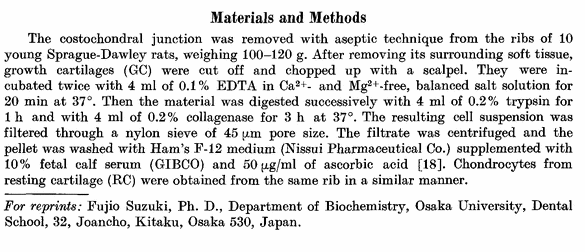
Screen Shot 2013-02-12 at 2.49.26 AM

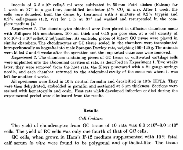
Screen Shot 2013-02-12 at 2.49.40 AMAnalysis & Interpretation:

The reason why this article/study/paper is important is to show why it is more important to focus on the cartilage of the plate, then the bone area. We have sen in the past ideas like using Low Intensity Pulses Ultrasound (LIPUS) to possibly increase height, but from my own limited research so far it seems that any method or technique to increase the rate of bone growth or bone density will not do anything for our intention to grow taller ie. increase the length of the long bone in our body. Remember that commercial pills and supplements that are purported to be able to increase bone growth are true, but they don’t increase bone in the way we want. The bone growth happens inside the bone, often making the medullary cavity thinner and smaller, making the cortical bone layer thicker. Bone growth pills are sold as a way to treat or prevent bone degeneration in older people who might get osteoporosis.

LIPUS has been shown through maybe 1-2 PubMed studies to increase the rate of bone healing in distraction osteogenesis surgeries, but without the initial distraction, bone growth will not lengthen bone, at least within physically mature humans with no epiphyseal cartilage to widen and lengthen.

From the abstract, we learn that overall, the epiphyseal cartilage derived chondrocytes are special, diffierent from what we would find in other cartilage explants, like from the resting cartilage. When grown in the lab (in vitro) we find that the cartilage cells themselves already have bone creating properties.

It seems epiphyseal cartilage will always ultimately turn into bone in some way, whether through calcification, vascularization, etc. This suggest that when we are looking for a method to legnthen bone, we don’t really have to worry about the process of osteogenesis, but only have to worry about chondrogenesis. If we get the chondrocyte and cartilage formation correct from using growth plate derived cells, the osteogenesis and bone formation seems to automatically occur given time, and the right nutrients getting in, when the cultured cartilage or chondrocytes are finally implanted back into the subject.

Another thing that might be important to take away from the introduction is that maybe not all the hypertrophic chondrocytes in the hypertrophic zone of the growth plate completely die out through degeneration. Some researchers have suggested that the hypertrophic chondorcyte can actually differentiate even further going down deep into the calcification/ossification layer and turn into osteocytes/osteoblasts which start forming bone cells and bone elements.

So the big take-way from this article is to focus less on bone creation, osteogenesis, but focus more attention on cartilage formation, chondrogenesis.


Studying Changes In The Growth Plate By Restricting The Blood Vessel Supply, Ischaemia

Ever since I came across the study “SURGICAL STIMULATION OF BONE GROWTH BY A NEW PROCEDURE – PRELIMINARY REPORT” by ALBERT B. FERGUSON, M.D where it showed that it might be possible to induce increased longitudinal growth by severing the blood supply to the metaphysis, epiphysis, or the growth plate in growing children I have been curious over what other experiments have been done with a similar procedure and what were the results and effects seen in the growth plate. This study does that.

However I would guess that extensive study and reading of the study will only help contribute in adding to my personal understanding on how the blood vessels contribute to the growth plate in general. I have always believed that the more I know about the overall system, no matter how obscure or unrelated, it will only help contribute to the overall understanding of the system we are studying.

Note: The Actual PDF had many pictures which I have removed. For a more comprehensive view of the study, click the title link below.

Analysis & Interpretation:

The first thing to notice about this study is that unlike the other study being cited, the blood vessel being actually disrupted is going to the growth plate, not to the metaphysis. The old idea proposed fy Furgeson was that you can possibly stimulate higher than average levels of longitudinal growth by disrupting the inter-medullary blood vessels that contribute nutrients to the metaphysis of the tubular/long bone. This time the experimenters are disrupting the very blood vessels to the epiphysis, which would indicated that only decreased/stunted longitudinal growth is the likely outcome.

For the actual experimental part, the researchers took rabbits and drilled one small hole in the right medial side of the tibial epiphysis, then put a small spatula inside. The spatula is spinned around to cut & disrupt the blood vessles inside the epiphysis. The hole is drilled deep into the middle of the epiphysis. Then the hole is filled with some type of plastic to prevent re-vascularization. The lateral side of the right tibia was left as a control. The left tibia was treated similarly except for putting something on the other side for support.

Here is what the discussion has said about the blood vessels from the side, or the epiphyseal vessels. They are damn important. The proliferative chondrocytes need them to undergo further division. If any disruption of the vessels are done, lesions are developed which can cause breaks in the growth plate causing blood vessels to breakthrough causing vascularization, resulting in bone bridges, which would effectively stop all further longitudinal growth.

It would seem that there is at least two group sof blood vessels carrying nutrients to the growth plates, one coming from the side or epiphysis and the other coming from the middle, from the metaphysis. As for the source coming from the metaphysis the main roles and functions of it include carrying Vitamin D and Calcium as well as phosphates using the corpuscles which would cause calcification of the matrix, the removal of the cells that have disintegrated, and the use of laying down layers of bone material on the walls of the empty cavities in the matrix. The blood vessels from the metaphysis seem to hold no nutritional value to the hypertrophic chondrocytes but is what actually causes them to die. The level of alkaline phosphatase was also decreased in the experimental part where the blood vessels from the metaphysis was selectively removed. It would then suggest that only when you notice the hypertrophic chondrocytes releasing the compound alkaline phosphatase would the chondrocytes actually start to go through the process of dying through disintegration.

The researchers concluded that what will actually keep the hypertrophic chondrocyte alive and prevent them from dying is to prevent the process of calcification, not the presence of metaphyseal vascularization. When we compare the calcification rate of the matrix after vascularization in the normal way compared to say Vitamin D or A defficiency leading to Rickets, we can see that the whole process is very fast. The way that we should be remembering the growth plate ossification process is…

Calcification starts everything. The calcification leads to the blood vessels breaking in and getting the The hypertrophic chondrocytes starts releasing aklaline phosphatase signaling that they ready to die. They disintegrate, leaving the empty cavities to get coated with a layer of lamellae which will get filled then by osteoblasts which convert the pockets into real bone material.

The researchers end the study with this easy to take away lesson for the readers who wish to just take the basic idea away….

“The nutritional dependence of the proliferative cells on the epiphysial vessels has been established whereas the metaphysial vessels were seen to take part in calcification and ossification at the metaphysis.”

Interpretation & Implications:

This post has only helped reaffirm the idea that it may be possible to increase the lengthening of the bone in people with open growth plates through the disruption of the blood vessels that are reaching the growth plate from the metaphysis. Also, we definitely don’t want o damage the blood vessels that are supplying the blood and nutrients needed by the cells at the top of the growth plate process, around the reserve zone area.


From paper study “THE VASCULAR CONTRIBUTION TO OSTEOGENESIS, Changes in the Growth Cartilage Caused by Experimentally Induced Ischaemia

J. TRUETA, OXFORD, ENGLAND, and V. P. AMATO, SLIEMA, MALTA

From the Nuffield Orthopaedic Centre, Oxford.

In the two preceding papers of this series the vascular pattern close to the growth cartilage, and the relationship between vessels, cartilage cells and calcification, were studied under normal, or experimentally unaltered, conditions (Trueta and Morgan I 960, Trueta and Little 1960) and the findings were used as a control for the experimental work to be described in this and succeeding papers.

This study reports the changes in the epiphysial cartilage caused by the suppression of its blood supply. The changes caused by the interruption of the epiphysial blood flow will be described first and a description of the changes produced by interrupting the metaphysial circulation will follow.

METHOD

Forty six-weeks-old chinchilla rabbits, averaging 1,100 grammes in weight, were used. Operations were done on both tibiae under general anaesthesia (Nembutal and ether). In the right tibia a small hole was drilled on the medial side of the tibial epiphysis, avoiding so far as possible any direct damage to the growth plate. A small flat spatula was introduced through the drill hole until it reached approximately to the centre of the epiphysis when it was moved in a circular sweep in a plane parallel to the epiphysial cartilage. The cavity thus produced was packed with a strip of polythene film to prevent revascularisation. The lateral side of the epiphysis was left undisturbed to serve as a control. In the left tibia a similar technique was used but the block was placed on the metaphysial side of the growth cartilage and at a sufficient distance to prevent direct damage to it. All the forty rabbits were subjected to this procedure and out of the eighty tibiae operated upon seventy-eight were available for analysis : one tibia became infected and another fractured. The investigation proceeded in the following way: Group A-Twelve rabbits were killed at intervals of 1 , 2, 3, 4, 5, 6, 9, 12, 1 5, 18, 2 1 and 24 days after operation in order to study the changes in the epiphysial cartilage caused by ischaemia. Group B-A second operation was performed on all the remaining rabbits to remove the polythene sheath at intervals of 2, 4 and 8 days after the first operation. The rabbits were killed and injected at intervals of 2, 4, 6, 12, 16, 18, 21 and 24 days after the second operation in order to study the revascularisation that occurred.

RESULTS

INTERRUPTION OF THE EPIPHYSIAL BLOOD SUPPLY

The first changes occurred in the epiphysis itself. As early as the second day after operation many trabeculae were seen to be dead, with empty lacunae. By superimposing the stained histological preparations on the unstained sections it was seen that the dead trabeculae corresponded exactly to areas deprived of vascularity. These two changes-that is, the signs of bone death and the presence or absence of blood vessels-when seen in the terminal bone plate covering the growth cartilage, proved that the blood supply of the epiphysial plate had been cut off. When revascularisation was allowed to occur the evidence was still available, for the dead trabeculae could be seen enveloped by new bone (Fig. 1). The thick sections showed that the new blood supply in most instances was entering the epiphysis from the site of the operation. The newly established circulation was seen to correspond exactly with the areas where new bone was being laid down on the scaffolding of dead trabeculae in the terminal bone plate.

When the operation scar was placed far from the plate and the penetration was not deep enough into the central portion of the epiphysis there were only moderate changes attributable to interference with the circulation. The terminal bone plate appeared normal and all the vessels filled with dye. A transient widening of the growth plate was seen sometimes when it was compared with the undisturbed side of the plate. This widening was due both to a moderate increase in the number of cells of the hypertrophic layer and also to the increase in the size of the individual cells; apparently the proliferative layer remained unaltered although it is possible that accelerated division had taken place but, because of the great rapidity of this division, could not be detected, but could have produced the moderate increase in the number of hypertrophic cells. The widening of the growth cartilage was accompanied by a denser network of blood vessels in the region of the terminal bone plate and it was always more noticeable towards the periphery. The intercellular matrix took on a darker stain.

These effects were transient and could not be seen after a few days. Occasionally patchy damage was detectable in the plate. The thick sections showed this to correspond exactly to the area of ischaemia. The upper cells of the growth plate, referred to in a preceding paper of this series (Trueta and Little 1960) as the germinal cells, showed metachromasia; also a varying number of cells appeared dead, leaving empty V-shaped spaces consisting of intercellular matrix or matrix interspersed with dead or dying cells (Fig. 2). This minor degree of damage appeared capable of repair by a bulging inwards of the surrounding columns, so that the gap became closed off. The columns in such plates appeared fewer and farther apart, and the cytoplasm of many cells was granular (Fig. 3). The matrix was less well stained, appearing pale in colour.

In areas where the damage involved a larger number of columns the gap was too wide to become sealed off. Growth continued normally in the surrounding columns and resulted in a pulling out of the infarcted portion of the plate (Fig. 4). Long, empty, or partly empty tubes of cartilaginous matrix stretched from the epiphysis to the metaphysis, and as no further calcification took place this area was left behind in the metaphysis. If this stretching out process went too far, eventually a break occurred, usually at the metaphysial end, and blood vessels invaded the area (Fig. 5). New bone was laid down along these capillaries (Figs. 6 and 7) so that the end-result was usually a bone bridge between metaphysis and epiphysis. The shortest interval for the whole sequence of events to develop after this type of injury was found to be eight days from the operation. In some of the experiments in which the blood flow was allowed to return, the invasion of the blood vessels took place from the epiphysial to the metaphysial side, but the ultimate result-a bone bridge between the epiphysis and metaphysis-was the same.

In massive lesions, where the main source of supply to the whole epiphysis was successfully cut off, rapid death of the whole of the central part of the plate occurred. The cells became metachromatic, losing their polarisation and their morphological characteristics, and the matrix became very pale throughout (Figs. 8 and 9). The area that suffered least was a limited rim of the peripheral part of the cartilage which receives its blood supply from vascular anastomosis around the plate. The capillaries from the metaphysial side invaded the devitalised area on a wide front and new bone was very soon laid down in their wake. Epiphysiodesis occurred within a short while, usually ten days from the vascular interruption, and growth ceased altogether.

Direct injury to the terminal bone plate-In certain instances the epiphysial bone plate and the upper part of the growth plate, which is so intimately connected to it, were injured at the time of operation. Irreparable damage always resulted and an invasion of the blood vessels took place early, followed by partial epiphysiodesis (Figs. 10 and 1 1). As the medial side of the tibia was always used and as this type of injury was most likely to affect the peripheral end of the plate, a varus deformity developed.

Lesions of this area were often complicated by damage to the perichondral ring as will be mentioned later.

INTERRUPTION OF THE METAPHYSIAL BLOOD FLOW

After division of the main vessels at the metaphysial side of the growth plate this increases appreciably in width within twenty-four hours, entirely as a result of the accumulation of the cells of the hypertrophic zone. As stated by Trueta and Morgan (1960) the number of cells in the columns of the upper tibial epiphysis of the rabbit at six weeks varies from thirty-five to fifty, according to the mitotic activity of the proliferative part of the columns at the time the count is made. An average increase of approximately ten to sixteen cells occurs every twenty-four hours in the area of total suppression of the metaphysial blood flow. After six days the average width of the growth cartilage is increased by sixty to ninety hypertrophic cells (Figs. 12 and 13) and after eight days it may be as much as 100 cells or more above the normal average. After this extraordinary increase in width of the growth plate, the rate of expansion becomes limited and the metaphysial end of the columns of cartilage cells, now placed deep into the metaphysis, press against each other often at a depth of six or eight cells before the end. This permanency of the hypertrophic cells is accompanied by a total lack of calcification of the intercolumnar matrix. A close investigation of the morphology of these hypertrophic cells shows that along the whole length of the columns they do not exhibit any appreciable modification, so that individually the hypertrophic cells at the top of this section resemble those placed at its end, often fifty or more cells apart (Fig. 14). No degenerating cells are seen under these experimental conditions at the ends of the columns. The significance of this finding will be shown later. It is of particular interest to make a comparative study of the part of the growth cartilage placed over a metaphysial area of ischaemia and that which corresponds to a metaphysial area of normal blood flow. It is as ifall the activities so energetically engaged in the normal had been suspended, including calcification, preliminary ossification, bone reabsorption and final bone formation. Large zones of calcified cartilage and preliminary bone (Fig. 15) remain at a level in the metaphysis where, normally, remodelling is taking place (Fig. 16).

A somewhat surprising feature is the apparent survival of osteocytes in the metaphysial zone over the area of vascular suppression, suggesting that either the osteocytes, if they were dead. had no time to disintegrate or even change their stalnlng characterlstlcs or else that sufficlent blood flow still remained in the neighbourhood to provide the required transudates for their survival. Of these possibilities the latter seems the most likely, because in the injected specimens some of the per-fusing mass was scattered along vessels in the supposedly avascular area. The reason for this .l was the integrity of a number of perforating meta-physial vessels which, as explained elsewhere (Trueta and Morgan 1960) provide some of the blood, while the main blood flow from the nutrient artery is interrupted by the polythene film. In general, osteoclasts were absent and were seen only in reduced numbers in the proximity of the new, or the few spared, blood vessels. Another constant finding was the lack of alkaline phosFIG. 14 phatase concentration, characteristic of the

Detail of a “ tongue “ of epiphysial cartilage hypertrophic cartilage cells near the zone of showing the healthy hypertrophic cells with no empty columns at their sides, and the degenerating caIcli Icatlon. cells at the distal end of the columns. ( x 45.) Changes of the growth cartilage accompanying metaphysial vascular regeneration-Even without removal of the polythene film vascular proliferation from the perforating vessels invaded the ischaemic metaphysial area in less than twenty-four hours after the main blood flow had been blocked. Soon calcification and ossification proceeded along the now very elongated tubes surrounding the hypertrophic cells up to the normal level ofcalcification, with the usual eight or twelve hypertrophic cells left untouched, except that the last two or three were surrounded by calcified matrix, as in the normal (Fig. 17).

When the circulation was allowed to return freely by removal of the vascular block. the expanded growth plate calcified at a strikingly rapid rate from the periphery to the centre, each column starting at the farthest end in the metaphysis. The capillary loops entered at the bottom of the columns and in a few days travelled up-one loop to each column-to the level they would normally have reached had the vessels not been interrupted. New bone was laid down by the osteoblasts, appearing as a lining inside each tube. The trabeculae thus formed were thinner and more uniform than those of the control side and remodelling lagged behind. Alkaline phosphatase activity was increased and appeared to be related to the vascular progression. The last part of the growth cartilage to be calcified and ossified was always that in the centre, being the last to be reached by the incoming vessels. It often persisted as a long finger-like process of varying thickness and consisted of orderly columns of hypertrophied cells which on occasion numbered as many as 100 or more (Fig. 14). In thick sections a clear division between the old and the new blood vessels was seen, a division which exactly corresponded to the width of the finger-like process (Figs. 18 and 19). The vascular loops were always orientated in the direction of the columns, for, as has been stressed above, they only gained entrance to each column from its distal end. The widened growth cartilage did not tend, as a rule, to be broken up into islands of columns and cells but. on the contrary, calcification and ossification always proceeded very regularly. Occasionally. however, in the animals with the main metaphysial vessels obstructed for eight days or more an impenetrable barrier became established round the columns and their distal ends remained firmly closed. The vascular loops in these cases short-circuited the barrier by getting in between the long but normal-looking columns and those which appeared disorganised and unhealthy, and proceeded to lay down bone by the normal mechanism of vascular invasion of the hypertrophic cartilage.

In some instances if the central part of the epiphysial cartilage remained unossified for too long, permanent damage was caused by its tendency to get peeled off from the terminal bone plate, covering it at its epiphysial side. Thus a severe lesion of the germinal and proliferative cells was caused in breaking these away from their natural surroundings, particularly the epiphysial vessels ; they degenerated and soon were invaded by new epiphysial vessels which, by progressing towards the metaphysis, ended by establishing connections with the metaphysial vessels (Figs. 20 and 21). New bone was laid down and soon a central epiphysiodesis was established.

In the diaphysis the site of operation was always visible by its increased bone density; and its progress towards the shaft depended on whether or not a bony bridge had been established across the plate. Damage to the perichondral ring-The operation occasionally damaged the perichondral ring of the ossification groove (Lacroix 1951) and a large chondroma or osteochondroma resulted. It often spread down the metaphysis, and vessels from this region grew into it (Figs. 22 and 23). Normal moulding with organised growth did not take place and osteoclasts were absent from this region. There was a disorderly vascular invasion of the mass of cartilage cells, and bone was laid down as in the cartilage anlage before the growth cartilage is arranged in parallel columns ofcells (Fig. 24). The bone trabeculae were therefore irregular and the orderly laying down of bone characteristic of metaphysial bone formation was totally absent.

Bone bridging and partial epiphysiodesis-When a bone bridge became established between epiphysis and metaphysis it resulted in growth disturbances which followed a regular pattern according to the size and location of the bone bridge. Superimposition of thin and thick sections showed that a bone bridge was always immediately preceded by a vascular invasion across the area of damaged cartilage. If the two circulations could be kept apart no bridging occurred.

The effect upon growth after the establishment of a bone bridge was of interest and will be analysed elsewhere. It will suffice to mention that bone bridges of equivalent sizes caused greater deformities if placed at the periphery than at the centre of the growth plate, particularly if the perichondral ring was seriously affected, as in the experiments reported by Trueta and TrIas (1957).

DISCUSSION

The results of this investigation throw light on the respective role of each of the two systems of vessels (epiphysial and metaphysial) carrying the blood to the epiphysial cartilage. The technique used allowed the production of isolated disturbance to either of the two systems and offered the opportunity of studying the part each plays in the process of growth. It was established in a preceding work (Trueta and Morgan 1960) that the metabolic requirements of the reproductive cells of the epiphysial cartilage, and perhaps of all the cells in the columns, are supplied through the epiphysial vessels described as forming a ceiling under the roofofthe bone plate. There is very little doubt that these vessels are responsible for carrying the blood to the cells actively engaged in reproduction. The fact that any extensive damage to the epiphysial vessels causes irreparable lesions to the columns ofcartilage cells does not exclude the possibility that temporary ischaemia may be followed by an augmented vascularity responsible for causing an increase in the width of the growth cartilage. It is interesting that this is due to an increase in size of the hypertrophic cells and in their number, which in the absence of vascular impairment at the metaphysial side of the plate suggests an accelerated cell division at the proliferative segment of the column. The existence of the tubes empty of proliferative and hypertrophic cells in the middle of normal columns suggests that death of individual columns may be compatible with normal or nearly normal growth.

Once the responsibility of the epiphysial vessels for nourishing the epiphysial cartilage is established, it is interesting to consider the depth to which the transudates from these vessels may reach. In a normal epiphysial growth cartilage the proliferative cells begin to enlarge. and to become hypertrophic by the middle of its width. These new cells acquire the characteristics of the giant cells with vacuoles, oedema, etc. But no further changes occur to them as they become progressively separated from the epiphysial source of nutrition until they reach the area of matrix calcification. In the normal average cartilage this occurs from eight to twelve cells from the first showing hypertrophy. Only two or three cells farther down, degeneration sets in and, with the proximity of the metaphysial vascular loops, the final removal of the remains ofthe cartilage cells takes place. The nutritional dependence ofall the cells in the tubes on the epiphysial vessels is further shown by the disorganisation and death which occurs when the tops ofthe columns are pushed away from their normal position after the suppression of the main metaphysial blood flow, which is responsible for the growth cartilage enlargement.

The main role of the metaphysial vessels-carrying calcium and vitamin D in the serum and phosphates in the red corpuscles-was seen to be the calcification of the matrix, the removal of the degenerate cells and the laying down of lamellar bone along the inner side of the empty tubes. That the blood carried by these vessels is of no nutritional importance to the hypertrophic cells was shown by their healthy appearance once the majority of the metaphysial vessels were divided; with this no further calcification of the matrix occurred and the hypertrophic cells took a new lease of life, which suggests that the primary cause of their death is the presence of the metaphysial vessels-with normal blood-in their neighbourhood.

Alkaline phosphatase activity was also suspended by the arrest of the metaphysial blood flow, suggesting that with its production the hypertrophic cartilage cells must be on their way towards ‘ ‘ degeneration.” The long columns of hypertrophic cells resembled those which are characteristic of rickets (Fig. 25), thus pointing out that that which keeps the hypertrophic cells alive is not the absence of the metaphysial vessels but the absence of calcification. The vascular pattern in rickets will be studied elsewhere. Provided that-as in these experiments-there was no deprivation of vitamins A and D, calcification to the normal level occurred with extraordinary rapidity shortly after revascularisation was allowed. The columns of hypertrophic cells disintegrate and the tubes round them calcify up to the normal level, which suggests that vascular invasion depends on calcification-which will weaken the cell first-and that calcification can only occur close to a vessel which carries the appropriate blood, and when the matrix is ready to calcify. The method of metaphysial vascular suppression has allowed the study of the rate of new cellular additions in the growth cartilage in twenty-four hours. The evidence suggests that, in the rabbit’s tibia, from ten to sixteen new cells are added to each column in a day. That would mean about one-third to half of a millimetre in growth in length in twenty-four hours as an average, which is near the findings obtained in this centre by the measurement of growth on fine grain radiographs.

CONCLUSIONS

In this work the role of the blood vessels surrounding the epiphysial growth plate has been studied. The nutritional dependence of the proliferative cells on the epiphysial vessels has been established whereas the metaphysial vessels were seen to take part in calcification and ossification at the metaphysis. As it does not seem likely that the blood circulating in the two systems of vessels had a different constitution, particularly in hormones and vitamins, it seems permissible to assume that it is the characteristics, particularly in shape and number, of such vessels that make growth the orderly process it is, with the repeated birth of a cell at the top of a column and burial at the bottom end. But, despite this undeniable role of the vessels, growth depends on the ability of the cartilage cell to form a matrix which, in due course, will be avid for apatite crystals.

The Effect Of Starvation, Infection, Or Septicaemia Inflammation On Growth Cartilage Plates

From the department of National Institute of Health website PDF HERE

This study I found I felt was important in showing how different types of maladies, whether external in source or internal will affect the growth plates.

Analysis & Interpretation:

The results are obvious but the mechanics are not well known. The researchers found that from stuff like a traumatic episode, or an illness, or from starvation the growth of the skeleton in adolescent lab rats slowed down, but the effect was not so damaging in the long term if the external malady is removed. The researchers conclude with “withdrawal of the somatotrophic hormone of the anterior pituitary gland may initiate the changes in starvation, and possibly also during septicaemia and other illness.” All the signs from studying the metaphysis of the rats showed that chondroplasia was reduced. There was more vascularization of the cartilage and reduced ossification later in the process.

From the discussion, the researchers speculated that “The thinning of the cartilage plate suggests that the normal balance between rates of cartilage growth and bone formation is disturbed, and that osteogenesis is, for a time at any rate, outstripping the provision of the cartilaginous scaffolding upon which the new bone is laid down…For whereas in health the distension and vacuolation of the cartilage cells causes the intercellular matrix to be pressed thin before calcification occurs, in the experimental animals extensive calcification precedes these changes and indeed seems to prevent them from occurring at all”

From careful reading of the study, here is what I have figured out on the detail on how normal cartilage cell go through their life cycle. The cell comes from the reserve zone and eventually reaches the point where both its nucleus and itself increases in size. This seems to come about from these air pockets that develop in the cytoplasm of the cell. The fact that the nucleus grows in size is a bit strange. The nucleus will go through the process of enlarging in size, and then disintegrate and then die completely. The whole structure itself will also increase in size, which we have been calling hypertrophy before. In normal healthy people, I would guese either during or after the nucleus disappears completely, the other sutff inside the cell membrane also disintegrate. The expanding of the cell means that the cell out edges push the extracellular matrix of the cartilage to be thinner. The cell eventually goes away leaving hollow cavities in the cartilage. The stuff like osteoblasts that come from the other side of the bone, from the metaphysis will rise up and fill in the empty cavities building the bone cells which will go in the place of where the cartilage cells used to be at.

From the testing, it seems that chronic starvation has similar effects on the growth plate cartilage as septicaemia. The researchers noted that the growth plate width was smaller, being much thinner. This seems to be from smaller chondrocyte columns. Before in healthy cartilage, the process of calcification would not occur in the cartilage until after the chondrocytes have hypertrophied and then degenerated and died out leaving empty cavities ready for calcification, the growth plate here had calcification going on already in the cartilage that still had the cells who hadn’t even gotten a chance to expand/hypertrophy yet. This means that the amount of longitudinal growth possible from hypertrophied has been stunted. The chondrocytes will have to push against the hardened extracellular matrix which now have calcium in them. In the case of the septicaemia, it seems that the effects are seen after just 24 hours. With healthy cartilage, there is a gradual easily seen change from the first bone formation and where the metaphysis elements first first get into the empty cavities. With starvation and septicaemia, it seems that the line that divides when new bone is formed and the cartilage is far more sharply defined and abrupt with little space in between. The results from chronic illness showed “The growth cartilage plate was very narrow and inactive”. The researchers would conclude that unlike chronic starvation, with acute starvation, from testing the trabecular bone in the metaphysis of growing children, you can detect when the child suffered either starvation, illness, septicaemia, etc. because in that band of region the bone density will be much higher than in the other areas. The child will still be able to get longitudinal growth after say starvation or illness pass but in that area of the bone the signs of something happening is from bone density increases which means that calcification was more serious and that the normal cartilage cell to bone material was disrupted or stunted in some way.


EFFECTS OF STARVATION, SEPTICAEMIA AND CHRONIC ILLNESS ON THE GROWTH CARTILAGE PLATE AND METAPHYSIS OF THE IMMATURE RAT

BY ROY M. ACHESON

Moyne Institute of Preventive Medicine, Trinity College, Dublin

The effects of a short-lasting period of total starvation, and of pneumococcal septicaemia treated with penicillin, upon the skeletal development of the 25-day-old albino rat have been the subject of a recent experiment (Acheson & Macintyre, 1958; Macintyre, Acheson & Oldham, 1958). Daily records were taken of weight and length of the experimental animals and of their litter-mate controls, and assessments were made of skeletal maturity by radiographing the rats once a week. It was found that the traumatic episode, whether illness or starvation, caused an abrupt slowing of skeletal growth, but that the effect upon skeletal maturation was not so marked. The present paper describes the histological appearances of the tissues in the region of the growth cartilage plate of some of the animals which succumbed during the traumatic episode and of others from a similar experiment carried out more recently.

FINDINGS

The normal growth cartilage plate and metaphysis

The growth cartilage plate is a unipolar structure, that is to say, it grows in one direction only. The site of growth is in the reserve layer, where mitosis
occurs, and this is situated in immediate relation to the bony epiphysis. As each new cell forms it pushes away its predecessor, thus forming columns of cartilage cells, first of increasing maturity and later of advancing degeneracy. The cells passing through this cycle make up the serial and columnar layers of the growth cartilage plate. The process of degeneration of the cartilage cells has two distinct characteristics: first the nucleus enlarges, disintegrates, and finally disappears, and secondly, the cell itself becomes vacuolated and greatly enlarged. As a consequence the vessels and osteoblasts of the metaphysis are invading a hollow scaffolding. The uprights of this scaffolding are pressed thin by the vacuolation of the cells between them, but maintain their pliability until their contact with the bone-forming tissue is imminent, when they become calcified lamellae. The dominant cells at the metaphyseal margin are osteoblasts, which are marshalled in their thousands against the calcified lamellae, where they form bone. During rapid growth, which is characteristic of the healthy young animal, calcification does not penetrate far, and much of the cartilaginous matrix between the metaphysis and the reserve layer of the growth cartilage plate is uncalcified. There is, however, an appreciable distance between the earliest new bone and the osteogenic elements which are most advanced into the cartilage. Capillaries can be traced between the delicate newly calcified lamellae, reaching up as far as the degenerate vacuolated cartilage cells. Nowhere does this process of invasion appear to be held back or restricted; in fact the osteogenic tissues give the appearance of growing freely into empty spaces created by the degeneration of the cartilage.

The growth cartilage plate and metaphysis in septicaemia and acute starvation

The changes in the normal pattern which occur in response to septicaemia and to starvation are similar, and will be described together. There is a pronounced decrease in the depth of the growth cartilage plate, which is mostly due to a reduction in the size of the columnar layer. Distended and degenerate cells are no longer to be seen at the metaphyseal margin, nor is the delicate intercellular matrix which characterizes normal growth any longer evident. As a consequence, the calcified cartilage, which penetrates as far as the serial layer of the plate, has lost its filigree appearance, and has become stout and thick; calcification is also visible in many of the septae between the cells of the columnar layer. The effect of this increased penetration of calcification is that whereas in health only degenerate or empty cells are being surrounded by calcification, with slowed growth due to septicaemia or starvation, calcium salts are being laid down in a matrix which has not yet been pressed thin by vacuolation, and cartilage cells which only show the earliest evidence of degeneracy become enmeshed in a calcified network. In septicaemia these appearances manifest themselves within 24 hr. of the animal showing obvious signs of illness. Changes at the chondro-metaphyseal boundary, and in the metaphysis itself, are less dramatic and slower to develop. The line of demarcation between cartilage and newly forming bone is sharper than in health; and the new bone gradually comes nearer to the cartilage, and as this happens the number of osteoblasts becomes reduced. In contrast the number of osteoclasts and chondroclasts increase, and many of these are to be seen at calcified intercellular septa which seem to act as barriers to free capillary and osteoblastic penetration of the cartilage. The newly formed bony trabeculae are much thicker than in healthy animals of the same age, and frequently the transverse as well as the longitudinal septae become ossified.

The growth cartilage plate and metaphysis in chronic illness

One male rat recovered from its initial septicaemia, but a few days later developed an otitis media from which it died aged 36 days, when its litter-mate control, also a male, was sacrificed. Throughout its illness the sick rat was fed on a full laboratory diet which was supplemented with milk given by hand from a dropper. Thus, the considerable interference which took place with its developmental processes cannot be ascribed to starvation in this case. The growth cartilage plate was very narrow and inactive and a deep blue coloration with haematoxylin suggested extensive calcification (P1. 4, figs. 12, 13), a suggestion which was supported by the radiographic appearances (P1. 4, figs. 14, 15). The animal was dead for about 8 hr. before the bones were fixed, so that the changes in cell structure may, in part, be the result of post-mortem degeneration: nevertheless the general acellularity of the metaphysis is unlikely to be entirely due to this cause.

DISCUSSION

Measurements of the animals subjected to starvation or septicaemia had previously shown that growth stopped almost immediately after exposure to these adverse circumstances (Acheson & Macintyre, 1958). Histological studies now indicate that narrowing and increased calcification of the growth cartilage plate accompany the slowing of growth, and that later there is a decrease in the rate of osteogenesis in the metaphysis. The thinning of the cartilage plate suggests that the normal balance between rates of cartilage growth and bone formation is disturbed, and that osteogenesis is, for a time at any rate, outstripping the provision of the cartilaginous scaffolding upon which the new bone is laid down. The altered pattern of calcification whereby calcium salts are deposited deeper and deeper along the interstitial matrix and through the septa of the growth cartilage plate is likewise explicable in terms of slowed cartilage growth and maturation. For whereas in health the distension and vacuolation of the cartilage cells causes the intercellular matrix to be pressed thin before calcification occurs, in the experimental animals extensive calcification precedes these changes and indeed seems to prevent them from occurring at all.

Osteogenesis continues fairly normally for a while and, as a result, new bone is brought up to the very margin of the cartilage, but then osteoblasts become fewer,
and further osteogenesis only proceeds with the help of numerous chondroclasts, which permit capillary penetration by eroding the hardened cartilage. Finally, however, if the general systemic disturbance continues, the osteoblasts vanish, and the whole process of skeletal development is brought almost to a halt. These histological appearances in experimental animals are consistent with findings in the living child. Increase in stature is a measure of the chondroplasia
in the tibiae, femora and the vertebrae; osteogenesis in the epiphyses can be studied in radiograms where it shows up as a series of shape changes in the shadow of the bony epiphysis (in this context it is usually called ‘skeletal maturation’) (Acheson, 1954, 1957). Study of these two processes has shown that when a child is sick, or when it lives in a poor home, increase in stature suffers a more serious setback than does skeletal maturation (Acheson & Hewitt, 1954; Hewitt, Westropp & Acheson, 1955; Falkner, 1958). Using similar radiographic methods it has been found that in the rat also longitudinal growth seems much more susceptible to interference than skeletal maturation (Acheson & Macintyre, 1958). Thus, the clinical and histological evidence go to support the suggestion already made by Park and his collaborator Follis (Follis & Park, 1952; Park, 1954) that chondroplasia and osteogenesis are dissociable. The nature and degree of dissociation would seem to depend upon the duration and severity of the adverse experience.

Pathogenesis of lines of increased density in radiographs of growing bones

Although Stettner (1920, 1921) and Harris (1926, 1981) both realized that a line of increased density in the radiogram of the metaphysis indicated that a child had
suffered a period of arrested or slowed growth, Follis & Park (1952) were the first to suggest that a dissociation between chondroplasia and osteogenesis was the immediate cause of such lines. They differentiate between a ‘transverse stratum’ of thickened bone, and a ‘growth retardation lattice’ of calcified cartilage, both of which are radio-opaque. The first, they believe, is due to continued osteoblastic activity when cartilage growth has slowed, the second to ‘the continued growth of the cartilage’ with ‘osteoblastic and vascular failure’ (Follis & Park, 1952). They state (loc. cit.) that ‘transverse strata in bones may be the result of illnesses of a most temporary and relatively mild nature’, whereas ‘lattice formation is the result of a growth disturbance of a number of days or weeks’ such as ‘the severe pneumonias following whooping cough’. This hard and fast differentiation between the two is almost certainly artificial. The formation of a calcified lattice (the penetration of calcium salts deep into the cartilage) followed immediately upon systemic disturbance in the rats discussed in this paper; Harris (1933) commented upon similar changes in puppies which were starved for 72 hr. It is a little more than an exaggeration of the physiological calcification of cartilage which is an essential step in normal bone formation; and the thickened trabeculae illustrated in PI. 2, fig. 6, and P1. 3, fig. 11, are evidently the result of ossification occurring on the bulky cartilaginous matrix of the growth retardation lattice. These thickened trabeculae show up very clearly in the radiogram of the metaphysis as a dense shadow and, in animals which survived the systemic disturbance, radiographs taken after recovery revealed a classical ‘line or arrested growth’ in the diaphysis. In cases where the systemic disturbance is protracted and osteoblastic activity diminishes, the retardation lattice will have less and less bone formed on it, and eventually will itself become the principal reason for a dense shadow in an X-ray of the metaphysis. It seems, however, that even in the most unfavourable conditions cartilage growth does not come to a complete halt. Study of serial radiograms of children in prolonged coma due to tuberculous meningitis show that a certain amount of new bone is still being formed at the metaphysis (Acheson, 1958, and unpublished data). In the experimental animal, Winters, Smith & Mendel (1927) and Quimby (1951) found that immature rats, whose weight was held constant for several weeks, continued to enlarge their skeletons a little, and Follis & Park (1952) observed some growth occurring in the ribs of chronically ill children, which post-mortem were found to have a pronounced ‘growth retardation lattice’.

There is a considerable amount of evidence to suggest that the pars anterior of the pituitary gland undergoes atrophic structural changes during starvation which involve, in particular, the acidophil cells (Jackson, 1917; Meyer, 1917; Sedlezky, 1924; Stefko, 1927; Kylin, 1987) and that in such circumstances, there is some withdrawal of the somatotrophic and other hormones (Kylin, 1987; Werner, 1939; Mulinos & Pomerantz, 1940; Stephens, 1941; Vollmer, 1948). Furthermore, it has been shown that anterior pituitary extract, given as a supplement to normal feeding, after the starvation of young rats, improves the quality of recovery (Quimby, 1951; Fabry & Hruza, 1956).

It is well known that normal cartilage growth cannot take place without adequate secretion of somatotrophic hormone (Asling, Simpson, Li & Evans, 1950, 1954; Ray, Simpson, Li, Asling & Evans, 1950; Ray, Asling, Walker, Simpson, Li & Evans, 1954; Simpson, Asling & Evans, 1950), so it may be postulated that the slowing of chondroplasia in the starved rat is due to the withdrawal of the somatotrophic hormone, and that a similar mechanism is brought into action during septicaemia and other illness. The phenomenon may, in fact, be looked upon as an example of what Hubble (1957) has called endocrine homeostasis.

SUMMARY

The changes evoked by acute starvation, pneumococcal septicaemia or chronic otitis media, in the growth cartilage plates and metaphyses of immature rats are described. There appears to be immediate slowing of chondroplasia, with more extensive calcification of the cartilage than is normal, followed later by a reduction of osteoblastic activity. The pathogenesis of lines of arrested growth, often visible in the radiogram of the metaphysis of the growing child, is discussed in the light of these findings. It is suggested that withdrawal of the somatotrophic hormone of the anterior pituitary gland may initiate the changes in starvation, and possibly also during septicaemia and other illness.

A Bone And Tissue Lengthening Device Using Intramedullary Nail, Hydraulic Cylinder, And Piston

Me: This is a device that is commonly used by orthopaedic surgeons to lengthen the limbs for people who decide to use the limb lengthening surgery for cosmetic reasons ie. become taller. This is known as the internal method since the metal rod is implanted into the bone’s intermedullary cavity. Some places like Dr. Betz of Germany specialize in the internal nail method. On the Make Me Taller Boards this method is discussed extensively.

The device found below is a derivation of the general intramedullary nail method. If you go to the overview section of the google patents you would see that many other companies and corporations have invented and patented similar internal devices.

From Google Patents HERE

Bone and tissue lengthening device

Alan R. Spievack

A device for lengthening bone in an human or animal by incrementally extending the distance between discrete separated portions of the bone to permit continued bone growth between the separated portions comprising an intramedullary nail having distal and proximal portions both of which are secured with the medullar canal of the bone. A hydraulic cylinder comprises the proximal portion of the nail and a piston comprises the distal portion of the nail. An implantable supply of operating fluid communicates with the cylinder. A ratcheting mechanism, between the piston and cylinder, limits their relative movement. A shock absorber mechanism permits limited lost motion between the piston and cylinder and ratcheting release mechanisms are employed to permit the piston and cylinder to reverse directions.

[Note: The picture on the right was clipped from the patent webpage link above. It is not mine.]

Analysis and Interpretation: This post was done to show that there are indeed innovations and steps being moved forward in the limb lengthening area of study. Distraction Osteogenesis, the process where a bone is broken through an induced fracture and then made to expand through pulling, is not just for cosmetic reasons. I highly doubt that the original creator of this overall technique would be so thrilled to know that his surgical breakthrough was being used so extensively to make people taller. We as agroup care about our height and how tall we are. Almost all of us want a relatively painless, non-invasive way to grow taller even when we are adult. This inventions which I won’t go into the technical details shows that there are indeed many biomedical and orthopedic device companies around the world which do come out with some innovative ideas on how to make limb lengthening surgery better, easier, faster, and with less pain.

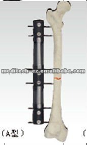
Screen Shot 2013-01-24 at 2.19.24 PMIt is a little painful for me to say this but it seems that if a person really, REALLY wanted to grow taller, they actually do have so me good options. One member from the Make Me Taller forums was featured on ABC News Apotheosis would choose to go through limb lengthening surgery twice, to go from 5′ 6″ to 5′ 9″ and then again to go from 5′ 9″ to 6′ 2″ to gain a total of 8 inches through surgery. On the 2nd surgery, he would take the internal intramedullary nail approach so that the lengthening is not visible to outersiders. The Chinese Surgeon Bai Helong would claim HERE that his external fixator device would be even simpler, less invasive since the device he uses (pictured on the left) only punctures the body in two main areas, is NON-PAINFUL (which I think is bullshit), cost a little over $10,000 (around the time of the article, about 6 years ago), and very fast for healing, around 6 months. These types of conditions suggest that maybe, just maybe the limb lengthening surgery approach is not that crazy, long, or painful to go through. A person who recently finished high school or college may decide to spend the next entire year in getting this type fo surgery to start a completely new life out.

Quoted from the article…

“We’ve not had a single failure since 1995, and now it’s not painful,” insists the doctor, who charges 75,000 yuan (11,000 dollars) for the surgery.”

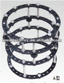
exfixIn the 90s when people first started to hear about the idea of limb lengthening surgery, images of very large, ugly looking scary external fixators like from the movie Gattaca where Ethan Hawke portrays a guy who was not tall enough to become in a space program but he accepted to subject himself to getting surgery and wearing the external fixators to get that 3-4 extra inches.

This patent shows that more and more, the surgery to be done to make our height dreams come true is becoming better, faster, easier to do, and with less complications.

The truth is that due to what the orthopedic surgeon Gavriil Abramovich Ilizarov from the Soviet Union, who pioneered the technique did we already do have the technology to grow taller, even when we are in our 40s. The method is not as bad as some people may believe. It is true that we do have our bones initially broken apart, but that is for a certain reason and the break is not as bad as others like journalists would make it seem.

A clip of the patent application is posted below….

Can You Increase Height And Grow Taller Using Ayurvedic Urea?

Me: This is one of the most craziest of ideas for potentially increasing ones height and even though I am open to all ideas from east and western science and medicine, this idea is a little extreme for me to accept. As we all know Urea is the main component of human Urine. From my studies on ayurvedic medicine years ago, I know that the consumption of the human or cow urine has been practiced quite extensively in certain cultures, but specifically the Indian culture.

Analysis: While the main link of Urea is to the human urine, urea is also leaked out of the human body through the pores from sweating. If the Indian medicine therapy of Ayurveda through urea is believe to increase height, then we must find some evidence in the medical literature to see what are it’s effects when injected or taken orally. It is stated, “Urea is synthesized in the body of many organisms as part of the urea cycle, either from the oxidation of amino acids or from ammonia. In this cycle, amino groups donated by ammonia and L-aspartate are converted to urea, while L-ornithine, citrulline, L-argininosuccinate, and L-arginine act as intermediates. Urea production occurs in the liver and is regulated by N-acetylglutamate. Urea is found dissolved in blood (in the reference range of 2.5 to 6.7 mmol/liter) and is excreted by the kidney as a component of urine. In addition, a small amount of urea is excreted (along with sodium chloride and water) in sweat.” Urea itself has no color, no smell, is solid and is really just a nitrogen derived compound. Urea converts to amino acids and amino acids convert to Urea. The Urea molecule can convert to carbon dioxide and ammonia molecules and is use extensively in fertilizers. If human ingested it in high amounts, it will actually break apart the covalent bonds in proteins. At this point, I would say that the idea of trying urea to grow taller has no validity. On the grow taller website, the people say that Ayurvedic Urea costs up to $10,000 for the real Urea. How am I supposed to know this expensive chemical compound was not the product of human urine waste or chemical plant waste?

From this article I found from googling “drink urine” into google from the website The Independent HERE…the people claim that drinking one’s urine can cure the cold and cancer. Other highlights are…

  • Advocates claim it has antibacterial, antifungal, antiviral and anticancer properties.
  • Research in the 1990s claimed that drinking urine could cure jet lag.
  • It is highly sterile. The Aztecs used it to prevent wounds becoming infected.

There is some science to the practice called Auto-urine therapy or Urotherapy. The technical general term is Urophagia (wiki article).

From the Grow Taller Pyramid Secret website HERE


Ayurvedic Urea to Grow Taller

October 27, 2012 in How To Grow Taller

I have received hundreds of emails about people asking me about Ayurvedic Urea. Apparently, Ayurvedic Urea is a product that promises people to grow taller without doing anything. I have tried researching about it, and I am going to reveal some information about this product to you! Whether or not it really works, you guys decide for yourself! It is soon going to be launched in 2014!

What is Ayurvedic Urea?

Ayurvedic Urea is a powder form product that when consumed, is proven to help one grow taller significantly! Depending on how much one wants to grow taller, the duration and dosage will apply.

Does it guarantee growth?

Apparently, many online forums and past clients have told me that they really grew taller from Ayurvedic Urea, and that they really became a lot taller!

How much does it cost?

It costs a bomb! It can range from US$10,000- US$100,000 depending on where you can get it, how much you are planning to grow, and whom you purchase it from!

Who can use Ayurvedic Urea to grow taller?

People from ages 13-35 who wants to grow taller can try taking Ayurvedic Urea! It is not advised for people above 50 years old, and people with HIV or dwarfism to take it as there will be no effect on them!

Where can I find Ayurvedic Urea?

You can Ayurvedic Urea in remote parts of Nepal and Africa!

How long must I wait to see results?

Result is said to be visible within 1-2 weeks of usage! A person can generally grow 1 inch in 10-14 days!

What are some negative effects of Ayurvedic Urea?

There are no found negative effects or long term disadvantages to it! However, people who consume Ayruvedic Urea may experience extreme fatigue and is advised to sleep and rest more! Therefore, try to reduce your activity level to the minimum if you are consuming this!

Where can I order it?

You can order it online! However choose the right supplier as you might get scammed easily!

Why is it so expensive?

Ayurvedic Urea is very rare. The government in Nepal and Africa is regulating the sale of it, and only a certain amount of it can be sold every year! Therefore you have to grab fast!

How do you use it?

Basically, you will have to consume about 0.5kg-1kg of Ayurvedic Urea a day (morning, afternoon, night) , depending on the dosage. Instructions will be given when you purchase them! Also, avoid drinking and smoking when you are on Ayurvedic Urea!
In conclusion, those of you who want to purchase this item must think critically, and be ready to fork out a huge sum of money! It can be said that this product costs more than the traditional limb lengthening surgery! Do not purchase immediately before researching and doing the necessary measures to protect yourself from scammers.

With that, we wish all of you the best of luck in your growing taller journey!

From the Wikipedia article on Urea for Medicinal Use

Urea or carbamide is an organic compound with the chemical formula CO(NH2)2. The molecule has two —NH2 groups joined by a carbonyl (C=O) functional group.

Urea serves an important role in the metabolism of nitrogen-containing compounds by animals and is the main nitrogen-containing substance in the urine of mammals. It is a colorless, odorless solid, although the ammonia that it gives off in the presence of water, including water vapor in the air, has a strong odor. It is highly soluble in water and practically non-toxic (LD50 is 15 g/kg for rat). Dissolved in water, it is neither acidic nor alkaline. The body uses it in many processes, the most notable one being nitrogen excretion. Urea is widely used in fertilizers as a convenient source of nitrogen. Urea is also an important raw material for the chemical industry. The synthesis of this organic compound by Friedrich Wöhler in 1828 from an inorganic precursor was an important milestone in the development of organic chemistry, as it showed for the first time that a molecule found in living organisms could be synthesized in the lab without biological starting materials (thus contradicting a theory widely prevalent at one time, called vitalism).

The terms urea and carbamide are also used for a class of chemical compounds sharing the same functional group RR’N—CO—NRR’, namely a carbonyl group attached to two organic amine residues. Examples include carbamide peroxide, allantoin, and hydantoin. Ureas are closely related to biurets and related in structure to amides, carbamates, carbodiimides, and thiocarbamides.Related chemicals

History

Urea was first discovered in urine in 1727 by the Dutch scientist Herman Boerhaave, though this discovery is often attributed to the French chemist Hilaire Rouelle.[3] In 1828, the German chemist Friedrich Wöhler obtained urea by treating silver isocyanate with ammonium chloride.[4][5][6]

AgNCO + NH4Cl → (NH2)2CO + AgCl

This was the first time an organic compound was artificially synthesized from inorganic starting materials, without the involvement of living organisms. The results of this experiment implicitly discredited vitalism: the theory that the chemicals of living organisms are fundamentally different from inanimate matter. This insight was important for the development of organic chemistry. His discovery prompted Wöhler to write triumphantly to Berzelius: “I must tell you that I can make urea without the use of kidneys, either man or dog. Ammonium cyanate is urea.” For this discovery, Wöhler is considered by many[who?] the father of organic chemistry.

Physiology

Urea is synthesized in the body of many organisms as part of the urea cycle, either from the oxidation of amino acids or from ammonia. In this cycle, amino groups donated by ammonia and L-aspartate are converted to urea, while L-ornithine, citrulline, L-argininosuccinate, and L-arginine act as intermediates. Urea production occurs in the liver and is regulated by N-acetylglutamate. Urea is found dissolved in blood (in the reference range of 2.5 to 6.7 mmol/liter) and is excreted by the kidney as a component of urine. In addition, a small amount of urea is excreted (along with sodium chloride and water) in sweat.

Amino acids from ingested food that are not used for the synthesis of proteins and other biological substances are oxidized by the body, yielding urea and carbon dioxide, as an alternative source of energy.[7] The oxidation pathway starts with the removal of the amino group by a transaminase, the amino group is then fed into the urea cycle.

Ammonia (NH3) is another common byproduct of the metabolism of nitrogenous compounds. Ammonia is smaller, more volatile and more mobile than urea. If allowed to accumulate, ammonia would raise the pH in cells to toxic levels. Therefore many organisms convert ammonia to urea, even though this synthesis has a net energy cost. Being practically neutral and highly soluble in water, urea is a safe vehicle for the body to transport and excrete excess nitrogen.

In water, the amine groups undergo slow displacement by water molecules, producing ammonia and carbonate anion. For this reason, old, stale urine has a stronger odor than fresh urine.

In humans

The handling of urea by the kidneys is a vital part of human metabolism. Besides its role as carrier of waste nitrogen, urea also plays a role in the countercurrent exchange system of the nephrons, that allows for re-absorption of water and critical ions from the excreted urine. Urea is reabsorbed in the inner medullary collecting ducts of the nephrons,[8] thus raising the osmolarity in the medullary interstitiumsurrounding the thin ascending limb of the loop of Henle, which in turn causes water to be reabsorbed. By action of the urea transporter 2, some of this reabsorbed urea will eventually flow back into the thin ascending limb of the tubule, through the collecting ducts, and into the excreted urine.

This mechanism, which is controlled by the antidiuretic hormone, allows the body to create hyperosmotic urine, that has a higher concentration of dissolved substances than the blood plasma. This mechanism is important to prevent the loss of water, to maintain blood pressure, and to maintain a suitable concentration ofsodium ions in the blood plasmas.

The equivalent nitrogen content (in gram) of urea (in mmol) can be estimated by the conversion factor 0.028 g/mmol.[9] Furthermore, 1 gram of nitrogen is roughly equivalent to 6.25 grams of protein, and 1 gram of protein is roughly equivalent to 5 grams of muscle tissue. In situations such as muscle wasting, 1 mmol of excessive urea in the urine (as measured by urine volume in litres multiplied by urea concentration in mmol/l) roughly corresponds to a muscle loss of 0.67 gram.

In other species

In aquatic organisms the most common form of nitrogen waste is ammonia, whereas land-dwelling organisms convert the toxic ammonia to either urea or uric acid. Urea is found in the urine of mammals and amphibians, as well as some fish. Birds and saurian reptiles have a different form of nitrogen metabolism, that requires lesswater and leads to nitrogen excretion in the form of uric acid. It is noteworthy that tadpoles excrete ammonia but shift to urea production during metamorphosis. Despite the generalization above, the urea pathway has been documented not only in mammals and amphibians but in many other organisms as well, including birds,invertebrates, insects, plants, yeast, fungi, and even microorganisms.[citation needed]

Uses

Agriculture

More than 90% of world production of urea is destined for use as a nitrogen-release fertilizer. Urea has the highest nitrogen content of all solid nitrogenous fertilizers in common use. Therefore, it has the lowest transportation costs per unit of nitrogen nutrient. The standard crop-nutrient rating of urea is 46-0-0.[10]

Many soil bacteria possess the enzyme urease, which catalyzes the conversion of the urea molecule to two ammonia molecules and one carbon dioxide molecule, thus urea fertilizers are very rapidly transformed to the ammonium form in soils. Among soil bacteria known to carry urease, some ammonia-oxidizing bacteria (AOB), such as species of Nitrosomonas, are also able to assimilate the carbon dioxide released by the reaction to make biomass via the Calvin Cycle, and harvest energy by oxidizing ammonia (the other product of urease) to nitrite, a process termed nitrification.[11] Nitrite-oxidizing bacteria, especially Nitrobacter, oxidize nitrite to nitrate, which is extremely mobile in soils and is a major cause of water pollution from agriculture. Ammonia and nitrate are readily absorbed by plants, and are the dominant sources of nitrogen for plant growth. Urea is also used in many multi-component solid fertilizer formulations. Urea is highly soluble in water and is, therefore, also very suitable for use in fertilizer solutions (in combination with ammonium nitrate: UAN), e.g., in ‘foliar feed’ fertilizers. For fertilizer use, granules are preferred over prills because of their narrower particle size distribution, which is an advantage for mechanical application.

The most common impurity of synthetic urea is biuret, which impairs plant growth.

Urea is usually spread at rates of between 40 and 300 kg/ha but rates vary. Smaller applications incur lower losses due to leaching. During summer, urea is often spread just before or during rain to minimize losses from volatilization (process wherein nitrogen is lost to the atmosphere as ammonia gas). Urea is not compatible with other fertilizers.

Because of the high nitrogen concentration in urea, it is very important to achieve an even spread. The application equipment must be correctly calibrated and properly used. Drilling must not occur on contact with or close to seed, due to the risk of germination damage. Urea dissolves in water for application as a spray or through irrigation systems.

In grain and cotton crops, urea is often applied at the time of the last cultivation before planting. In high rainfall areas and on sandy soils (where nitrogen can be lost through leaching) and where good in-season rainfall is expected, urea can be side- or top-dressed during the growing season. Top-dressing is also popular on pasture and forage crops. In cultivating sugarcane, urea is side-dressed after planting, and applied to each ratoon crop.

In irrigated crops, urea can be applied dry to the soil, or dissolved and applied through the irrigation water. Urea will dissolve in its own weight in water, but it becomes increasingly difficult to dissolve as the concentration increases. Dissolving urea in water is endothermic, causing the temperature of the solution to fall when urea dissolves.

As a practical guide, when preparing urea solutions for fertigation (injection into irrigation lines), dissolve no more than 30 kg urea per 100 L water.

In foliar sprays, urea concentrations of 0.5% – 2.0% are often used in horticultural crops. Low-biuret grades of urea are often indicated.

Urea absorbs moisture from the atmosphere and therefore is typically stored either in closed/sealed bags on pallets or, if stored in bulk, under cover with a tarpaulin. As with most solid fertilizers, storage in a cool, dry, well-ventilated area is recommended.

Chemical industry

Urea is a raw material for the manufacture of many important chemical compounds, such as

  • Various plastics, especially the urea-formaldehyde resins.
  • Various adhesives, such as urea-formaldehyde or the urea-melamine-formaldehyde used in marine plywood.
  • Potassium cyanate, another industrial feedstock.

Explosive

Urea can be used to make urea nitrate, a high explosive that is used industrially and as part of some improvised explosive devices.

Automobile systems

Urea is used in SNCR and SCR reactions to reduce the NOx pollutants in exhaust gases from combustion from diesel, dual fuel, and lean-burn natural gas engines. The BlueTec system, for example, injects water-based urea solution into the exhaust system. The ammonia produced by the hydrolysis of the urea reacts with the nitrogen oxide emissions and is converted into nitrogen and water within the catalytic converter.

Other commercial uses

  • A stabilizer in nitrocellulose explosives
  • A component of animal feed, providing a relatively cheap source of nitrogen to promote growth
  • A non-corroding alternative to rock salt for road de-icing, and the resurfacing of snowboarding halfpipes and terrain parks
  • A flavor-enhancing additive for cigarettes
  • A main ingredient in hair removers such as Nair and Veet
  • A browning agent in factory-produced pretzels
  • An ingredient in some skin cream,[12] moisturizers, hair conditioners
  • A reactant in some ready-to-use cold compresses for first-aid use, due to the endothermic reaction it creates when mixed with water
  • A cloud seeding agent, along with other salts
  • A flame-proofing agent, commonly used in dry chemical fire extinguisher charges such as the urea-potassium bicarbonate mixture
  • An ingredient in many tooth whitening products
  • An ingredient in dish soap
  • Along with ammonium phosphate, as a yeast nutrient, for fermentation of sugars into ethanol
  • A nutrient used by plankton in ocean nourishment experiments for geoengineering purposes
  • As an additive to extend the working temperature and open time of hide glue
  • As a solubility-enhancing and moisture-retaining additive to dye baths for textile dyeing or printing

Laboratory uses

Urea in concentrations up to 10 M is a powerful protein denaturant as it disrupts the noncovalent bonds in the proteins. This property can be exploited to increase the solubility of some proteins. A mixture of urea and choline chloride is used as a deep eutectic solvent, a type of ionic liquid.

Urea can in principle serve as a hydrogen source for subsequent power generation in fuel cells. Urea present in urine/wastewater can be used directly (though bacteria normally quickly degrade urea.) Producing hydrogen by electrolysis of urea solution occurs at a lower voltage (0.37V) and thus consumes less energy than the electrolysis of water (1.2V).[13]

Urea in concentrations up to 8 M can be used to make fixed brain tissue transparent to visible light while still preserving florescent signals from labeled cells. This allows for much deeper imaging of neuronal processes then previously obtainable using conventional one photon or two photon confocal microscopes.[14]

Medical use

Urea-containing creams are used as topical dermatological products to promote rehydration of the skin. Urea 40% is indicated for psoriasis, xerosis, onychomycosis, ichthyosis, eczema, keratosis,keratoderma, corns, and calluses. If covered by an occlusive dressing, 40% urea preparations may also be used for nonsurgical debridement of nails. Urea 40% “dissolves the intercellular matrix”[15] of the nail plate. Only diseased or dystrophic nails are removed, as there is no effect on healthy portions of the nail. This drug is also used as an earwax removal aid. Urea can also be used as a Diuretic.

Certain types of instant cold packs (or ice packs) contain water and separated urea crystals. Rupturing the internal water bag starts an endothermic reaction and allows the pack to be used to reduce swelling.

Like saline, urea injection is used to perform abortions.

Urea is the main component of an alternative medicinal treatment referred to as urine therapy.

The blood urea nitrogen (BUN) test is a measure of the amount of nitrogen in the blood that comes from urea. It is used as a marker of renal function.

Urea labeled with carbon-14 or carbon-13 is used in the urea breath test, which is used to detect the presence of the bacteria Helicobacter pylori (H. pylori) in the stomach and duodenum of humans, associated with peptic ulcers. The test detects the characteristic enzyme urease, produced by H. pylori, by a reaction that produces ammonia from urea. This increases the pH (reduces acidity) of the stomach environment around the bacteria. Similar bacteria species to H. pylori can be identified by the same test in animals such as apes, dogs, and cats (including big cats).

Chemical properties

Molecular and crystal structure

The urea molecule is planar in the crystal structure, but the geometry around the nitrogens is pyramidal in the gas-phase minimum-energy structure.[20] In solid urea, the oxygen center is engaged in two N-H-O hydrogen bonds. The resulting dense and energetically favourable hydrogen-bond network is probably established at the cost of efficient molecular packing: The structure is quite open, the ribbons forming tunnels with square cross-section. The carbon in urea is described as sp2 hybridized, the C-N bonds have significant double bond character, and the carbonyl oxygen is basic compared to, say, formaldehyde. Urea’s high aqueous solubility reflects its ability to engage in extensive hydrogen bonding with water.

By virtue of its tendency to form a porous frameworks, urea has the ability to trap many organic compounds. In these so-called clathrates, the organic “guest” molecules are held in channels formed by interpenetrating helices composed of hydrogen-bonded urea molecules. This behaviour can be used to separate mixtures, e.g. in the production of aviation fuel and lubricating oils, and in the separation ofparaffins.

As the helices are interconnected, all helices in a crystal must have the same molecular handedness. This is determined when the crystal is nucleated and can thus be forced by seeding. The resulting crystals have been used to separate racemic mixtures.

Reactions

Urea reacts with alcohols to form urethanes. Urea reacts with malonic esters to make barbituric acids.

Safety

Urea can be irritating to skin, eyes, and the respiratory tract. Repeated or prolonged contact with urea in fertilizer form on the skin may cause dermatitis.

High concentrations in the blood can be damaging. Ingestion of low concentrations of urea, such as are found in typical human urine, are not dangerous with additional water ingestion within a reasonable time-frame. Many animals (e.g., dogs) have a much more concentrated urine and it contains a higher urea amount than normal human urine; this can prove dangerous as a source of liquids for consumption in a life-threatening situation (such as in a desert).

Urea can cause algal blooms to produce toxins, and its presence in the runoff from fertilized land may play a role in the increase of toxic blooms.[21]

The substance decomposes on heating above melting point, producing toxic gases, and reacts violently with strong oxidants, nitrites, inorganic chlorides, chlorites and perchlorates, causing fire and explosion.[citation needed]


From the Wikipedia article on Urine Therapy

In alternative medicine, the term urine therapy or urotherapy, (also urinotherapy or uropathy) refers to various applications of human urine for medicinal or cosmetic purposes, including drinking of one’s own urine and massaging one’s skin with one’s own urine. While there is currently insufficient evidence for the therapeutic use of urine, many chemical components of urine have wide-scale use, such as urea and urokinase.[1][2][3][4][5]

Many of the earliest human cultures used urine as a medicine.History

Hinduism

A Sanskrit text called the Damar Tantra, not part of core Hinduism, contains 107 stanzas on the benefits of “pure water, or one’s own urine”.[10] In this text, urine therapy is referred to as Shivambu Kalpa.[10]This text suggests, among other uses and prescriptions, massaging one’s skin with fresh, concentrated urine. In the Ayurvedic tradition, which is related to the Hindu scriptures called the Vedas. urine therapy is called amaroli which when practised requires some dietary requirements such as mixing it with water to “cure cancers” and other “diseases” along with “raw food and certain fruits like banana, papaya and citrus fruits” which are claimed to be “very good in the practice of amaroli”.[11][12][13][14] One of the main aims of this system is to “prevent illness, heal the sick and preserve life”.[13][15]

Other cultures

The French customarily soaked stockings in urine and wrapped them around their necks in order to cure strep throat.[2] Aristocratic French women in the 17th century reportedly bathed in urine to beautify their skin.

In Sierra Madre, Mexico, farmers prepare poultices for broken bones by having a child urinate into a bowl of powdered charred corn. The mixture is made into a paste and applied to the skin.[17]

As in ancient Rome, urine was used for teeth-whitening during the Renaissance, though they did not necessarily consume their own urine.

The homeopath John Henry Clarke wrote, “…man who, for a skin affection, drank in the morning the urine he had passed the night before. The symptoms were severe, consisting of general-dropsy, scanty urine, and excessive weakness. These symptoms I have arranged under Urinum. Urinotherapy is practically as old as man himself. The Chinese (Therapist, x. 329) treat wounds by sprinkling urine on them, and the custom is widespread in the Far East. Taken internally, it is believed to stimulate the circulation”.[18]

Modern claims and findings

Urine’s main constituents are water and urea; the latter of which has some well-known commercial and other uses. Urine also contains small quantities of thousands of compounds, hormones and metabolites,[5][19] including corticosteroids.[20] Pregnant mare’s urine has high amounts of estrogens, which are isolated and sold as Premarin. There is no scientific evidence of a therapeutic use for untreated urine.[1][2][3][4][5]

It has been claimed that urine is similar to other body fluids, like amniotic fluid or even blood, but these claims have no scientific basis.[4]

Urinating on jellyfish stings is a common folk remedy, but has no beneficial effect and may be counterproductive, as it can activate nematocysts remaining at the site of the sting.

People who use Amanita muscaria as an intoxicating drug will sometimes drink their own urine in order to prolong its effects, especially when there are shortages of the fungus.[21][22]

Use as anti-cancer agent

Urine and urea have been claimed by some practitioners to have an anti-cancer effect. It has been hypothesized that because some cancer cell antigens are transferred through urine, through “oral autourotherapy” these antigens could be introduced to the immune system that might then create antibodies.[23]

Auto-urine drinking and meditation

Drinking one’s morning urine (‘amaroli’) was an ancient yoga practise designed to promote meditation. The ancient Hindu and yoga texts that mention auto-urine drinking, require it be done before sunrise and that only the mid-stream sample be used.[33] The pineal hormone melatonin and its conjugated esters are present in morning urine in significant quantities, the pineal gland secreting melatonin maximally at about 2 am, this secretion being shut off by the eyes’ exposure to bright sunlight.[33] Melatonin, when ingested or given intravenously, amongst other effects, provokes tranquility and heightened visualisation.[33] There are high concentrations of melatonin in the first morning urine, but not in a physiologically active form.[33] Mills and Faunce at Newcastle University Australia in 1991 developed the hypothesis that ingestion of morning urine into low pH gastric acid would cause deconjugation of its esters back to the active form of melatonin. This, they suggested, might restore plasma night-time melatonin levels. Thus, they argued, oral pre-dawn consumption of auto-exogenous melatonin, by either re-setting of the sleep-wake cycle or enhancement of the physiological prerequisites for meditation (decreased body awareness (i.e. analgesia) and claimed slowed brain wave activity, as well as heightened visualization ability), may be the mechanism behind the alleged benefits ascribed to ‘amaroli’ or auto-urine drinking by ancient texts of the yogic religion.[33] Obvious experimental difficulties (particularly in constructing a double-blind clinical trial) mean that this is a difficult hypothesis to reliably test to any requisite evidence-based standard.


Increase Height And Grow Taller Through Intermittent Fasting

Me: One very interesting idea for possible height increase that was brought to my attention was the possiblity of using intermittent fasting to grow taller. From a quick scientific point of view, there is some validity. Fasting is wher eyou don’t eat. Intermittent fastinf measn you go through the cycle of eating and not eating over and over again. When you fast, you get hungry. When you first start out the hunger cycle, the peptide/ protein ghrelin is released into the human system. If we remember, ghrelin is what is known as a GnRH, or Gonadotropin releasing hormone analogue. From the wikipedia article on Ghrelin HERE

Ghrelin is a 28 amino acid hunger-stimulating peptide and hormone that is produced mainly by P/D1 cells lining the fundus of the human stomach andepsilon cells of the pancreas.[1] Ghrelin levels increase before meals and decrease after meals. It is considered the counterpart of the hormone leptin, produced by adipose tissue, which induces satiation when present at higher levels. In some bariatric procedures, the level of ghrelin is reduced in patients, thus causing satiation before it would normally occur.[2]

Ghrelin is a potent stimulator of growth hormone from the anterior pituitary gland.[3] The ghrelin receptor is a G protein-coupled receptor, known as thegrowth hormone secretagogue receptor. Ghrelin binds to the GHSR1a splice-variant of this receptor which is present in high density in the hypothalamus, pituitary as well as vagal afferent cell bodies and vagal afferent endings throughout the gastro-intestinal tract.[4][5]

Ghrelin plays a significant role in neurotrophy, particularly in the hippocampus, and is essential for cognitive adaptation to changing environments and the process of learning.[6][7] Ghrelin has been shown to activate the endothelial isoform of nitric oxide synthase in a pathway that depends on various kinases including Akt.[8]

Me: Ghrelin does indeed stimulate the growth hormone. Plus, we have seen that too much food from constant eating is probably not good for height increase. Sure, we all realize that malnitrition will stunt a kids’s growth, but would too much food stunt a kids’s growth? I think it does. If we remember that we come from a paleolithic ancestry, our ancestor’s diet habits involved long time intervals where we had no food to times of abundance, like when a buffalo was finally hunted and killed after a week of planning and work.

From my studies on the ESWT, LIPUS< PEMF, and DC/AC E&M field stimulation, It is almost always that having a non-linear, more cyclical signal transduction, like the sinusoidal tapping of loading and clamping from the LSJL technique is better than a consistent rate of external stimuli intensity. We are seeing over and over again that a quick external application of stimuli in cyclical fashion helps promote cell division and proliferation. If we decide to cycle our eating habits and go hungry in a cyclical rhythmic way, it might make sense that the human body might be able to actually grow taller. It thus might be more beneficial for height increase to go through a few cycles of fasting to cause hunger, causing ghrelin release, causing more growth hormone release. Plus, when a person is hungry, they feel weak so they go to sleep. Since sleep is when the most height increase happens, maybe intermittent fasting might be a good idea. The main arguement for the idea of fasting is that reduction of insulin to the brain and that the cells in the body are given a chance to repair themselves since it does take a lot of cellular energy and work to process food and that leads to wear and tear.

From wikipedia….

Mechanism of action

Ghrelin has emerged as the first identified circulating hunger hormone. Ghrelin and synthetic ghrelin mimetics (the growth hormone secretagogues) increase food intake and increase fat mass.15][16] by an action exerted at the level of the hypothalamus. They activate cells in the arcuate nucleus[17][18] that include the orexigenic neuropeptide Y (NPY) neurons.[19] Ghrelin-responsiveness of these neurons is both leptin- and insulin-sensitive.[20] Ghrelin also activates the mesolimbic cholinergic-dopaminergic reward link, a circuit that communicates the hedonic and reinforcing aspects of natural rewards, such as food, as well as of addictive drugs, such as ethanol.[21][22][20] Indeed, central ghrelin signalling is required for reward from alcohol.[23] and palatable/rewarding foods.[24][25] There is also strong evidence that ghrelin has a peripheral appetite modulatory effect on satiety by affecting the mechanosensitivity of gastric vagal afferents, making them less sensitive to distension resulting in over eating.[5]

….Ghrelin through its receptor increases the concentration of dopamine in the substantia nigra, a region of the brain where dopamine cell degeneration leads to Parkinson’s disease. Hence ghrelin may find application in slowing down the onset of Parkinson’s disease.[48]


From a website called Grow Taller Pyramid Secret

On the sidebar of the Grow Taller Pyramid Secret website you have a facebook widget that shows how many people “like” the Grow Taller Guru , who I will assume is Lance Ward. I would guess that this website is a affiliate site to sell a E-product. From the website….

So, your question would be. How does Intermittent Fasting help one to grow taller?

Firstly, intermittent fasting or skipping meals can help reduce brain insulin level. Insulin is a peptide hormone, produced by beta cells of the pancreas, and is central to regulating carbohydrate and fat metabolism in the body. Insulin causes cells in the liver, skeletal muscles, and fat tissue to take up glucose from the blood. Therefore with reduced insulin level, a person’s body have lesser fat, and this can aid the process of growth and development a lot more efficiently, which is necessary for growing taller.

Secondly, intermittent fasting can increase growth hormone production significantly! It is known and studied that people who ate only 2 big meals in a day, and fasted, tend to be healthier and stronger, and in some cases taller! For example.

The Maasai people of Africa generally eat two meals a day – in the morning and at night. The Masai are exceptionally tall people and some of the healthiest that Weston Price observed in his study of traditional peoples in the early 1930s.

Clearly, their bodies don’t lack nutrients. They eat plenty of food at meal time. They aren’t calorically or nutritionally deprived. They merely condense their intake between longer intervals.

Intermittent fasting does not significantly reduce calories. In fact, the object of intermittent fasting is not caloric restriction. Regular large, high nutrient meals satisfy a person’s nutritional needs and provide the building blocks needed to grow. Not only does fasting not stunt growth it increases growth hormone. An increase in growth hormone can actually boost a child’s height making them grow taller than they otherwise would.

Another reason for why intermittent fasting could actually be a good strategy to grow taller is that it helps in cell repair and waste elimination. During a grow taller program, through intense exercises and stretching, many muscle tear and tissue damaged is being done. Intermittent fasting will help the recovery period and help to ensure that the muscle and bones do grow well. Intermittent fasting also helps cleanse the body off toxins and ensures the body’s renewed revival state! Therefore, some people say that intermittent fasting is like a body cleansing period where your body will feel more rejuvenated and refreshed than ever before!

From ThePrimalParent.com website HERE

Fasting in Paleolithic Times
It is a little narrow minded to assume that children have been eating three squares since the dawn of time. Life isn’t so easy outside of our opulent kitchens where refrigerators, boxes, and cans unnaturally prolong the shelf life of food. Hunter gatherers actually have to leave their camps to kill or gather foods, collect materials for a fire, return to the camp to light the fire, and then wait for their catch to cook. I’m guessing this ordeal takes a little longer than it takes to open a box of cereal and pour pasteurized milk over it.

J. Stanton just published a great article this week about hunter gatherers skipping breakfast and says that skipping lunch is probably even quite normal.

The guide standing next to the Masai man is six feet tall.

The Maasai people of Africa generally eat two meals a day – in the morning and at night. The Masai are exceptionally tall people and some of the healthiest that Weston Price observed in his study of traditional peoples in the early 1930s. This was before modern foods had been introduced into the diet of the Masai.

Clearly, their bodies don’t lack nutrients. They eat plenty of food at meal time. They aren’t calorically or nutritionally deprived. They merely condense their intake between longer intervals.

Intermittent fasting does not significantly reduce calories. In fact, the object of intermittent fasting is not caloric restriction. Regular large, high nutrient meals satisfy a person’s nutritional needs and provide the building blocks needed to grow. Not only does fasting not stunt growth it increases growth hormone. An increase in growth hormone can actually boost a child’s height making them grow taller than they otherwise would.

The effect of boosting or at least maintaining average height of mammals while fasting was observed in one of the most famous studies done on rats and intermittent fasting called Apparent Prolongation Of The Life Span Of Rats By Intermittent Fasting. The study was performed with baby rats.

“The fasting was begun at the age of 42 days and was continued until the rats died.”

Rats are not fully grown until 6-7 months of age and have an average life span of 2-3 years.

“In some cases, the average femoral lengths of the fasted rats at death were greater than, or equal to, those of the controls and, in other cases, the rats were only a little smaller. In short, intermittent fasting seems to make it possible to increase the life span to some extent without stunting the rats.”

A Tradition of Fasting Through Illness

Traditionally, people fast when they’re sick, often called a cleanse. The same trend is observed in animals both domesticated and wild. The healing mechanism is called autophagy.  Alice Villalobo describes how autophagy benefits our cells.

“Scientists have observed that cell debris—proteins and organelles—gets encapsulated by tiny rearrangements of membranes and moved into empty spaces called vacuoles. The transportation of the cell debris is a pathway now called “cytoplasm-to-vacuole targeting,” or the Cvt pathway. Autophagy is the sequestration of the cargo material, bulk cytoplasm or specific organelle within double-membrane structures and its delivery to the vacuole for further degradation.”

It almost seems abusive to prohibit the process of autophagy in children by denying them the right to fast. While children may not need to fast for as long as do adults, the benefits and safety of short, intermittent fasting has its roots in the history of traditional cultures, illness, and even in religion.