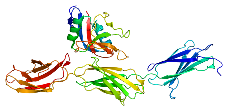
Increase Height And Grow Taller Using Chitosan

Another compound I have heard people claim could possibly increase height was over the compound Chitosan. When I first googled “chitosan grow taller” the first link was to the site GrowTall.com

The website states that chitosan is a type of “fiber” that is made from crustacean shells from a chemical process. Besides being found in shrimp, lobsters, it seems that chitosan can also be found from teh plastic part of the squid we remove which can not be eaten. The first thing that comes to my head is to remember that a lot of high increase pills have a compound made from seashells as well based on calcium, usually calcium carbonate. From high school biology we remember that human digestive systems can’t digest fibers unlike most plant eating animals like cows which have 4 stomachs. The chitosan would go into the intestine area, bond with ingested fats, and come out when we go #2 in the bathroom.

The website suggest that chitosan might be useful for weight loss, can be used in wound treatment (ie closure) and that it should not be used by children and/or pregnant women due to growth retardation. This claim would mean that chitosan would actually make growing children shorter than they would actually be, which is the opposite of what we are looking for. As fror doasge, 3-6 grams of the compound can be taken each day with food. It seems chitosan has the ability to remove the body of certain minerals which again shows that it is a compound that is more likely to retard and stunt growth that promote it. The fact that it is stated that it can be used for woud healing may indicate it does have some angiogenic properties.

As for medicinal or therapeutic uses, some studies suggest it can possibly lower cholesterol as well as treat kidney failure. For the kidney failure, the chitosan is said to combine with an toxic compounds and this causes the toxins to be excreted out when the chitosan as a fiber is excreted. From a very syperficial level, that is a reasonable scientific guess as why researchers may think it helps with kidney failure.

As for the claim that it is used to treat wounds and help in wound healing/closure, the idea is that along with the plasma, the chitosan may be able to bind to whatever usually like blood and plasma that comes along to close wounds and help built new tissue. That other idea is that chitosan may be able to kill some bacteria (like Strep) and yeast (like Candida)

Some studies claim it helps prevent or treat tumors, which would suggest that it is more catabolic in nature which is against what most height increase products would claim.

From the website GrowTall.com…

Animal studies suggest that some forms of chitosan may help to prevent bone loss;… however, because chitosan also interferes with mineral absorption, the net effect in humans might actually be to increase bone loss…

Safety Issues 

There is significant evidence that long-term, high-dose chitosan supplementation can result in malabsorption of some crucial vitamins and minerals including calcium, magnesium, selenium, and vitamins A, D, E, and K.33,34 In turn, this appears to lead to a risk of osteoporosis in adults and growth retardation in children. For this reason, adults taking chitosan should also take supplemental vitamins and minerals, making especially sure to get enough vitamin D, calcium, and magnesium.

The overall conclusion on this resource is that chitosan is more like to remove important minerals for bone instead od prevent bone loss. If this is the case, for children who are still growth, that can indeed interfere with with the endochondral ossification process. Let’s  not forget that while a lot of our research now is on cartilage and cartilage regeneration, for us to have the height we already do, it does involve the fact that calcium and otehr hard deposits have been layered and grown on top of each from the natural height growing process. Bone and mineral loss is a problem for people still growing.

However, the effects on people who are finished done growing who wish to do exercises to lengthen long bones may be the exact opposite. I do note that there has been some weak positive correlation with the idea that weaker bones with lower BMD (bone mass density) may mean that they are easier to change and be melleable (if only a little) to some bone modeling loading. If chitosan can remove the minerals in the bones that make them so hard, it might indeed assist in some techniques we have talked before like the shinbone method, in inducing microfractures, and the LSJL method.

As for the conclusion of this source on Chitosan, I say that there is a slight chance that Chitodan may help people who are already finished with the natural growth process grow taller if they take it long enough and but also did some type of bone remodeling exercises.

A post on the prospective affects by Chitosan was looked at already by HeightQuest.com in “Height increase with Chitosan?” .

Analysis:

His main point is to get the chitosan to actually go into the blood stream for some effect instead of getting it completely passed through the digestive tract without it ever getting used. The difficulty is to get the chitosan content into the bone marrow. From one study he quotes “Effect of dietary supplementation of chitosan and galacto-mannan-oligosaccharide on serum parameters and the insulin-like growth factor-I mRNA expression in early-weaned piglets.” Chitosan might help because it will “encouraging epiphyseal bone marrow stem cells to become a more rounded pro chondrogenic shape

The last resource I would raise is another result found from Google on a forum thread from Soompi HERE. A member named Jaeho would write…

Also, my mom ordered Royal Jelly and something called SmarTall. SmarTall is apparently the kids’ version of Royal Jelly. My brother and sister have been taking SmarTall for 2 days.

I also searched Chitosan online, but all the sites say it’s a weight loss pill and that it’s a scam… BUT in Korea, it’s marketed as a miracle product that cures all sorts of health problems. I can’t find anything on SmarTall though.

Anyway, I asked my mom why she bought SmarTall and she said it was like Royal Jelly for children… but it’s for height too? Well, the name gives it away… lol. From a first guess

Analysis: 

It would seem that chitosan is being marketed in east asian countries like Korea for being a sort of miracle pill that has multiple therapeutic benefits. It’s claims to help people loss weight makes it very attractive to the Korean people who are well known by being very appearance conscious. There is no claim that chitosan has any effect on height but there is another product that is Royal Jelly derived called SmarTall being sold (at least back in 2006) in Korea which is given to kids to help them grow taller. The conclusion here is that Chitosan has no height increasing properties.

If we type in the term “chitosan height increase’ into google instead what we find are studies which do suggest that chitosan does have some height and size increasing ability but those are just for vegetables, specifically grains.

A link to a webpage on Vanderbilt University HERE shows that people have already looked into chitosan quite extensively and summarized its properties and possible effects. They conclude with….

“Although companies selling chitosan claim that the product is very effective and claim to have medical research about how well their product works, the medical studies show that when taken alone, chitosan has not been proven to help a person lose weight, increase their HDL cholesterol, or decrease their LDL cholesterol. Instead, chitosan can cause unwanted gastrointestinal cramps and constipation”

Conclusion: 

Chitosan may be harmful to growing children in terms of growth retardation from its ability to absorb important minerals the body and bones need when the person is still growing. After the person is finished growing, there might be a small chance that a person can use chitosan’s mineral absorbing properties to make bones less hard and easier to model so it might be possible to use it with some bone loading technique to lengthen bone.

Using BMP-6 To Differentiate Adipose Derived Adult Stem Cells Into Chondrocytes And Cartilage Regeneration

Me: This post is to show that another option of growth factors we can use to turn the adipose derived adult stem cells into chondorcytes is to use BMP-6. Remember that in the long bones of adults, the marrow is considered yellow and mostly made of fatty acids. The study showed that TGF-Beta1 has also been shown to work, causing the expresion of cartilage specific genes an proteins like aggrecan and type II collagen. There was actually 5 growth factors all looked at in this study. They are looking at the chondorgenic potential of these on ADAS cells in alginate beads
  • 1. TGF-Beta 1
  • 2. TGF-Beta 3
  • 3. IGF-1
  • 4. BMP-6
  • 5. Dexamethasone
The two main points from the study are.
1. BMP-6 up-regulated AGC1 and COL2A1 expression by an average of 205-fold and 38-fold, respectively, over day-0 controls, while down-regulating COL10A1 expression by approximately 2-fold.
2. BMP-6 is a potent inducer of chondrogenesis in ADAS cells, in contrast to mesenchymal stem cells, which exhibit increased expression of type X collagen and a hypertrophic phenotype in response to BMP-6.
So for future reference, we realize that if we wanted to inject the BMP-6, we would put it in the bone marrow part, not the epiphysis part since the MSCSs only caused COL10 expression and hypertrophic phenotype. Any height increase method we create will have growth factors added in certain areas in certain combinations in certain sequences,
From PubMed study link HERE
Arthritis Rheum. 2006 Apr;54(4):1222-32.

Potent induction of chondrocytic differentiation of human adipose-derived adult stem cells by bone morphogenetic protein 6.

Estes BT, Wu AW, Guilak F.

Source

Department of Surgery, Duke University Medical Center, Durham, North Carolina 27710, USA.

Abstract

OBJECTIVE:

Recent studies have identified an abundant source of multipotent progenitor cells in subcutaneous human adipose tissue, termed human adipose-derived adult stem cells (ADAS cells). In response to specific media formulations, including transforming growth factor beta1 (TGFbeta1), these cells exhibit significant ability to differentiate into a chondrocyte-like phenotype, expressing cartilage-specific genes and proteins such as aggrecan and type II collagen. However, the influence of other growth factors on the chondrogenic differentiation of ADAS cells is not fully understood. This study was undertaken to investigate the effects of TGFbeta1, TGFbeta3, insulin-like growth factor 1, bone morphogenetic protein 6 (BMP-6), and dexamethasone, in various combinations, on the chondrogenic potential of ADAS cells in alginate beads.

METHODS:

The chondrogenic response of alginate-encapsulated ADAS cells was measured by quantitative polymerase chain reaction, 3H-proline and 35S-sulfate incorporation, and immunolabeling for specific extracellular matrix components.

RESULTS:

Significant differences in chondrogenesis were observed under the different culture conditions for all outcomes measured. Most notably, BMP-6 up-regulated AGC1 and COL2A1 expression by an average of 205-fold and 38-fold, respectively, over day-0 controls, while down-regulating COL10A1 expression by approximately 2-fold.

CONCLUSION:

These findings suggest that BMP-6 is a potent inducer of chondrogenesis in ADAS cells, in contrast to mesenchymal stem cells, which exhibit increased expression of type X collagen and a hypertrophic phenotype in response to BMP-6. Combinations of growth factors containing BMP-6 may provide a novel means of regulating the differentiation of ADAS cells for applications in the tissue-engineered repair or regeneration of articular cartilage.

PMID: 16572454   [PubMed – indexed for MEDLINE]    Free full text

Using Microspheres With TGF-Beta1 And Chitosan To Differentiate Adipose Derived Stem Cells Into Chondrocytes And Repair Cartilage Defects

Me: This seems to suggest that we can create these hybrid microspheres which are just basically encapsulations or coatings of some collagenous fibrous material with TGF-Beta1 and/or Chitosan put inside and put it into the intermedullary cavity to get what little stem cells inside to differentiate into chondrocytes since the adult bone marrow is not red but yellow which is fatty acids and anything that is derives from fatty acids.

I have written in another article over the ability of BMP-6 to turn adiposed derived adult stem cells into chondrogenic in phenotype unlike for mesenchymal stem cells. It seems that the winner in this study was the hybrid microsphere with TGF-beta 1 and chitosan inside. This helps me further in deriving a height increase method. I know now that we can create microspheres for injection close to the bone marrow. Personally I would use the TFG-Beta 1 with Chitosan first which has a lesser chondrogenic effect and then added the BMP-6 encapsulations for further chondrogenesis.

From PubMed study link HERE

Joint Bone Spine. 2010 Jan;77(1):27-31. Epub 2009 Dec 22.

Cartilage regeneration using adipose-derived stem cells and the controlled-released hybrid microspheres.

Han Y, Wei Y, Wang S, Song Y.

Source

Department of orthopaedics, Xijing Hospital, The Fourth Military Medical University, West Road Changle, Xi’an, China. yishenghan@homtail.com

Abstract

OBJECTIVE:

This study was to evaluate the effect of hybrid microspheres (MS) composed of gelatin transforming growth factor-beta (TGF-beta1)-loaded MS and chitosan MS on the enhancement of differentiation of adipose-derived stem cells (ASCs) into chondrocytes in pellet culture in vitro and the reparative capacity of pellet from ASCs and the hybrid MS-TGF used to repair cartilage defects in vivo.

METHODS:

The morphology of the controlled-released MS was observed with scanning electron microscopy (SEM) and mechanical property was also tested in this study. In vitro TGF-beta1 release was evaluated by an enzyme-linked immunosorbent assay. The protein expression of Collagen II was tested by Western blot. In addition, a preliminary study on cartilage regeneration was also performed in vivo.

RESULTS:

When chondrogenic differentiation of ASCs in both MS was evaluated, the protein expression of Collagen II became significantly increased for the hybrid MS-TGF, as compared with the gelatin MS-TGF. Mechanical result showed that the hybrid MS was superior to the gelatin MS. Observation of histology in vivo demonstrated that the pellet from ASCs and the hybrid MS-TGF promoted cartilage regeneration in the defects of articular cartilage much better than other groups.

CONCLUSION:

Our study demonstrated that the pellet from ASCs and the hybrid MS-TGF can provide an easy and effective way to construct the tissue engineered cartilage in vitro and in vivo.

Copyright 2009. Published by Elsevier SAS.

PMID: 20022784   [PubMed – indexed for MEDLINE]

High Impact Sports Improves Bone Strength And Bone Geometry

Me: It is important to note that only two sports were compared side by side, specifically soccer and swimming for the first study. What we see is that when the swimmers and soccer players are compared to control groups, the loading from weight by the soccer players resulted in an increase in bone mineral density (BMD) which results in an increase in bone strength, and increased thickness of the cortical area. Apparently the bone strength of swimmers seems to be even lower than the controls, which makes sense since we have seen astronauts who go into space will increase in height from decompression of spine but will drop dramatically bone mineral density, bone loss, and bone strength. This might suggest that the viscoelastic nature of water might do a similar effect on the human body allowing for it to expand longitudinally in the water but probably goes back to normal when the swimmer gets out of the water just like how the astronaut gets back to their normal height after spending time back on earth. We know the loading from soccer leads to stronger bones and thicker bones. However we are not clear whether it would lead to longer bones as well, which is what we have been trying to achieve.

For the second study, it seems that with old age, the medullary cavity decreases in apposition and increase in size. Exercise can help increase the endocortical apposition and cortical area from the inside thus resulting in slight shrinkage of the cavity. With boys, during puberty and post puberty, the periosteal apposition is higher than the cortical resorption so for males, the cortical area is larger. With females from the tennis study, the periostral apposition is also higher from exercise and loading before puberty but the cavity seems to increase from endocortical resorption. What is important to note is that the rising estrogen levels in the pubertal tennis players will result in less bone sensitivity to loading. 

From PubMed study link HERE

J Bone Miner Metab. 2011 May;29(3):342-51. Epub 2010 Oct 21.

Bone geometry and strength adaptations to physical constraints inherent in different sports: comparison between elite female soccer players and swimmers.

Ferry B, Duclos M, Burt L, Therre P, Le Gall F, Jaffré C, Courteix D.

Source

Laboratoire Interuniversitaire de Biologie des APS, EA 3533, PRES Clermont Université, Université Blaise Pascal, 24 avenue des Landais, BP 80026, 63177 Aubiere Cedex, France.

Abstract

Sports training characterized by impacts or weight-bearing activity is well known to induce osteogenic effects on the skeleton. Less is known about the potential effects on bone strength and geometry, especially in female adolescent athletes. The aim of this study was to investigate hip geometry in adolescent soccer players and swimmers compared to normal values that stemmed from a control group. This study included 26 swimmers (SWIM; 15.9 ± 2 years) and 32 soccer players (SOC; 16.2 ± 0.7 years), matched in body height and weight. A group of 15 age-matched controls served for the calculation of hip parameter Z-scores. Body composition and bone mineral density (BMD) were assessed by dual-energy X-ray absorptiometry (DXA). DXA scans were analyzed at the femoral neck by the hip structure analysis (HSA) program to calculate the cross-sectional area (CSA), cortical dimensions (inner endocortical diameter, ED; outer width and thickness, ACT), the centroid (CMP), cross-sectional moment of inertia (CSMI), section modulus (Z), and buckling ratio (BR) at the narrow neck (NN), intertrochanteric (IT), and femoral shaft (FS) sites. Specific BMDs were significantly higher in soccer players compared with swimmers. At all bone sites, every parameter reflecting strength (CSMI, Z, BR) favored soccer players. In contrast, swimmers had hip structural analysis (HSA) Z-scores below the normal values of the controls, thus denoting weaker bone in swimmers. In conclusion, this study suggests an influence of training practice not only on BMD values but also on bone geometry parameters. Sports with high impacts are likely to improve bone strength and bone geometry. Moreover, this study does not support the argument that female swimmers can be considered sedentary subjects regarding bone characteristics.

PMID: 20963459        [PubMed – indexed for MEDLINE]

Changes in bone geometry during growth

Growth in the external size of a long bone, its cortical thickness and the distribution of cortical bone about the neu- tral axis is determined by the absolute and relative behavior of the periosteal and endocortical bone surfaces along the length of the bone8,13. Before puberty, periosteal apposition accounts for most of the increase in cortical area, this is part- ly offset however by the enlarging marrow cavity due to endocortical resorption. The net result is an enlarged corti- cal area located further from the neutral axis, leading to increased resistance to bending4. Late in puberty, periosteal apposition continues and is now accompanied by endocorti- cal apposition14, leading to an increase in cortical thickness.

The temporal sequence of events in boys tracks that of girls before puberty. Sexual dimorphism occurs during puberty and is characterized by boys exhibiting greater perisoteal expansion late in and post-puberty, and the absence of any endocortical contraction. Thus, in boys, the net result is the attainment of a greater cortical area that is located further from the axis of rotation compared to girls13.

The skeleton’s temporal sequence of events due to growth are not only surface-specific but also region-specific with more rapid maturation of distal than proximal regions. Distal segments of the appendicular skeleton mature before the proximal segments14. Similarly, contraction of the medullary cavity occurs in a distal to proximal pattern8,14.

The effect of additional loading on bone geome- try during growth

If additional loading does enhance the effect of growth then it would follow that exercise during childhood would result in an increase in periosteal but not endocortical apposition. Late in puberty, and in the immediate years following puberty the predominant effect would be narrowing of the medullar cavity due to endocortical apposition. This maturity-dependent preferential change in cortical surfaces with mechanical loading has been demonstrated in animals15-17. Younger animals showed greater periosteal expansion, while older animals showed greater medullar cavity narrowing. Reduced mechanical loading through limb immobilization or weightlessness also leads to preferential changes at the cortical surfaces: younger animals show a greater periosteal response (inhibition of bone formation), while older animals showed a greater endocortical response (increased resorption)7,16,18.

The results of human studies however are equivocal; for instance, consistent with this proposal is the finding that pre- pubertal female gymnasts had a larger total bone area (periosteal expansion) of the forearm despite a smaller stature19. While the playing arm in adult tennis players resulted in no detectable change to the total bone area of the radius, it did however result in thicker trabeculae20. Exercise also led to medullary contraction (but no periosteal expan- sion) at the tibia in adult military recruits21. In contrast, load- ing in pre-pubertal female gymnasts and non-athletic boys resulted in increased cortical area at the mid-femoral shaft due to endocortical contraction, not periosteal expansion2.

The aforementioned inconsistencies in the literature are likely to partly reflect the limitations imposed by two-dimen- sional measures (i.e., X-ray) of a three-dimensional struc- ture (i.e., bone). Radiographs and dual energy X-ray absorp- tiometry (DEXA) provide a two-dimensional projection of bone in the coronal plane which integrates periosteal and endocortical changes in the medio-lateral, not antero-poste- rior direction. Predicting changes using two-dimensional projections makes the flawed assumption that the bone is cylindrical and that the osteogenic response is uniform. These measurements in one plane do not provide informa- tion about changes that may occur cross-sectionally because of bone modelling. The cortical bone could be contracting in one plane but expanding in the other to resist bending moments. For this reason analysis of the cross sectional bone geometry is imperative. Furthermore, inferences from one or two measures at a site may not provide an accurate representation of changes that occur along the length of the bone8,22. Measuring techniques (MRI or CT) that provide a cross sectional view in the transverse plane is required for a more accurate assessment of surface specific changes in long bones. MRI is useful (particularly in children) because of the ability to collect images along the whole length of the bone without any radiation exposure.

In a recent study, MRI was used to compare the side-to- side differences in bone traits in the arms of competitive female tennis players during different stages of maturation8. The key findings were that loading did magnify the structural changes produced during growth. Prior to puberty, loading

magnified periosteal apposition along the length of the shaft; at the mid-humerus loading resulted in increased endocorti- cal resorption (medullary expansion). During the post-puber- tal period loading magnified the effect of endocortical appo- sition (medullary contraction), which makes an important contribution to cortical thickness in females. In fact, endo- cortical apposition accounted for most of the greater side-to- side difference attained in the post-pubertal years.

Most of the structural changes due to loading occurred early in the pre-pubertal years because adaptive changes in response to loading were sufficient to reduce the strains in bone that may lead to microdamage if not decreased23,24. The only additional benefit achieved from tennis training later in puberty was contraction of the medullary cavity. The rising estrogen levels during puberty are thought to lower the bone (re)modeling threshold on this surface, and thus sensitize bone next to marrow to the effect of mechanical loading25. Interestingly, medullary contraction did not confer any addi- tional increase in the structural rigidity of the bone.

Is Growth Differentiator Factor 5 GDF5 Gene The Most Influential Gene Towards Height?

Me: This article on the link between height and the susceptibility for the risk of osteoarthritis seems to show that the most common area in the genome which show height variation effects is around the GDF5 producing gene area. I must do more research on the GDF5 gene and GDF5 in general.

What I do want to note is that the study and article was published in 2008, before the news of HMGA2 or LIN28B or the world wide gene project was published in Nature. The study looked at over 35,000 people and looked at over 2 million genetic variants. The thing is that this gene was fond to have less than 1% influence over the entire genetic influence. The effect was only about 4 milimeters on average, which ranged up to over 1 cm in height difference.

The most important part of the sciencedaily articles is….

The variants most strongly associated with height lie in a region of the human genome thought to influence expression of a gene for growth differentiation factor 5, called GDF5, which is a protein involved in the development of cartilage in the legs and other long bones. Rare variants in the GDF5 gene have been associated with disorders of skeletal development, and more common variants recently have been tied to susceptibility to osteoarthritis of the hip and knees in Asian and European populations.

It says that GDF-5 is involved in the development of cartilage in the legs and other long bones. This might be a real breakthrough in our understanding of which genes control over height. The lack of the gene producing its proteins leads to a moderate decrease in height and increased susceptibility to osteoarthritis.

From the article on Science Daily

Genetic Connection Between Short Stature And Arthritis Uncovered

ScienceDaily (Jan. 17, 2008) — Common genetic variants linked to arthritis may also play a role in human height, a new study shows.

The new study confirms observations by health professionals of a connection between decreased height and increased risk of osteoarthritis, the most common form of arthritis. Researchers speculate that both extremes of height may be associated with osteoarthritis for different reasons. Shorter bones and/or less cartilage may render the joints more susceptible to damage, while longer bones may produce greater levels of damaging stress on the joints.

The findings are exciting for several reasons, said Gonçalo Abecasis, assistant professor in the School of Public Health. For one, there are many genes that control height, but only a few associated with osteoarthritis, he said. The international study was co-led by the University of Michigan School of Public Health.

“In this case the gene we picked also is important in osteoarthritis and it’s actually quite hard to find genes for osteoarthritis,” said Abecasis, who co-directed the study with Karen Mohlke of the University of North Carolina. “One of the things we were excited about is you could study (height) in many people, and once you’ve done that you have a short list of genes that you can then study for what they do in terms of osteoarthritis.”

The findings also add to the general understanding of height.

“It is useful to know all genes responsible for height variation, so we are reassured if our baby is shorter than others because he has a collection of “short” alleles on his DNA, and not because he has something wrong, like a metabolism disorder,” said Serena Sanna, co-author who worked on the paper as a post-doctoral student in Abecasis’ group and who is now at the National Research Council di Cagliari in Italy. Anne Jackson, a research specialist at U-M, is also a co-author.

To arrive at their findings, researchers from the United States and Europe analyzed the genomes of more than 35,000 people. If there were average height differences for individuals with certain genetic variants, this indicated that something in that genomic region containing the variants likely influenced height. In this particular study, researchers initially examined the effects of more than 2 million genetic variants.

The new variant accounts for less than 1 percent of the genetic basis of height, and is associated with an average difference in height of about 0.4 centimeters, or a little more than an eighth of an inch. The range went from 0.3 cm to 1.4 cm, depending on the population and whether an individual had one or two copies of the so-called taller version of the variant. A variety of factors, including genetics, diet and prenatal environment, interact to determine how tall someone grows. It is currently thought that genetic factors are responsible for at least 80 percent of the variation in height among people.

The variants most strongly associated with height lie in a region of the human genome thought to influence expression of a gene for growth differentiation factor 5, called GDF5, which is a protein involved in the development of cartilage in the legs and other long bones. Rare variants in the GDF5 gene have been associated with disorders of skeletal development, and more common variants recently have been tied to susceptibility to osteoarthritis of the hip and knees in Asian and European populations.

The researchers speculate that genetic variants that reduce production of the GDF5 protein may affect the amount of cartilage in the spine, the proportion of limbs and/or the angles of joints, resulting in both a modest decrease in height and increased susceptibility to osteoarthritis.

The completion of the map of human genetic variation, or HapMap, has fueled a surge in this type of genome-wide association study, with most of the growth coming in the past 10 months. Researchers around the globe have now associated more than 60 common DNA variants with the risk of more than 20 common diseases or related traits.

The journal Nature Genetics will publish the findings online Jan. 13.

The research received major support from National Human Genome Research Institute, National Institute on Aging, National Institute of Diabetes and Digestive and Kidney Diseases, and the National Heart, Lung and Blood Institute, all of which are part of the National Institutes of Health.

Share this story on FacebookTwitter, and Google:

Gonadotropin Releasing Hormone (GnRH) AKA Luteinizing Hormone Releasing Hormone (LHRH) Can Delay Puberty

Me: I found from this study that it may be possible to slow down or delay the closure of the growth plates at least for growth hormone deficiency adolescents with using LHRH. As stated by the study, “These results indicate that delaying puberty with LHRH-A in GHD children during treatment with GH increases final height“. Of course keep in mind that the LHRH was used in combination with GH. The test was done with a control, a group only subjected to GH, and the last group who was treated with LHRH and GH.

What I found fascinating is how the GnRH releases the right type of hormones in the anterior pituitary…At the pituitary, GnRH stimulates the synthesis and secretion of the gonadotropins, follicle-stimulating hormone (FSH), and luteinizing hormone (LH). These processes are controlled by the size and frequency of GnRH pulses, as well as by feedback from androgens and estrogens. Low-frequency GnRH pulses lead to FSH release, whereas high-frequency GnRH pulses stimulate LH release.”

The results are…”We observed a significant decrease in the rate of BA maturation in the group treated with GH+LHRH-A (1.5 ± 0.2 yr) compared with the group treated with GH alone (4.2 ± 0.5 yr) during the 3 years of LHRH-A therapy (P < 0.05)…These results indicate that delaying puberty with LHRH-A in GHD children during treatment with GH increases final height

Interpretation: it would seem that children treated with just GH treatment might have only increase grow velocity but did not slow the bone maturation process down. With the LHRHa, we did slow it down. This tells us a critical clue on what can be done for future research and potential ideas.


From the Wikipedia article on GnRH aka LHRH HERE we learn first what is the Luteinizing hormone releasing hormone and how it functions.

Gonadotropin-releasing hormone (GnRH), also known as Luteinizing-hormone-releasing hormone (LHRH) and luliberin, is a trophic peptide hormone responsible for the release of follicle-stimulating hormone (FSH) and luteinizing hormone (LH) from the anterior pituitary. GnRH is synthesized and released from neurons within the hypothalamus. The peptide belongs to gonadotropin-releasing hormone family.

Neurohormone

GnRH is considered a neurohormone, a hormone produced in a specific neural cell and released at its neural terminal. A key area for production of GNRH is the preoptic area of the hypothalamus, which contains most of the GnRH-secreting neurons. GnRH neurons originate in the nose and migrate into the brain, where they are scattered throughout the medial septum and hypothalamus and connected by very long >1-millimeter-long dendrites. These bundle together so they receive shared synaptic input, a process that allows them to synchronize their GnRH release.[1]

GnRH is secreted in the hypophysial portal bloodstream at the median eminence. The portal blood carries the GnRH to the pituitary gland, which contains the gonadotrope cells, where GnRH activates its own receptor, gonadotropin-releasing hormone receptor (GnRHR), a seven-transmembrane G-protein-coupled receptor that stimulates the beta isoform of Phosphoinositide phospholipase C, which goes on to mobilize calcium and protein kinase C. This results in the activation of proteins involved in the synthesis and secretion of the gonadotropins LH and FSH. GnRH is degraded by proteolysis within a few minutes.

Control of FSH and LH

At the pituitary, GnRH stimulates the synthesis and secretion of the gonadotropins, follicle-stimulating hormone (FSH), and luteinizing hormone (LH). These processes are controlled by the size and frequency of GnRH pulses, as well as by feedback from androgens and estrogens. Low-frequency GnRH pulses lead to FSH release, whereas high-frequency GnRH pulses stimulate LH release.

There are differences in GnRH secretion between females and males. In males, GnRH is secreted in pulses at a constant frequency, but, in females, the frequency of the pulses varies during the menstrual cycle, and there is a large surge of GnRH just before ovulation.

GnRH secretion is pulsatile in all vertebrates, and is necessary for correct reproductive function. Thus, a single hormone, GnRH1, controls a complex process of follicular growth, ovulation, and corpus luteum maintenance in the female, and spermatogenesis in the male.

Activity

The GnRH neurons are regulated by many different afferent neurons, using several different transmitters (including norepinephrine, GABA, glutamate). For instance, dopamine appears to stimulate LH release (through GnRH) in estrogen-progesterone-primed females; dopamine may inhibit LH release in ovariectomized females.[2] Kisspeptin appears to be an important regulator of GnRH release.[3] GnRH release can also be regulated by estrogen. It has been reported that there are kisspeptin-producing neurons that also express estrogen receptor alpha.[4]


Next the PubMed study link HERE

Near Final Height in Pubertal Growth Hormone (GH)-Deficient Patients Treated with GH Alone or in Combination with Luteinizing Hormone-Releasing Hormone Analog: Results of a Prospective, Randomized Trial

  1. M. Veronica Mericq, Martha Eggers, Alejandra Avila, Gordon B. Cutler Jr. 2 and Fernando Cassorla

Author Affiliations

  • Institute of Maternal and Child Research (M.V.M., M.E., A.A., F.C.), University of Chile, Santiago, Chile; and DEB, NICHD (G.B.C.), National Institutes of Health, Bethesda, Maryland 20892
  • Address correspondence and requests for reprints to: M. Verónica Mericq, IDIMI, University of Chile, Casilla 226-3, Santiago, Chile.

Abstract

To study the effects of delaying puberty in GH-deficient (GHD) children, we studied 21 GHD (9 boys, 14 girls), treatment-naive, pubertal patients in a prospective, randomized trial. Their chronological age was 14.3 ± 1.6 yr, and their bone age was 11.3 ± 1.1 yr (mean ± SD) at the beginning of the study. Four patients who developed hypogonadotropic hypogonadism were subsequently excluded from the study. Patients were randomly assigned to receive GH + LH-releasing hormone analog (LHRH-A) (n = 7), or GH alone (n = 10). GH and LHRH-A treatment started simultaneously in each patient. GH (Nutropin) was administered at a dose of 0.1U/kg·day sc, until patients reached a bone age (BA) of 14 yr in girls and 16 yr in boys, and LHRH-A (Lupron depot) was administered at a dose of 300 μg/kg·every 28 days in during 3 yr. We defined GH deficiency as patients with a growth velocity less than 4 cm/yr, BA delay more than 1 yr in relationship to chronological age, GH response to two stimulation tests less than 7μ g/L, associated with low serum insulin-like growth factor I and insulin-like growth factor binding protein 3 levels. Statistical analysis was performed by ANOVA or Kruskall Wallis when variances were not homogeneous. We observed a significant decrease in the rate of BA maturation in the group treated with GH+LHRH-A (1.5 ± 0.2 yr) compared with the group treated with GH alone (4.2 ± 0.5 yr) during the 3 years of LHRH-A therapy (P < 0.05). This delay in BA maturation produced a significant gain in final height in the group treated with GH+LHRH-A, which reached −1.3 ± 0.5 SDscore compared with −2.7 ± 0.3 SD score (P < 0.05) in the group treated with GH alone. These results indicate that delaying puberty with LHRH-A in GHD children during treatment with GH increases final height.

  • Received August 31, 1999.
  • Revision received October 12, 1999.
  • Accepted October 25, 1999.