Ischemia is reduced blood supply to an organ(in our case we would be interested in cartilage and bone).
“Ischemic osteonecrosis is a process that involves impaired outflow of blood from the marrow space, increased intramedullary blood pressure, and reduced blood flow circulation in the bone marrow. This results in the death of osteocytes and other marrow tissues.” from Ischemic Osteonecrosis. Now obviously osteonecrosis is not what we want but there’s a difference between doing cardio to induce an oxygen debt and choking yourself to death. It’s a matter of severity. If we can induce healthy, moderate ischemia(“Ischemia is a restriction in blood supply to tissues”) then that may be enough for neo chondrogenesis. Maybe via something like occlusion bands? It looks like intramedullar pressure is not what can induce possible chondrogenic adaptation but likely enhanced blood flow as a result of transient ischemia.
They’ve already kind of studied the effects of knee loading on osteonecrosis and found that it protects against it in “Knee loading protects against osteonecrosis of the femoral head by enhancing vessel remodeling and bone healing“
But we know that osteonecrosis increases intramedullary pressure so we can study this to see if it can prove that theory that increase intramedullary pressure can induce neo-chondrogenesis.
“Intramedullary pressures in osteonecrosis can be 5 times greater than normal because of backup pressure.”
“Shock wave therapy is based on the principles of ultrasonic lithotripsy and success in treating orthopedic neuralgias. Inducing micro-trauma by treatment of ischemic jawbones with an extra-oral ultrasonic wand can induce new circulation and bone regrowth”
“Where there is more bleeding, more bone will follow.” <-it’s probably not the ischemia that can induce anabolic effects but the overcompensation that occurs when blood flow returns to normal. Sort of like the oxygen debt and exercise. It may be the changes in blood flow that have the beneficial effects rather than the restriction itself.
Femoral Head Deformation and Repair Following Induction of Ischemic Necrosis
“Ischemic necrosis of the femoral head can be induced surgically in the piglet. We used this model to assess femoral head deformation and repair in vivo by sequential magnetic resonance imaging and by correlating end-stage findings with histologic assessments.
Ischemic necrosis of the femoral head was induced in ten three-week-old piglets by tying a silk ligature around the base of the femoral neck (intracapsular) and cutting the ligamentum teres. We used magnetic resonance imaging with the piglets under general anesthesia to study the hips at forty-eight hours and at one, two, four, and eight weeks. Measurements on magnetic resonance images in the midcoronal plane of the involved and control sides at each time documented the femoral head height, femoral head width, superior surface cartilage height, and femoral neck-shaft angle. Histologic assessments were done at the time of killing.
Complete ischemia of the femoral head was identified in all involved femora by magnetic resonance imaging at forty-eight hours. Revascularization began at the periphery of the femoral head as early as one week and was underway in all by two weeks. At eight weeks, magnetic resonance imaging and histologic analysis showed deformation of the femoral head and variable tissue deposition. Tissue responses included (1) vascularized fibroblastic ingrowth with tissue resorption and cartilage, intramembranous bone, and mixed fibro-osseous or fibro-cartilaginous tissue synthesis and (2) resumption of endochondral bone growth{Obviously resumption of endochondral bone growth in adults would be what we want}. At eight weeks, the mean femoral head measurements (and standard error of the mean) for the control compared with the ligated femora were 10.4 ± 0.4 and 4.8 ± 0.4 mm, respectively, for height; 26.7 ± 0.8 and 31.2 ± 0.8 mm for diameter; 1.1 ± 0.1 and 2.3 ± 0.1 mm for cartilage thickness; and 151° ± 2° and 135° ± 2° for the femoral neck-shaft angle. Repeated-measures mixed-model analysis of variance revealed highly significant effects of ligation in each parameter (p < 0.0001).
Magnetic resonance imaging allows for the assessment of individual hips at sequential time periods to follow deformation and repair. There was a variable tissue response, and histologic assessment at the time of killing was shown to correlate with the evolving and varying magnetic resonance imaging signal intensities. Femoral head height on the ischemic side from one week onward was always less than the initial control value and continually decreased with time, indicating collapse as well as slowed growth. Increased femoral head width occurred relatively late (four to eight weeks), indicating cartilage model overgrowth concentrated at the periphery.”
“At higher magnification, the tissue was vascularized fibrocartilage.”<-this is good because fibrocartilage indicates possibly new cartilage(neo growth plates).
“Increased vessels within the lateral epiphyseal cartilage were frequently seen, many of which eventually became associated with ectopic foci of endochondral ossification.”<-ectopic means abnormal which could mean new growth plates.
“Greater cartilage height was observed in the ligated group compared with contralateral, control femoral heads at two weeks (F = 9.6, p = 0.003), four weeks (F = 33.4, p < 0.0001), and eight weeks (F = 55.7, p < 0.0001).”
Quantification of Angiogenesis in Otosclerosis
“The determinants of clinical versus histologic otosclerosis{“Otosclerosis is a condition where one or more foci of irregularly laid spongy bone replace part of normally dense enchondral layer of bony otic capsule in the bony labyrinth.”} are unknown, but angiogenesis is associated with active disease. We hypothesized that quantification of angiogenesis in otosclerotic human temporal bones could reveal significant differences between clinical and histologic cases.
Study Design: We reviewed all otosclerosis specimens meeting criteria from the temporal bone collection of the Massachusetts Eye and Ear Infirmary and 10 normal controls.
Methods: Digital images were taken at predilection sites, followed by computer‐assisted analysis. Canalicular area (CA), the aggregate of vascular spaces within bone, microvessel density (MVD), area, and depth were the main measures. Evidence of a direct connection between local vessels and the vasculature of the otosclerotic focus was also recorded for each specimen.
Results: The average area (mm2) and depth (number of sections containing otosclerosis) of clinical lesions was significantly greater than histologic lesions. Total microvessel counts were significantly greater in clinical versus histologic lesions, and both clinical and histologic lesions contained significantly greater numbers of microvessels than the normal otic capsule. CA was also significantly higher in clinical lesions. MVD was slightly but not significantly higher in clinical lesions. Importantly, a direct connection between named vessels and the otosclerotic vasculature was significantly more frequent in clinical lesions.
Conclusions: Computer‐assisted quantification revealed significantly greater measures of angiogenesis in clinical versus histologic otosclerosis. Direct connection to adjacent vessels may support angiogenesis in this disease. Sustained angiogenesis may be an important determinant of clinical otosclerosis.”
“otosclerosis resulted from instability in embryonic cartilage rests called “globuli interossei.” Because these rests are remnants of incomplete endochondral ossification, one possibility is that otosclerotic bone represents resumption of arrested endochondral ossification in the globuli interossei.”
“Indirect evidence for resumed endochondral ossification within the globuli interossei exists in otosclerotic temporal bones: otosclerotic bone demonstrates a woven pattern on polarized light microscopy identical to immature woven bone formed at sites of endochondral ossification“
“Angiogenesis is critical for the conversion of cartilage to bone to the extent that animal models of endochondral ossification are used to assess candidate angiogenesis inhibitors”
“The development of otosclerosis requires angiogenesis, which may reflect resumed endochondral ossification of the globuli interossei.”
Quantifying the effect of ischemia on epiphyseal growth in an extremity replant model
“Warm ischemia (21°C) of 0, 2, 4, 6, or 8 hours was produced in a modified hindlimb preparation of 35 10-week-old Lewis rats by amputation. Subsequent microvascular anastomoses of each hindlimb to a syngeneic animal was done after which fluorochrome bone labels were administered 5 minutes after operation and on day 14 after operation. Epiphyseal plate growth (that between bone labels) was analyzed histomorphometrically and statistically. Epiphyseal plate growth was found to have a linear inverse relationship to ischemia time. Overgrowth occurred at all ischemic periods except 8 hours, and vascular pedicle patency decreased as ischemia time progressed.”
“Early clinical reports suggested that epiphyseal growth after replantation might be an all-or-none phenomenon, with overgrowth not uncommon”
“warm ischemia in a young rat hindlimb replant model. In general, at time periods of total warm ischemia between 0 and 4 hours, epiphyseal overgrowth (up to three times that of control hindlimbs) and excellent vascular pedicle patency may be expected. At 6 hours of total warm ischemia some minor epiphyseal overgrowth and good to fair vascular patency can be expected. At 8 hours of total warm ischemia and later, normal or retarded epiphyseal growth and poor vascular patency rates are expected.”
That 8 hours did not induce overgrowth likely means that the growth stimulation is not due to the ischemia itself but due to the compensation afterwards.
Ischemia’s effects may be due to hypoxia.
Effect of Hypoxia on Gene Expression of Bone Marrow‐Derived Mesenchymal Stem Cells and Mononuclear Cells
“MSC have self‐renewal and multilineage differentiation potential, including differentiation into endothelial cells and vascular smooth muscle cells. Although bone marrow‐derived mononuclear cells (MNC) have been applied for therapeutic angiogenesis in ischemic tissue, little information is available regarding comparison of the molecular foundation between MNC and their MSC subpopulation, as well as their response to ischemic conditions. Thus, we investigated the gene expression profiles between MSC and MNC of rat bone marrow under normoxia and hypoxia using a microarray containing 31,099 genes. In normoxia, 2,232 (7.2%) and 2,193 genes (7.1%) were preferentially expressed more than threefold in MSC and MNC, respectively, and MSC expressed a number of genes involved in development, morphogenesis, cell adhesion, and proliferation, whereas various genes highly expressed in MNC were involved in inflammatory response and chemotaxis. Under hypoxia, 135 (0.44%) and 49 (0.16%) genes were upregulated (>threefold) in MSC and MNC, respectively, and a large number of those upregulated genes were involved in glycolysis and metabolism. Focusing on genes encoding secretory proteins, the upregulated genes in MSC under hypoxia included several molecules involved in cell proliferation and survival, such as vascular endothelial growth factor‐D, placenta growth factor, pre‐B‐cell colony‐enhancing factor 1, heparin‐binding epidermal growth factor‐like growth factor, and matrix metalloproteinase‐9[extracellular matrix remodeling], whereas the upregulated genes in MNC under hypoxia included proinflammatory cytokines such as chemokine (C‐X‐C motif) ligand 2 and interleukin‐1α. Our results may provide information on the differential molecular mechanisms regulating the properties of MSC and MNC under ischemic conditions.”

There are some anabolic genes in here(table 2).
Anabolic Pathways(table 4).
Spontaneous regeneration can occur after osteonecrosis
Spontaneous regeneration of the mandible following hemimandibulectomy for medication-related osteonecrosis of the jaw
“oral function including mobility of the tongue and buccal mucosa may influence spontaneous regeneration of the mandible”

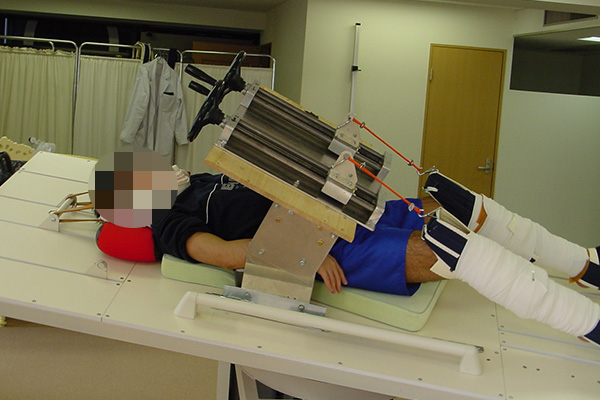
 It looks like it applies a rotational/twisting force on the leg combined with some kind of pressure.
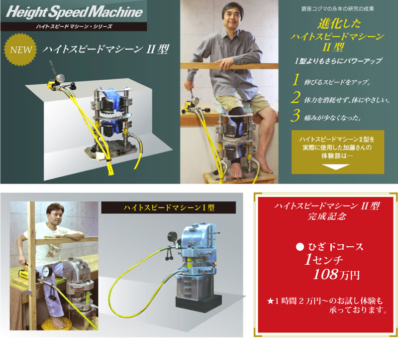