Sky’s Mistake, Why He Never Increased In Height

These days I am doing a lot of reading and research on sky’s old easyheight website and seeing what his methods were. Overall, I am starting to understand his approach and idea.

Sky was trying to use exercise and stretching methods to lengthen either the backbone/ lumbar area, the thigh bone, or the shin bone. He started the testing and work around 2003 and the site was around until 2011. He did not gain anything in terms of extra height. For all his passion, enthusiam, and belief, he didn’t grow taller. Why?

Here is my most educated guess. He was trying to increase his height using only exercise and stretching.

The big problem with Sky’s approach is that ever since humans evolved to the species that they are now, there has been estimated that there are over 100,000,000,000 humans. Of that 100 billion, that spans a time range of about 5 million years. In those 5 million years, there have been obviously many people who were “short” or have been considered short within the society they grew up and raised in. All these people probably wished at some point to be able to increase their height at some point.

Many of them when they were younger would have probably tried to increase their height using some “common sense” exercises. The biggest ideas are stretching, pulling the long bones, using gravity as a downward force like using inversion tables and hanging on a bar (even Michael Jordan did that). For the average person, not a real medical professional, those exercises seem reasonable in that they might be able to help increase height a little. The truth is that those exercises will increase height at least a little, but the increase is only temporary.

And throughout the 5 million years people have existed, not one person or group of people have found one method to be able to increase in height, at least through the ordinary exercise way. That was sky’s mistake. Why did he think he could discover a way to make the long bones elongate when so many other people before him couldn’t.

Let’s look at some of the most barbaric and tough civilizations that have existed.

1. Spartans- this society existed in creating super soldiers. Anything that resulted in stronger, bigger, and better fighting warriors was prized. They used to throw newly born babies down a cliff if the baby did not look healthy. This civilization would have trained their men and soldiers insanely hard to grow stronger. Even a person of average intelligence could ahve guessed that they probably also tried experiments to see if they could make their people taller too, for itimidation and war reasons. They failed to make their soldiers taller through all the training and exercises.

2. Aztecs- this civilization has gained a lot of infamy for taking slaves and war captures and sacrificing them by cutting out their hearts. The civilization was very brutal in the treatment of their bodies. I would guess that these people tried also to possibly grow taller from manipulating their bodies. THey were also a rather violent group of people who was almost always as war with other tribes. Being taller would have helped them look more intimidating and given them more of an advantage in battles. The failed to make their people bigger.

3. Japanese- During the 20th century the Japanese went into world war against the allied nations. If you have ever watched the old documentaries you coudl see that the young japanese children were forced to learn calisthenics, martial arts, stretch, and gymnastics to make their bodies taller and stronger so that they can be sent to the fornt lines and fight the othe nations. The Japanese knew during those times that they were much shorter than some other nations, especially the Americans who were the tallest nation during the 30s and 40s, and they were trying to get their young children to do exercises to match the height and size of the Americans they were fighting against. They never succeeded in creating a program to actually make the kids taller.

4. Chinese martists artists – In China’s 3000 year old tradition there has been thousands if not millions of martial arts experts, masters, etc. If you would guess anyone had better understanding of their own bodies and how to possibly grow taller, you would guess these types of people would have the best chance. So far, I have not heard of one martial arts master or ancient mythical fighter who was able to grow taller using some secret hidden technique they learned from a system of fighting. No one succeeded

5. Soviet scientists – The soviets were very famous for trying all sorts of bizzarre experiments on their own people to test the limits of what the human body was capable of. They used their own soldiers to take mind altering drugs and try out ancient meditation practices to see fi their military can learn to harness the old claims of ESP and psionic powers. It was one of these soviet scientists name Ilizarov through testing children and adult through brutal and painful experiments that discovered the world’s first real way to lengthen bones which gave us a real height increasing technique. Those types of experiments Ilizarov did to create his device would have never been allowed by the American medical community for how brutal and inhumane they treated their human patients and test subjects. Today, that type of human body experimentation would have been completely outlawed and he would be thrown in jail. I am almost positive that the Soviets also took their most brilliant of anatomists and asked them to look for ways to make their soldiers and people taller, again to show to the Americans that their way of thinking and society, running on the Communist ideas was better than the capitalist ways. Again, these brilliant soviet scientists could not find a way to increase the human height, at least just from exercise routines. One of them did succeed however in building a device with coupled with bone distraction surgery can increase a person’s height.

6. Chinese scientists – Like the soviets, the chinese scientists would have also tried at some point to try to develop some system to make their countrymen’s height increase. We have no idea what type of biological or chemiscal testing is being done by the chinese government right now. They could be testing on subjects for psionic and ESP powers just like the soviets did 40 years ago. They could also be testing exercise and surgical methods on subjects to try to make people taller too. So far, nothing in the chinese news have stated anything of big breakthroughs on the science of height increase yet.

My entire point from all these examples is to show that the way Sky was approaching the problem was exactly the same way billions of people before him who desired to achieve height increase also did. He thought a simple easy form of exercise routine which focused on tensile loading of the human leg long bones would be able to stretch the body out. Did sky realize that the human femur has a maximum tensile strength of 150 MPa? That strength of femur cortical bone is just at strong as steel. That’s right, steel!! It would make no sense that a person who can barely exert 500 lb/in^2 of power or even be able to rip up a phone book can possibly be able to use 30 lb ankle weights from repetitive kicking allow the bones of such high tensile strength to create enough microfractures to cause elongation.

Sky’s approach was wrong. We have seen that people from the past who were more dedicated than us, with more intelligence, more education, more time to do experiments and test on real humans, and more resources all fail in their endeavor to increase people’s height. Not one civilization in human history has been able to create a way to grow taller until the soviets through experimentation did create the ilizarov device, but they still had to use surgery and cut the bones in half first. There was no way sky using the small loads and the low rates that he did could have succeeded.

The Effect On Height By Proteoglycans

When I was doing my research to learn more about the detail of the growth plates and it’s subsections, I read that the chondrocytes creates a waste or output which is collagen and proteoglycan. I have already looked at the effects and possibilities of Collagen Type II on height and height increase in a previous article. Now in this post, I wanted to explore the affect and effects that proteoglycan has on human growth and height.

First, What is proteoglycans?

From the Medical Dictionary website (source HERE)…

proteoglycan /pro·teo·gly·can/ (pro″te-o-gli´kan) any of a group of polysaccharide-protein conjugates present in connective tissue and cartilage, consisting of a polypeptide backbone to which many glycosaminoglycan chains are covalently linked; they form the ground substance in the extracellular matrix of connective tissue and also have lubricant and support functions.

(Dorland’s Medical Dictionary for Health Consumers. © 2007 by Saunders, an imprint of Elsevier, Inc. All rights reserved.)

pro·te·o·gly·can (prt-glkn, -kn)

n.

Any of various mucopolysaccharides that are bound to protein chains in covalent complexes and occur in the extracellular matrix of connective tissue.
(The American Heritage® Medical Dictionary Copyright © 2007, 2004 by Houghton Mifflin Company. Published by Houghton Mifflin Company. All rights reserved.)

proteoglycan [pro″te-o-gli´kan]

any of a group of glycoproteins found primarily in connective tissue and formed of subunits of glycosaminoglycans (long polysaccharide chains containing amino sugars) linked to a protein core like bristles on a bottle brush. Hydrated proteoglycans form the highly viscous fluid of mucus and the matrix of the intercellular ground substance of connective tissue. Called also mucopolysaccharide.
(Miller-Keane Encyclopedia and Dictionary of Medicine, Nursing, and Allied Health, Seventh Edition. © 2003 by Saunders, an imprint of Elsevier, Inc. All rights reserved.)

——->

For a further in depth detail, From Wikipedia (source HERE)…

Proteoglycans are proteins that are heavily glycosylated. The basic proteoglycan unit consists of a “core protein” with one or more covalently attachedglycosaminoglycan (GAG) chain(s). The point of attachment is a Ser residue to which the glycosaminoglycan is joined through a tetrasaccharide bridge (For example: chondroitin sulfate-GlcA-Gal-Gal-Xyl-PROTEIN). The Ser residue is generally in the sequence -Ser-Gly-X-Gly- (where X can be any amino acid residue), although not every protein with this sequence has an attached glycosaminoglycan. The chains are long, linear carbohydrate polymers that are negatively charged under physiological conditions, due to the occurrence of sulfate and uronic acid groups. Proteoglycans occur in the connective tissue.

Proteoglycans can be categorised depending upon the nature of their glycosaminoglycan chains. Proteoglycans can also be categorised by size (kDa).

Types

Types include:

Glycosaminoglycans Small proteoglycans Large proteoglycans
chondroitin sulfate/dermatan sulfate decorin, kDa=36

biglycan, kDa=38

versican, kDa=260-370, present in many adult tissues including blood vessels and skin
heparan sulfate/chondroitin sulfate testican, kDa=44 perlecan, kDa=400-470
chondroitin sulfate neurocan, kDa=136

aggrecan, kDa=220, the major proteoglycan in cartilage

keratan sulfate fibromodulin, kDa=42

lumican, kDa=38

Certain members are considered members of the “small leucine-rich proteoglycan family” (SLRP).[3] These include decorin, biglycan, fibromodulin and lumican.

Function

Proteoglycans are a major component of the animal extracellular matrix, the “filler” substance existing between cells in an organism. Here they form large complexes, both to other proteoglycans, tohyaluronan and to fibrous matrix proteins (such as collagen). They are also involved in binding cations (such as sodium, potassium and calcium) and water, and also regulating the movement of molecules through the matrix. Evidence also shows they can affect the activity and stability of proteins and signalling molecules within the matrix. Individual functions of proteoglycans can be attributed to either the protein core or the attached GAG chain and serve as lubricants.

Synthesis

The protein component of proteoglycans is synthesized by ribosomes and translocated into the lumen of the rough endoplasmic reticulum. Glycosylation of the proteoglycan occurs in the Golgi apparatus in multiple enzymatic steps. First a special link tetrasaccharide is attached to a serine side chain on the core protein to serve as a primer for polysaccharide growth. Then sugars are added one at a time by glycosyl transferase. The completed proteoglycan is then exported in secretory vesicles to the extracellular matrix of the cell.

Proteoglycans and disease

An inability to break down proteoglycans is characteristic of a group of genetic disorders, called mucopolysaccharidoses. The inactivity of specific lysosomal enzymes that normally degrade glycosaminoglycans leads to the accumulation of proteoglycans within cells. This leads to a variety of disease symptoms, depending upon the type of proteoglycan that is not degraded.

ME: What is important to takeaway from this is that Proteoglycans (mucoproteins) are formed of glycosaminoglycans (GAGs) covalently attached to the core proteins. There are many types of glycosaminoglycans (GAGs). The most commonly well known GAGs are Hyaluronic acid, Dermatan sulfate, Chondroitin sulfate, Heparin and heparin sulfate, and Keratan sulfate (source HERE

From source http://www.cryst.bbk.ac.uk/pps97/assignments/projects/emilia/Proteoglycans.HTM

Structure of proteoglycans 

The GAGs extend perpendicular from the core protein in a bottlebrush- like structure.

The linkage of GAGs such as (heparan sulfates and chondroitin sulfates) to the protein core involves a specific trisaccharide linker :

Some forms of keratan sulfates are linked to the protein core through an N-asparaginyl bond.

The protein cores of proteoglycans are rich in Ser and Thr residues which allows multiple GAG attachment.


Role of proteoglycans and glycosaminoglycans

They perform numerous vital functions within the body.

GAG dependent functions can be divided into two classes: the biophysical and the biochemical.

The biophysical functions depend on the unique properties of GAGs : the ability to fill the space, bind and organize water molecules and repel negatively charged molecules. Because of high viscosity and low compressibility they are ideal for a lubricating fluid in the joints. On the other hand their rigidity provides structural integrity to the cells and allows the cell migration due to providing the passageways between cells.

For example the large quantities of chondroitin sulfate and keratan sulfate found on aggrecan play an important role in the hydration of cartilage. They give the cartilage its gel-like properties and resistance to deformation.

Aggrecan is one of the most important extracellular proteoglycans. It forms very large aggregates (a single aggregate is one of the largest macromolecules known ; it can be more than 4 microns long). Aggrecan molecules are non-covalently bound to the long molecule of hyaluronan (like bristles to the backbone in a bottlebrush). It is faciliated by the linking proteins. To each aggrecan core protein multiple chains of chondroitin sulfate and keratan sulfate are covalently attached through the trisaccharide linker .

The other, more biochemical functions of GAGs are mediated by specific binding of GAGs to other macromolecules, mostly proteins. Proteoglycans participate in cell and tissue development and physiology.

EXAMPLES OF GAG BINDING PROTEINS :

Secreted proteases and antiproteases

For example antithrombin III (AT III) binds tightly to heparin and certain heparan sulfates (so do its substrates). Thus they control the blood coagulation. In the absence of GAGs AT III inactivates proteases (such asthrombin, factors IXa and XIa) very slowly. In the presence of appropriate GAGs these reactions are accelerated 2000-fold.

GAGs are sufficiently long that both protease and protease inhibitor can bind to the same chain (thus the likelyhood of the two proteins binding to each other is increased enormously). GAGs also affect the protein conformation that contributes to improving AT III binding kinetics.

Polypeptide growth factors

Members of the FGF family, as well as several other growth factors, bind to heparin or heparan sulfate. Binding to endogenous GAGs entraps these molecules in ECM from which they may be later released. GAGs can alter the conformation, proteolytic susceptibility and biological activity of some of these proteins. The bound growth factor is resistant to degradation by extracellular proteases. Active hormone is released by proteolysis of the heparan sulfate chains. It occurs during the tissue growth and remodeling after infection.

Me: Tyler from HeightQuest wrote a similar post talking about the link between proteoglycans and height which questioned whether strengthening the intervertebral disks will increase height.
His results show that exercise seems to increase the size of the proteoglycans but not the numbers for certain areas. , and he seems to conclude that an increase in the size of proteoglycans will not lead to disk widening because the proteoglycans are proteins and proteins don’t make up the structure. There was an increase of collagen and proteoglycan on the inner material (nucleus pulpous) but not the external part (annulus fibrosis).
Conclusion: Here is what I am willing to conclude at this time. The decrease in proteoglycans will definitely result in lose of cartilage and height decrease, but the increase in proteoglycans probably will not increase the amount of cartilage available in the body or increase height. 

Increase Height And Grow Taller Using Collagen II

One of the compounds that appear over and over again in my search was collagen, both type I and type II. If we remember, collagen is one of the compounds that the chondrocyte actually excrete (the other being proteoglycan) when they are still in the proliferate zone in the multilayer hyaline cartilage growth plate.

For most people, when they hear the word “collagen” they might think of collagen injections people get for plastic surgery to make their face look smoother and younger. Since I am currently living in a location of the world very famous for its plastic surgery clinics, I am very familiar with the use of collagen for plastic surgery reasons.

The first question to answer is “what is collagen type 2?”

From the Wikipedia article on it (HERE)…

Type-II collagen is the basis for articular cartilage and hyaline cartilage.

It makes up 50% of all protein in cartilage and 85-90% of collagen of articular cartilage.

Type II collagen does form fibrils. This fibrillar network of collagen allows cartilage to entrap the proteoglycan aggregate as well as provide tensile strength to the tissue.

Treatment of Rheumatoid Arthritis

According to a study published in the journal Science, oral administration of type II collagen improves symptoms of rheumatoid arthritis. The authors conducted a randomized, double-blind trial involving 60 patients with severe, active rheumatoid arthritis. A decrease in the number of swollen joints and tender joints occurred in subjects fed with chicken type II collagen for 3 months, but not in those that received a placebo. Four patients in the collagen group had complete remission of the disease. No side effects were evident.

Me: What the study seems to say is that collagen type 2 might have helped regenerate some lost articular cartilage at the ends of long bones or just in joints. Since the articular cartilage is made of 80-90% collagen type 2, it would kind of make sense that taking some type 2 collagen orally may indeed lead to some articular cartilage regeneration. Unfortunately, one of the most well known and accepted facts in the medical community is that unlike almost all other tissues, cartilage are not supposed to be able to regenerate back. This means the study for the treatment of rheumatoid arthritis and the results don’t agree with the non-regeneration of cartilage axiom held by mos tof the medical community. 

Note: It seems that while articular cartilage is 80-90% collagen type 2, for cartilage in general, specifically the hyaline cartilage in growth plates, it is only 50%. This seems to suggest the rest, the other 50% of proteins that form cartilage is from the proteoglycans. If that is true, I hypothesis that to keep the proliferation zone of the growth plate around longer, we should be taking something to help increase the proteoglycan in our bodies, not the collagen type 2!

Tyler at HeightQuest.Com already wrote an article on the idea HERE.

He states that “”…and if microfractures occur while the bone is in this stretched state it stands to reason that the bone will maintain some of this state.  If we can increase the collagen content of bone, then we should be able to put more of a distraction force on the cortical bone.  Microfractures will maintain some of this distracted state.””

From the paper he highlights, he concludes that “”Collagen is increased by Mesenchymal Stem Cells.  To increase collagen content in bone or the hyaline cartilage growth plate line then one needs to increase expression of the genes that control mesenchymal stem cell lineage(such as COL2A1 for hyaline cartilage).””

He continues to suggest that the other types of collagen (1, 9, 10) can all help in increasing height because they cause the mesenchymal stem cells to differentiate more into chondrocytes that hypertrophy.


Can We Build A Better Limb And Leg Lengthening Device For Surgical Application?

I was again doing research for another article, specifically the one for the internal ISKD method and I started to wonder whether It was possible for the height increase researchers on here to develop a better biomedical device to be used to stretch out the long bones.

Here are all the types of medical devices out there right now, all used in the surgical method for limb lengthening. The resource used to find all this information below was from the Short Support Website located HERE

1. Albizzia is also called GEN for Gradual Elongation over intramedullary Nail and is a variation of Internal Lengthening Over Nails. The device is sping loaded. The rotation of the patient’s lower extremity creates the distraction with an audible “click.”

2. Bliskunov’s method

3. The Fitbone® device – the only device that uses a powered system to lengthen the legs. The bones of the leg (Tibia and/or Femur) are cut and the intramedullary telescoping nail is implanted in the bone marrow of each bone. Each end of the telescope nail is attached to each end of the cut bone. The nail is connected to an induction receiver that is placed just under the skin. An external control unit powers the telescope nail though the induction plate. A similar technique has been used to power artificial hearts.

4. Intramedullary Skeletal Kinetic Distractor (ISKD) – uses a kinetic clutch mechanism to lengthen the leg. One segment of a rod is screwed onto another and the whole rod is inserted into the patient’s bone. When the patient rotates his or her leg, the lower segment rotates over the upper one, like screwing a bolt out of a nut, and the rod lengthens, expanding the leg.

5. Lengthening Over Nails (LON) – a metal rod is inserted into the central cavity (intramedullary) of the lower legs (the tibia bone), and then the external fixator device is attached to the bone. As the limb is lengthened, one end of the bone slides over the rod and new bone is grown around it. When the bone is fully lengthened, the external device is removed and the rod is surgically attached to each bone segment. During bone strengthening, the rod provides support instead of the more uncomfortable and unwieldy External Fixator Device. At the end of the Strengthening phase, a second operation is performed to remove the metal rod.

6. Micro-wound – uses a fixative clip instead of an Ilizarov fixator. The fixator clip covers just one side of the leg and appears to be more comfortable than the Ilizarov fixator which completely surrounds the leg.

7. Precice™ – uses non-invasive adjustable intramedullary rods or bone plates to lengthen long-bones (e.g., femur, tibia). There are no open wounds. Lengthening is achieved through an External Remote Controller (ERC). The “ERC” is a portable, hand held unit that uses permanent magnets to automatically modify the length of the implant through the touch of a switch

8. Salamehfix – a hinged External Fixation System.  It is an arc system rather than a circular system and consists of three small arcs. The arcs are not the same diameter so the system can take the shape of leg. The entrance of screws and wires are in minimally painful regions so it’s more tolerable, provides for stable fixation and allows early full weight bearing. The small size of the system allows clothing to be worn over it with full mobility of nearby joints.

9. Original Ilizarov External Device

Me: It really is absolutely amazing to see what type of technology has been developed in the last 40 years ever since the Ilizarov fixator first came on the scene and allow people who had uneven limbs to get medical treatments fot their attention. Very quickly,  the short stature community realized the potential of this device and got the surgeons to use them to make people taller. 

Dr. Paley who has been doing the limb lengthening surgery for over 30 years now have come up with two big innovations on his own just from doing the surgeries and from experiences, seeing how they can be improved on.

In my opinion however ,the devices that are currently out can be made even better and improved on in the design. So far there is 9 different types of devices, but they all operate on the exact same type of principles. First, you cut open the bone you want to lengthen. Then you put something that can hold the two bones a small distance apart from each other. The bone ends starts to heal, and you slow move the bone parts further and further apart as the bones heal The bones thus increase. The muscles around them are also stretched out in the process. 

Getting back to the original issue, can we design a better device? I think we can. Here is my proposition.

The first principle to focus on is to make the device as non-invasive as possible. In my opinion, that means that after the bone has increased and healed it’s over, a quick trip to the doctor should mean the removal of the device and the bones will need little recovery after that.

The 2nd principle is to design a device that is strong but lightweight enough to hold up to the rigors of the limb bending and movement. The device can not collapse to the weight exerted on it by the human bones.

The 3rd principle is to create a design that can allow the full support of the leg. The leg MUST be completely fixed in place or the bones might turn or tiwst incorrectly leading to improper healing and the eventual bone regrowing into something crooked or stunted.

My idea for the device is a combination of the Microwound device used by Dr. Helong Bai and the Salamehfix device used by Prof. Ghassan Salameh. I will write up the new design in a future article. That however probably won’t come out for another 4-6 months, mainly because I want to study all of the devices that are out now and I haven’t done that yet.


A Step By Step Procedure And Guide To Explain The Original Ilizarov Method

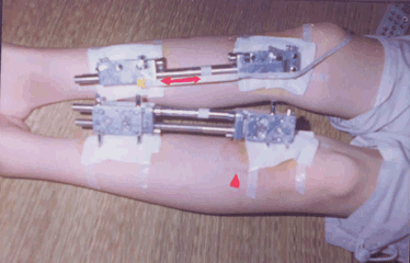
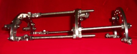
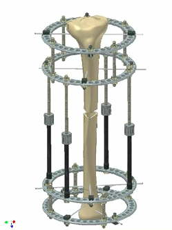
This will be a complete step by step explanation of how the original ilizarov method and device was used to increase patients limbs. Remember that the soviet orthopedic surgeon was only intending to use it to help people with pathologies like stunted limbs so that they can live a more normal life. I wanted to post a picture to the right of the beginning of this post to show what exactly are the benefits and the liabilities of using the external fixator ilizarov method. It is noted right now that the picture shown on the right was taken from a Video I saw on Youtube, specifically entitled “Finite Element Analysis of Ilizarov External Fixator” which is located HERE. The pictures is not mine, but seems to come from a presentation given by the School of Mechanical, Aerospace, and Civil Engineering at the University of Manchester.

From Youtube Video from HERE is named “Beauty” which was on Season 7 Episode 1 of National Geocraphic’s website series called “Taboo”, from time range 15:00-25:00.

Everyday the bones are pulled apart so that they grow longer as they heal. The patient was named Margarita Rosam(n)ova gained about inch or slightly more, I guess 3-4 cm more in height). The surgery was performed by Dr. Constantin Navukov (spelling?) . It was done at the Illarov Clinic located in Kurgan of Siberia. The procedure was stated to cost $26,000 and the complications are infection, disability, and maybe also amputation. This type of surgery, as in all other types of surgery, there is no complete guarantee of success.

Note: during the entire operation, the patient of course will be put under with the strongest possible painkillers. If they don’t do that, the surgeons you choose to use may not be the best quality.

0. The patient is wheeled into the operations room where the usually team of surgeons and anesthesiologists will be. Their lower legs are first shaved of all hair to keep the operated on areas smooth. The lower legs are then dabbed/swabbed with alcohol for infection protection reasons. Then the operation really begins.

1. First, long wire metal spokes are drilled into the patient’s leg with an electric power drill. The area and bone which is almost always worked on is the lower leg part where the tibia and fibula are. The metal spokes go right through the bone and protrude from the skin onto the opposite side. This process of making holes is called perforation. The holes will make allow the surgeons to manufacture the fracture and achieve the results they want.

2. Each spoke is attached to either the top or bottom of the patient’s tibia or fibula bones. If they are not placed in the utmost precision, they might damage tissue or nerves. Usually there will be around 8-12 long metal spokes drilled through the two entire long bones, tibia and fibula. The spokes are first drilled in one direction, and the next one is drilled in another direction, about 60-90 degrees away from the first spoke. The exact location and direction on where to drill the thin metal spokes/ rods are really based on the human anatomy and bone positioning, which I don’t know at this time. I would assume any orthopedic surgeon would know EXACTLY what are the exact areas to drill into.

3. 3 of the spokes are usually drilled to the top of the tibia, 2, spokes drilled around the upper middle area of the tibia, and 2 are drilled around the lower middle area of the lower leg bones, and 3 spokes are drilled to the bottom of the tibia. Again, the exact location and direction of on how and where the spokes are drilled are not know to me at this time. I would assume there must be at least 2 spokes that also go through the fibula as well so that the fibula can extend too but I am not sure about that part. I am not sure about the exact number of spokes/drills there are supposed to be. In some videos and pictures I see less spokes used like around 8 and in others I see a lot more like around 12.

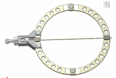
4. The surgeons then attach a metal brace called the Ilizarov frame, which is just two semicircular shaped metal pieces together, attaching the ends of the two pieces to form a thin circular ring which is about 2-3 mm thick and has a radial thickness of about 1 cm. In the metal semicircles, there are many holes on it equidistance positioned away from each other. The reason for the holes are for the poles and nuts to eventually go. Screw bolts or nuts or studs are used to close the ends.. The important thing is that the method allows the surgeons to keep all of the pieces and vitally important structures intact.

There is usually 8  semicircle metal pieces, that attach together to form 4 metal circles that will hold the spokes together and hold the legs into place.

5. The spokes are pruned at the ends for equal distance protruding from the leg. A rather larger piece of round plastic is pushing into the spokes, on both ends right next to there bone proferations are so they can hold the spokes into position. a piece of plastic goes right next to each bone/ skin protraction of spoke. The spokes is somehow attached to the circular metals by passing the thin spokes which look just like thick strong metal wires through a piece of strong plastic that is attached to the metal circle using screws which go through the holes.

The metal circles are then attached to each other through long and super long rod shaped screws. The shorter length screws are used to attach the circles on the edge of the lower leg regions to the circles closer to the middle. The really long rod shaped screws are the ones that hold the middle circles together. Of course, the ends of all screws are assumed to held into place with either strong plastic or lug nuts or bolts. If you can not picture what I am saying at this time, refer to the picture on the right.

6. With the frame in place, the surgeon then takes a chisel and a hammer. The chisel is inserted into the skin until it reaches contact with bone. While holding the chisel with one hand very steadily to one specific area in the tibia, the other hand swings the hammer at the tibia at a relatively strong force, in a tapping motion. The chisel acts similarly like a railway spike.

7. To avoid damaging the marrow in the inter medullary cavity, another surgeon works on the bone from the opposite side. The hammering in a steady but firm and strong tapping rate goes on usually for about 10 minutes until the chisel has made a clear fracture in the tibia, but only for the outer cortical bone area, not the inner cancellous (trabecular) bone part which should still be intact holding the marrow inside.

8. To check to make sure the proper fracture is made, the surgeons then get an X-Ray of the just operated on tibia bone. They want to make sure that both bones, the tibia and fibula are fractured.

9. Over the next few months in the futures, the gaps or fractures will be incrementally widened by turning screws on the long poles that have been used to hold the  inner metal circularly frames together. As the 4 screws are turned, the 4 poles used to hold circles together fixed in place will grow and extend longer. The bones will then grow longer to fill the gap.

10. The patient will be bed ridden for at least 4 months. The surgeons will come by on a regular basis to check up on the patient and to take measurements of the leg that is being lengthened. a baseline measurement of the leg is made. The frame will be slowly stretched. Usually the rods have only a set maximum amount of extension lengthening that is possible. Once the set lengthening is reached, the frames can be removed.

Eventually, the Frames are removed after about 4-6 months. The spokes are taken out.  There is still a lot of physical therapy that needs to be done. The newly formed leg bones may not be strong enough to hold up the patient so it takes a while. Since the spokes are pushing through the skin, the gradual lengthening and stretching of the bone also will touch the nerves and cause excruciating pain. The bone stretching may also tear the skin leaving scar marks.

Hairstyles And Hair Tips For Men And Women To Look Taller

This is one of those posts that is used to help you LOOK taller, and when it comes down to real life application, much easier to implement that any real height increasing procedures. It is too bad there really is not that much information that I could find that talks about how to look taller from using hair styles or tips. The general conclusion is to make the sides of the head short and keep the top of the head grown out and extended up.

From the Daily Makeover website HERE

The Shorter Haircut Can Make You Look Taller

If you’re petite and wish to look taller, consider ashort haircut. That may sound counterintuitive, but Scrivo explains: “A petite woman who wears her hair very long can seem even smaller in stature because the scale of the hair is not proportionate and overwhelming to her figure. If the hair were shorter, the focus would move up toward the face and visually lift up a petite frame.” Vanessa Hudgens is a great example. See how she looks taller with a shorter hair?

From WikiHow.com

Advice for Women

  1. Keep your hair short. Long hair can “drown” shorter women. A hairstyle that stops above the shoulder prevents a woman’s locks from overwhelming her entire stature.
  2. Put your hair up. If you prefer long hair, consider putting it back in high ponytails, buns, and other up-do’s. Hair that extends several inches above the head adds inches to your height. Moreover, keeping your hair back prevents you from “drowning” in it.
Add layers or highlights to your locks. Long, straight hair that hangs down in a single layer has the most overwhelming effect. Adding choppy layers or highlights breaks up the monotony of long hair, which may help prevent it from dwarfing you.

Advice for Men

  1. Add height to your hairstyle. Keep the sides of your hair neatly trimmed, but leave plenty of hair on top. The extra volume on top, when matched with the decreased volume on the side, adds inches to your overall height. This trick works especially well if you use gel to create a spiky style.