Increase Height And Grow Taller Using Low Intensity Pulsed Ultrasound, LIPUS

Now in my search for a solution to our height increase problem, I have looked through at least 30 different ideas people have thought up as a possible solution. Some of them make no sense, some of them are scams just trying to take your money, other methods are created with good intentions but just can’t work, and some have a chance to work but there has only been circumstantial evidence and testimonials which are hard to believe. Most of us want to see it with our own eyes to believe in something.

As for the latest technologies that might be coming along that can solve our problems, the 3 big ones are stem cells (probably through implants), gene therapy which is what I believe will ultimately happen if we can figure out how to integrate foreign DNA into our cells and mutate our bodies in a safe way for desired phenotypical traits, and the the joint loading modality (LSJL) which only a few people even know exists. There is some claims over hypnosis and mystic practices working too but that is vert hard to prove. Recently this new technology has come out that is really causing a lot of noise for height increase seekers, but also the dental community as well. It is the method called “Low Intensity Pulsed Ultrasound” or LIPUS for short.

Apparently I have been doing height increase research for so long and even I hadn’t heard about it until Harald of the Biomedical Growth Research Initiative told me that the two major technological developments that currently have the most potential were the LIPUS method and tissue engineering. I knew what tissue engineering did but I had not heard of LIPUS so I did some research. The stuff I found was very interesting. Then Harald explained to me that one of the ideas for his organization was to use LIPUS in some way to increase height, which made the method even more relevant.

Afterwards, I found information on Tyler’s HeightQuest.Com blog about LIPUS so I realized that the guys who are all at the cutting edge of height increase technology and innovation was suggesting that LIPUS could be very big deal. When I was doing research on the Lateral Synovial Joint Loading method on the HeightQuest.Com website, Tyler stated a lot about the potential of the LIPUS in being able to increase our height. That is why that I felt that I should devote quite a bit of time and effort into doing some extensive and serious research on the method and see just how it works, and how effective it will be.

So the first question would be “What is this technology called Low Intensity Pulsed Ultrasound?” . Wikipedia seems to have a clear and short answer to this question so I will paste their answer below (found HERE)


Low-intensity pulsed ultrasound (LIPUS) is a medical technology, generally using 1.5 MHz frequency pulses, with a pulse width of 200 μs, repeated at 1 kHz, at an intensity of 30 mW/cm2, 20 minutes/day.

Applications of LIPUS include:

  • Promoting bone-fracture healing.
  • Treating orthodontically induced root resorption.
  • Regrow missing teeth.[citation needed]
  • Enhancing mandibular growth in children with hemifacial microsomia.
  • Promoting healing in various soft tissues such as cartilage, inter vertebral disc.
  • Improving muscle healing after laceration injury.

Researchers at the University of Alberta have used LIPUS to gently massage teeth roots and jawbones to cause growth or regrowth, and have grown new teeth in rabbits after lower jaw surgical lengthening (Distraction osteogenesis) (American Journal of Orthodontics, 2002). As of June 2006, a larger device has been licensed by the Food and Drug Administration (FDA) and Health Canada for use by orthopedic surgeons. A smaller device that fits on braces has also been developed but is still in the investigational stage and is not available to the public.

It has not yet been approved by either Canadian or American regulatory bodies and a market-ready model is currently being prepared. LIPUS is expected to be commercially available before the end of 2012. The LIPUS foundation website currently announces that Lipus-Plasma application units are available for rental in the USA.

According to Dr. Chen from the University of Alberta, LIPUS may also have medical/cosmetic benefits in allowing people to grow taller by stimulating bone growth.

LIPUS has also been found to stimulate the proliferaton of chondrocytes.


Obviously the last two phrases on the wikipedia article are the most interesting since they state directly that the technology might have the application for stimulating bone growth by the promoting the proliferation of chondrocytes.

Not only this, this is incredible news for people who suffer from dental problems stemming from tooth decay. If this technology is as effective as advertized, it will change the dental industry. However, let’s learn more about what LIPUS does and how it does it.

From the UK website for National Institute For Health And Clinical Excellence located HERE the description for the device states…

Description – Low-intensity pulsed ultrasound aims to speed up fracture healing in broken bones by stimulating bone cells to grow and repair. This involves a short daily treatment using an ultrasound probe that is placed on the skin at the site of the fracture.

For the outline of the prodecure found in this sub-webpage HERE

2.2   Outline of the procedure

2.2.1  The aim of low-intensity pulsed ultrasound is to reduce fracture healing time and avoid non-union by delivering micro-mechanical stress to the bone to stimulate bone healing. This procedure is used to treat fresh fractures, fractures that are slower to heal than expected (delayed healing) and those that have failed to unite (non-union).

2.2.2  This is a non-invasive procedure. The ultrasound probe is positioned on the skin over the fracture and patients self-administer low-intensity pulsed ultrasound daily, usually for 20 minutes. If a patient’s limb is immobilised in a cast, then a hole is cut into the cast for the ultrasound probe. Coupling gel is used on the skin to aid conduction of the ultrasound signal. The operating frequency, pulse width, repetition rate and temporal average power of the ultrasound delivered can be varied.

In the Efficacy section, the experimental and testing results are provided…

2.3  Efficacy (RTC stands for randomized controlled trials)

2.3.1  A meta-analysis of 13 randomised controlled trials (RCTs) including a total of 563 patients (with a mixture of fresh conservatively or operatively managed and non-united fractures) treated by the procedure (n = 280) or a sham procedure (n = 283) reported a 34% (95% confidence interval [CI]: 21 to 44, 6 studies) overall mean reduction in healing time (confirmed by imaging) in patients treated by the procedure (follow-up not stated).

2.3.2  An RCT of 67 patients with closed or open grade I fractures of the tibial shaft (33 low-intensity pulsed ultrasound vs 34 sham) reported a mean healing time (defined as evidence of clinical and radiographic bridging of 3 cortices) of 96 days in the ultrasound group compared with 154 days in the sham group (p < 0.0001).

2.3.3  An RCT of 32 patients with fresh closed or open grade I fractures of the tibial shaft fixed with an intramedullary rod treated by the procedure (n = 15) or a sham procedure (n = 17) reported an average healing time (defined as radiographic bridging of 3 cortices assessed by a radiologist) of 155 days and 125 days respectively (p = 0.76).

2.3.4  An RCT of 21 patients with non-united fractures of the scaphoid treated by pedicle bone graft reported an average healing time (defined as clinical healing [solid and not causing tenderness or pain] and radiographic healing [complete bridging cortices]) of 56 days in patients who also received low-intensity pulsed ultrasound (n = 10) and 94 days in those who received sham therapy (n = 11) (p < 0.001).

2.3.5  An RCT of 30 patients with open tibial fractures or high-energy-induced complex tibial fractures treated by the procedure (n = 16) or a sham procedure (n = 14) reported an average time to full weight bearing of 9.3 weeks and 15.5 weeks respectively (p < 0.05).


Me: I managed to find a great scientific article that really goes into the explanation of the technology (located HERE). Let me take the most important parts of the article. 

“”….Low-intensity ultrasound is a biophysical form of intervention in the fracture-repair process, which through several mechanisms accelerates healing of fresh fractures and enhances callus formation in delayed unions and nonunions…Low-intensity pulsed ultrasound is currently applied transcutaneoulsy, although recent experimental studies have proven the efficacy of a trans-osseous application for both enhancement and monitoring of the bone healing process with modern smart implant technologies…”” (published by Elsevier Ltd. 2006)

Key Point: However, one of the fundamental concepts in orthopaedics is the understanding that the mechanical environment at the site of a fracture influences the pattern of fracture repair.

“….The healing of a fractured bone involves the spatial and temporal coordinated action of several different cell types, proteins and the expression of hundreds of genes working towards restoring its structural integrity….”

Basic science 

In vitro studies using cell cultures and research on experimental fractures in animal models have demonstrated a stimulatory biologic effect of low intensity ultrasonic energy on the intracellular activity, cytokine release and the bone healing process. In animal models, ultrasound appears to alter the time course or the sequence of gene expression of several genes, notably aggrecan, which is a proteoglycan involved in enchondral osteogenesis. Low-intensity ultrasound elevates intracellular calcium in cultured chondrocytes and stimulates endochondral bone formation in vitro. It also has direct effects on cell physiology by increasing the incorporation of calcium ions in cartilage and bone cell cultures and by stimulating the expression of numerous genes involved in the healing process. It alters potassium flux across the cell membrane in cultured thymocytes, and it modulates adenyle cyclase activity and TGF-b synthesis in osteoblastic cell
lines. In addition to modulating gene expression, ultrasound may enhance angiogenesis and increase blood flow around the fracture. Despite these well documented studies, the mechanism through which LIUS interacts with living tissue and stimulates bone healing remains unclear.

In addition to the above-mentioned molecular interactions, the acoustic pressure waves at the fracture site, facilitate fluid flow which, in turn, increases nutrient delivery and waste removal (acoustic streaming phenomenon), thus stimulating proliferation and differentiation of the fibroblasts, chondroblasts and osteoblasts.
In addition, the acoustic pressure waves produce micro-stress fields resulting in a mechanical response of the bone, analogous to the phenomena described by Wolf’s law. Small temperature fluctuations (<1 8C) appear at the fracture site as a result of the conversion of ultrasound energy to heat. Some enzymes, such as collagenase, are exquisitely sensitive to these small temperature variations, thus, ultrasound may also facilitate some enzymatic processes.

Trans-cutaneous application of ultrasound in the management of fresh fractures

Ultrasound increases soft callus formation and results in the earlier onset of endochondral ossification, suggesting that the most prominent effect is on the chondrocyte population….In a placebo-controlled study of bilateral mid-shaft fibular osteotomies in rabbits, Pilla et al. found that low-intensity pulsed ultrasound applied for 20 min/day significantly accelerated the recovery of torsional strength and stiffness.

The effect of ultrasound in distraction osteogenesis

Callus distraction is currently an established treatment for the management of defects larger than 3—4 cm in the long bones. However this technique carries the problem of the long time for healing and maturation of the newly formed bone and the burden to wear the external fixator for a very long time. The ossification process in distraction and maturation involves intramembranous bone as the dominant type of tissue formation while endochondral ossification normally is of minor importance.

The effects of low-intensity pulsed ultrasound on maturation of the distracted callus have been investigated in several animal studies, with controversial results. In a rabbit study, Shimazaki et al. found that bone mineral density, hard callus area, and mechanical test scores were greater in distraction callus treated with low-intensity pulsed ultrasound than in the control group. In a study of rats, Eberson et al. found that radiographicaly assessed healing occurred earlier in ultrasound treated bones than in control bones and that bone volume fraction and trabecular bone pattern, were higher in the ultrasound-treated bones. In a study of rabbits, Tis et al. found a greater hard callus area and less fibrous tissue in bones treated with low-intensity pulsed ultrasound than in control bones. Neither Eberson et al.
nor Tis et al. found a difference in bone mineral density or mechanical strength of distraction callus between ultrasound-treated bones and controls, although Eberson et al. observed a trend toward greater mechanical strength in ultrasound-treated bones. Uglow et al. found no substantial difference in bone mineral content, crosssectional area, or strength of distraction callus between ultrasound-treated bones and control bones of rabbits.

In a sheep metatarsal bone transfer model for the study of distraction osteogenesis, pulsed low-intensity ultrasound were applied transcutaneously after the distraction was complete and only throughout the maturation phase. Histologic analysis of the cortical defect zone showed approximately 32% more bone in the group stimulated by ultrasound. Although it presented seven times more intramembranous bone formation compared to endochondral in the control group, which is in accordance with results of another study, there was a three times higher rate of endochondral ossification in the specimens treated with ultrasound. Biomechanical tests showed significantly higher axial compression stiffness (1.4—2.7 times the control values) and significantly higher indentation stiffness of callus tissue in the healing zone of the treated bones. In all of the animal studies mentioned above, osteotomy and distraction were performed at the diaphysis, which consists of thick cortical bone. A recent investigation on rabbits showed that low-intensity pulsed ultrasound stimulates bone formation most effectively during the distraction phase.

In a randomised study (block randomisation) in humans with internal controls, the low-intensity pulsed ultrasound applied only during the consolidation phase (after distraction had ceased) on hemicallotasis after high tibial ostetomy, significantly enhanced the mineralisation of the callus. The bone mineral density in the metaphyseal segment adjacent to the distraction callus, in the previous study and also in animal studies collectively suggest that metaphyseal trabecular bone might be more susceptible than diaphyseal cortical bone to the mechanicalultrasonic stimuli.

Future clinical studies should address the question of whether additional low-intensity pulsed ultrasound treatment during the distraction phase can further shorten the period necessary for callus maturation. The distraction osteogenesis-specific mechanism that translates mechanical forces due to low-intensity pulsed ultrasound into bone formation need further clarification.

Me: Overall the paper was extremely insightful on the utility of the LIPUS technology. All I can say is that after reading the paper I can say with a 97% confidence that this technology is very amazing at what it can do. It really does have the potential to help people heal bones faster and to induce chondrocyte proliferation and bone formation. 

Note: Since this topic is a very large topic, I will stop right here and continue on this study on the device and technology in a later post. You can get to Part II by clicking HERE.

Growth Plate Regeneration By Robert Ballock Orthopaedic Surgeon

When I was in contact with Tyler from HeightQuest.Com he gave me two leads or ideas to look into so I could be better acquainted with what options and ideas are current available or in the process of being developed in our search for an increase in height. One of the ideas was the Joint Loading Modality done by Yokota which I wrote a very short review and analysis found HERE. The other idea he wanted me to do research on was the idea of “Growth Plate Regeneration” being developed by Robert Ballock from the Cleveland Clinic.

Let me do a little review and critical analysis on what I managed to find out about this method/technique. The first  thing I found from searching about the type of growth plate regeneration research done by Robert Ballock was either a paper submitted to the National Institute of Health (located HERE) or it was maybe a proposal for a grant or project.

However let’s get a summary of how Dr. Ballock describes his own research from his profile webpage found on the Cleveland Clinic website (located HERE) If you would like to contact him all his contact information is available on his profile webpage which you get get to again by clicking on the link above.

“”….I had the good fortune of being taken in by Drs. Michael Sporn and Anita Roberts in the Laboratory of Chemoprevention at the National Cancer Institute. Their laboratory had recently discovered TGF-beta and they were interested in exploring the role of this new peptide growth factor in regulating growth and differentiation in the skeleton.During these two years, I focused on the role of TGF-beta in the growth plate and developed the three-dimensional pellet culture model of growth plate chondrocyte differentiation that is now used by many laboratories throughout the world.This experience resulted in three publications, including first author papers in Developmental Biology and Journal of Cell Physiology….”

“”….I spent a third year in his laboratory at Johns Hopkins where I formulated the serum-free, chemically defined culture conditions for growth plate chondrocyte pellet cultures that are also now used by many laboratories world-wide.For example, Dr. Brian Johnstone and colleagues at CWRU used our pellet culture model and serum-free, chemically-defined conditions to demonstrate for the first time that mesenchymal stem cells could undergo chondrogenesis in vitro. This year at Johns Hopkins was marked by several other key observations, including the discovery that terminal differentiation of growth plate chondrocytes was a default pathway that could be accelerated by thyroid hormone, and that morphogenesis of columnar cartilage in the growth plate could be recapitulated in our serum-free three-dimensional pellet cultures by addition of thyroid hormone….”

“”….my research program began its focus on the molecular mechanisms of thyroid hormone action in the growth plate that continues to be the major thrust of our laboratory work today.We initially determined that the principal site of thyroid hormone action during skeletal maturation was regulation of the critical transition between cell proliferation and terminal hypertrophic differentiation in the growth plate.In addition to characterizing the expression of thyroid hormone receptors in this tissue, we also explored the interactions between thyroid hormone, vitamin D, and retinoic acid in regulating terminal differentiation of growth plate chondrocytes.Other work established an important link between thyroid hormone-induced terminal differentiation and upregulation of cell cycle proteins p21cip-1, waf-1 and p57 kip-1, indicating that growth arrest at the G1-S restriction point of the cell cycle is an obligatory step in the terminal differentiation process of growth plate chondrocytes….”

“”….we also began to investigate the role of obesity in the dysfunction of the growth plate that results in slipped capital femoral epiphysis, a potentially devastating hip condition in adolescents. These studies have demonstrated that peroxisome proliferator activated receptors (PPARs), which are upregulated in response to a high fat diet, are also expressed in growth plate chondrocytes and interfere with the normal transcriptional activation function of thyroid hormone receptors in these cells, eventually resulting in the inhibition of terminal differentiation and matrix mineralization that allows the subsequent mechanical failure of the growth plate to occur….”

“”….we initiated DNA microarray studies of growth plate chondrocytes in order to identify the direct downstream genetic targets of the thyroid hormone receptor in the growth plate.These studies resulted in the surprising identification of the gene encoding carboxypeptidase Z (CPZ) as a direct target of thyroid hormone action.CPZ is an enzyme that removes C-terminal amino acid residues, particularly arginines, from proteins, and has also been shown to modulate Wnt signaling.Further experiments in our laboratory have demonstrated that Wnt-4, which is the principal Wnt family member expressed in the mammalian growth plate, contains a C-terminal arginine, and that removal of this C-terminal arginine enhances the biological activity of Wnt-4 in inducing terminal differentiation of these cells….”

Me: As for what I can contribute to the effort or knowledge right now, there is little I can do that has not already been covered and researched by Tyler from HeightQuest.Com. He has already looked into the proposal ideas. And yes, it appears the first link was a grant proposal of some kind to get money to fund for a project. The project was supposed to have started in April of 2012 and end in March 2014. They appeared to have gotten some money, around $140,000 which in my opinion is really NOT a lot.  I’ll copy and paste the entire abstract right below here. I’ll highlight the areas which I feel are the most important and applicable to us height increase seekers. Again you can get to the paper submitted by clicking HERE.

Abstract Text:
DESCRIPTION (provided by applicant): The growth plate, also known as the epiphyseal plate or physis, is the area of growing tissue near the end of the long bones in children and adolescents that determines the future length and shape of the mature bone. Fractures through the cartilage growth plates of the long bones of children may result in growth arrest with subsequent leg length inequality and progressive deformity. This growth arrest is due to formation of a bony bar across the traumatic growth plate defect that acts as an tether to resist further longitudinal growth. If the bar is large or is located in the central portion of the growth plate, a complete growth arrest ensues. A bar located in the peripheral portion of the physis tethers growth asymmetrically, producing a progressive angular deformity of the limb. Once a physeal bar forms, surgical excision is technically difficult and resumption of further growth is quite variable. Previous studies of experimental growth plate injury have focused on the histological events in the growth plate defect leading to bar formation. However, our understanding of the factors that regulate the proliferation and differentiation of growth plate chondrocytes, as well as the principles of cartilage tissue engineering, have increased dramatically over the past decade. These advances now provide a unique opportunity to develop strategies for regeneration of normal physeal cartilage following serious growth plate injuries. Successful regeneration of growth plate cartilage architecture in vivo would have a transformational impact on the practice of pediatric orthopaedic surgery, providing for the first time not only the ability to replace growth plates irreversibly damaged by trauma, infection or irradiation, but also the possibility of restoring longitudinal growth in individuals beyond the age of skeletal maturity. Our hypothesis is that co-cultured chondrocytes and osteoblasts implanted into tibial bone defects in vivo will recapitulate the function of the normal growth plate and result in the reformation of columnar physeal architecture and resumption of longitudinal growth. This hypothesis will be tested by using a tissue engineering approach to determine the degree to which this optimized physeal construct replicates the function of the normal growth plate in vivofollowing implantation into a complete growth plate defect.PUBLIC HEALTH RELEVANCE: Our understanding of the factors that regulate the proliferation and differentiation of growth plate chondrocytes, as well as the principles of cartilage tissue engineering, have increased dramatically over the past decade. These advances now provide a unique opportunity to develop strategies for regeneration of normal physeal cartilage following growth plate injury. Successful regeneration of growth plate cartilage architecture in vivo would have a transformational impact on the practice of pediatric orthopaedic surgery, providing for the first time not only the ability to replace growth plates irreversibly damaged by trauma, infection or irradiation, but also the possibility of restoring longitudinal growth in individuals beyond the age of skeletal maturity.

Me: If you took the time to read the abstract, it might still be a little hard to understand what Dr. Ballock is talking about. The basic idea proposed is that for many children who are still growing, when they suffer an injury like a fracture on their growth plates, they can actually stunt their growth or growth irreuglarly afterwards because of how the growth plates cartilage form and heal themselves. To solve this problems, as well as give all height increase seekers around the world hope, is that they want to test this hypothesis and radical on implanting some cultured (made in the lab) chondrocytes (growth plate cells) and osteoblasts (bone cells) into the tibia fractures and defects and see if they can result in the growth plates in regrowing again and providing longitudinal growth (increase in bone length, thus increase in height). This technology is defenitely something every serious height increase seekers should stay up to date with. One might even be able to participate in the study and even get their height increase for free during the experimental trials and be paid some extra money for being a willing trial subject. Just something to think about. 

To be completely honest, it is actually really hard to find anymore information about Dr. Ballock or his research being done since professor doctors who do really advanced scientific research are not as easily found on the internet as a Kim Kardashian sex tape. To find the latest and most advanced innovations and scientific breakthroughs, you really have to dig through the science online databases like Pub Med. The problem is that I don’t have an account with PubMed and I am not sure I want to pay for something like a yearly fee to become some form of member. 

Chinese Orthopaedics Surgeon Bai Helong Says Limb And Leg Lengthening Surgery Is NOT Painful – Is He Lying?


OK. The main reason why I wanted to write about this article and about this subject, the Chinese surgeons and doctors who are willing to operate on people to increase their height is because it is one of the most controversial since the cosmetic procedure is technically banned in China but many Chinese doctors are still willing to do the surgery for the money. In some ways, China is like the former USSR and even Russia today. Many of the most radical medical technology creations and innovations seem to come from these countries because they don’t seem to care as much as people in the US over human safety protocols and often are willing to bend or break international rules and laws to make things happen. I am not condemning the actions of these doctors since it was Ilizarov, a Russian Doctor who developed the original idea on how to make limb lengthening surgery work for people with real legitimate medical conditions. I guess sometimes one does have to break the rules and try news things for real innovation to happen.

On the Make Me Taller boards I found a link to a rather well known distraction osteogenesis (aka limb and leg lengthening surgery) Chinese surgeon make some interesting claims that I found slightly ludicrous. The actual title to the Article is “Chinese Doctor Pioneers Height-Increase Surgery” . You can find the original article by clicking HERE.

As always, I will copy and paste the entire article right below, highlight the sections that I found the most fascinating ot most important, and then at the very bottom write a full review, analysis, or critique of the article and what it means for the application of height increase.


Chinese Doctor Pioneers Height-Increase Surgery

Updated Monday, April 12, 2010 11:26 am TWN, AFP -(Found on The China Post)

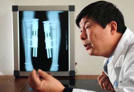
SHANGHAI — Orthopaedic surgeon Bai Helong hikes up his trousers, places his foot on his desk and marks the spot just below his hairless knee where he cuts into the legs of patients who want to be taller.

Over the past 15 years, Bai has given the gift of height to about 3,000 patients aged 14 to 55 — Chinese, Americans, Germans, Japanese — about half of whom went through with surgery simply because they did not like being short.

“I’m something of an authority in this field,” explains Bai, who uses a technique he developed himself at a modest private clinic in the suburbs of Shanghai.

He saws through both the tibia and the fibula below the knee — “without touching the bone marrow”, he says — to “make the dream come true” of those who say they suffer psychologically from being short.

One week later, the bones begin to regenerate. Heavy braces made of nickel and titanium, each weighing about half a kilo (one pound), are screwed into the inner part of the patient’s legs.

Every day for the following four months, Bai expands the braces to gradually stretch the leg.

“We need four months to get six to eight centimeters,” or two to three inches, the surgeon says. After that, for four more months, the bones get stronger and patients are allowed to begin to walk.

Leg-lengthening was first performed in the 1950s in the former Soviet Union, and then in China, but with sometimes catastrophic results.

In the past, the leg was cut in three places, affecting the delicate bone marrow, and pins were used to steady the bone. In some cases, one leg was left shorter than the other and infections were common.

Today, Bai says, the procedure is safe. Instead of stretching the leg by 1.0-1.5 millimeters a day as in the past, he aims to progress half as fast.

“We’ve not had a single failure since 1995, and now it’s not painful,” insists the doctor, who charges 75,000 yuan (US$11,000) for the surgery.

So who is willing to endure such a procedure, which involves months of total immobilization and a fair amount of discomfort?

“A small person can encounter all kinds of problems — in his or her marriage, family life, workplace,” Bai says.

“The person feels inferior, and experiences psychological problems. I even have met people who wanted to kill themselves.”

Dan Dan, a pretty 25-year-old Chinese woman who is studying Japanese, says she was unhappy when she stood 1.53 meters (five feet) tall. Four months after surgery at Bai’s clinic, she is smiling — and six centimeters taller.

“I wanted to improve my self-image. I am very happy,” says Dan Dan, grimacing as she walks at a snail’s pace on crutches through the halls of the clinic, her body contorted.

“I hope that within a year, I will be able to walk normally. Running, that’s another story,” she says.

Only Dan Dan’s mother is aware of what she is doing — her friends have no idea where she is.

“It seems pretty dangerous at the beginning — they cut through your bones, that is not really socially acceptable in China. It’s not like getting your eyelids done,” she explains, referring to a surgical procedure some Chinese women undergo to give them rounder, wider-looking eyes.

“I held off for a long time. I was really scared.”

Wang Lijun has not told her friends where she is either and as a result, they no longer contact her.

That was a price the 30-year-old was willing to pay during 13 months of treatment, which began in 2008 and took her from 1.52 meters to 1.60 meters — a height gain easily achieved with a pair of stilettos.

“It was my secret. I told no one,” says Wang, who now works on the administration side of Bai’s clinic and says she can run and jump “almost like before”.

“I had lost all of my self-confidence. I wanted a better life.”

Beyond the loss of her social circle, Wang says there were other sacrifices to make — the months of painful treatment, the dark, vicious-looking scars on her legs. Her next step? A plastic surgeon, perhaps.

Is Bai a miracle worker or a sorcerer’s apprentice with a screwdriver and a tire iron who is making a profit from the suffering of others?

The surgeon hits out at his critics, especially “those who oppose me without knowing what my work is all about”.

But as is often the case in China, Bai may be working in a grey zone.


Me: What is most interesting about the article is that this orthopaedic surgeon Bai Helong made a few claims in the article that made me really question whether he is being honest. The biggest claim is that his method, which he independently developed is NOT painful. I want to call him out on this bullshit. That is NOT fucking possible. The fact that he does have to cut a person’s long bones into separate pieces is A PAINFUL PROCESS. Even later in the article the author writes that the patients after surgery are getting around in an uncomfortable way. That means that they are in PAIN. I have read the diaries of other people who go through with this thing and there is no way that one can avoid pain. The Chinese surgeon is making it sound painless so that other people would consider using him as their surgeon than others who would scare them with stories of pain because they were just telling teh prospective patient the truth of the type of ordeal they have to go through.

The second issue I have with him was the extremely low price he put up. I think the article was written in before the last edited date which was in 2010 If I had to guess, I would say the article or the interview talk given by Bai Helong occurred in 2007 or 2008, maybe even earlier. He states that he charges $11,000 for the entire surgery. If you have ever done any research on the cost of limb lengthening surgery you would realize that the price eh gives is far , FAR lower than anything I have seen by any other surgeons who do the same type of surgery. The really low pricetag makes me suspicious and worried about his practices, since there are so many factors and surgical costs involved in this thing. Do remember the quote “If it seems to good to be true, it most likely is”.

The 3rd issue I have with Bai Helong is that apparently his device which works on the same principles as the Ilizarov Brace (and probably looks similar) is able to get 1.0-15. millimeters of bone expansion a day. That is not possible. The most advanced device that I know of which does distraction osteogenesis can do only 0.7 mm a day, not the 1.0-1.5 mm he stated. Going over the limit and trying to expand the human bone too quickly will lead to improperly boen healing and create many forms of complications. Not only that, Helong states that he is now developing a method that can double the speed of the leg expansion. REALLY?? How is that even possible?? I don’t believe he can do it, unless he has some form of magical healing potion he can pore into the separation space between the two separate bone pieces. 

For a better understanding of who this Chinese Othopaedic surgeon Bai Helong is, I went back on the Make Me Taller boards to find out some more info about him. You can find the link to the discussion about him by clicking HERE. I will post what I believe are the best posts that are informative and insightful.


What is first very interesting is that this Dr. Bai was featured on a Oprah Show with Lisa Ling (you can find the link by clicking HERE)

These are other profile’s comments.

–I went there in September 2004 when I was 18.  I stayed at the People’s 8th Hospital for 8 days and I talked to a lot of people that had gotten the surgery and were in the process of lengthening their legs or in the hardening process.  I also met with people that arrived after I did and were going to get the surgery.  I had made up my mind that I was going to get the surgery, but after I arrived I had to rethink it all over again and I ultimately decided not to get it.  It was a very difficult decision.  My biggest concern was amputation due to infection and at a very close second was disproportion because the femur in most people is usually ALWAYS longer than the tibia’s and fibula’s. 

I spent days and hours in one guys room.  I was closer to him than to anyone else.  After 8 years of completely losing contact with him I found him again and he told me that “this” is banned in China.  He did not specify if ONLY Microwound was banned in China or if Limb Lengthening is completely banned in China.  I think that he meant that Limb Lengthening is banned in China.  Apparently he was wrong if Doctors in Beijing, China are performing it.  

He told me that the practice that Dr.  Bai and Dr.  Yude had in the People’s 8th Hospital in Chongqing, China was shut down.  That is very unfortunate because I believe that this method is far superior to the Illizarov method.  The Illizarov method pierces the muscles and leaves a lot of scars.  It damages a lot of muscle tissue in the entire process.  Microwound ONLY pierces the thin layer of skin and maybe some fat in the shin and then goes through the bone. 

Does anyone have any reliable information as to what happened with Microwound? I went onto the web site and I have not gotten a response from and of the Doctors or staff.  Back in 2002 – 2004 I always got quick responses from Dr.  Ruan Yude.  I had a great experience with them.  Dr.  Ruan Yude waited for me at the Chongqing Airport and we both got into a taxi.  Once I arrived they did not pressure me AT ALL and they gave me as much time as I needed to decide again whether I wanted to get the surgery or not.  In the end I decided not to get it but it doesn’t mean that I didn’t want to do it.  If someone is going to get LL surgery they should go for both femurs and lower legs (tibia’s and fibula’s) along with UPPER and LOWER ARMS if they want to remain proportionate. 

Their website looks like it hasn’t been tended to in a while. 

So if anyone has information on this please respond. —

Dr. Bai is no longer in Chongqing and has moved to a hospital in Shanghai.

— In 2008 they moved from Chongqing to Shanghai.  There is no way the surgery costs 12,000 Yuan, that would come out to $1887 United States Dollars.  When I went to Chongqing, China in 2004 the price had just gone up from $9,000 to $10,000 USD.  their website is www. heightincreasing. net

I have recently contacted them by email and my eyes opened wide and they still have not shut when I found out that they are asking $35,000 for the surgery.  This price does not include the $1,000 a month hospital stay and $100 a month in food. 

Back in 2004 the $9,000 – $10,000 asking price included the surgery, one year hospital stay with care, one year’s worth of three meals daily, physical examination before surgery, care during lengthening and care after lengthening for an entire year from the day that the surgery was conducted. 

I don’t understand why this method is seldom talked about on this message board.  Why is it not recommended? Why is everyone going to Beijing over this one? I think that this place is the best because it does not pierce the muscles and cause possible permanent muscle and tendon damage.  I have seen it with my own eyes.  I have been there and talked for days and hours with people that had the microwound device implanted on their shins.  One guy that I was the closest to I have spoken to after 8 years and he has changed a lot.  He even deleted his email account right after he gave it to me back in September 2004.  I lost all contact with him for over 7 years until 2011 I found him on Facebook.  He gets very angry when I bring up LL and he refuses to talk about it and to explain to me in depth in detail how his body feels.  He won’t even send me pictures or go into any explanation which would a HUGE help for me.  After 8 years of recovery it would be enough time to know if one can actually be back to 100%. 

I also remember him telling me that he would remove the device back home.  He stayed in Chongqing for about 8 months from February 2004 to early October 2004.  A few months later he removed the microwound device by himself at his home. 

I got the emails of a few other guys that were lengthening in Chongqing, but they never responded back to my 2004 and 2005 emails. —

— Hi, sorry about the price info, it is actually 120,000 yuan (I miss typed) says on the question board.  Maybe this is the price for the Chinese citizens only.  And for those who’s considering micro-wound I think it is best to avoid it because I got a very mixed reiviews from chinese forum who underwent LL using this device.  And as I look through the question board, it mentions that Dr.  Ruan is retired and no longer doing this surgery, only Dr.  Bai is doing it using this device in Harbin but not in Shanghai.  He says that the website/clinic in shanghai is not legit, someone else is using his name to perform the surgery, then when I ask him how can I verify, he simply gave me his phone number from the website I mentioned and ask me to call him.  So, the guy from Shanghai might really be an impersonator. 

To plastikman: The device is not well known is because the doctors want to keep it a low profile and I am not sure why.  However, he does speak good thing about Dr.  Xia’s method and said you can recover faster when comparing to micro-wound.  Hope this helps. —

— To plastikman: it is an external device they goes on the boney part of the shins.  It pierces the thin layer of skin and then goes into the bone.  It completely avoids piercing through the muscle.  Most people that get microwound get it on the tibia’s and fibula’s.  I don’t have the exact numbers on me, but when I was in China in 2004 it seemed that probably 95% or more of everyone that gets Microwound exclusively gets it done on their tibia’s and fibula’s.  Touch your shin.  immediately as you work your way to the outside part of the shin you will feel muscle.  This device is implanted on the boney part so when they are installed they are facing in towards each other.  There are pictures on their web site: www.   heightincreasing.   net

to farewell: thank you so much for that information.  I don’t know how you find out this stuff, but thanks.  The whole thing about them moving to Shanghai and operating through a different doctor sounded VERY suspect, but my desperation may cause me to make huge mistakes when it comes to my irreplaceable body and difficult to make money.  The girl that works for this doctor told me in an email that the total cost is $35,000 plus room and board which would probably come out to $40,00 to $45,000.  I really felt that they were trying to rip me off when they gave me that price.  I tried to haggle it down to $20,000 and after three days they still have not responded back to me. 

I just checked back to this message board for the first time since June 18.  I guess that I was not too far off.  I offered them $20,000 and farewell says that the surgery actually costs 120,000 Yuan which comes out to $18,848 USD.  I had a feeling that I should have started off at $15,000 because I had a huge intuition that they were trying to really ripping me off bad. 

Farewell; how did you get Dr.  Bai’s contact information? I am very hesitant in dealing with this Shanghai hospital.  I also don’t understand why you claim that getting Dr.  Xia’s method allows faster recovery.  Microwound is a lot less invasive.  Does the piercing of muscle by getting the conventional Illizarov method have an affect? Most importantly do people getting LL recover back 100% to the way they were before LL? Are their bodies the same way post LL as if they had NEVER gotten the surgery? Can people run, jump and move just as well as they did before LL?

Also one more note about that guy that I met in Chongqing in 2004.  He also refuses to talk to me period; whether it is about Microwound or not.  Well I should not even mention Microwound because he will probably not even acknowledge its existence.  He just refuses to talk to me at all.  He was such a spiritual and nice guy that is surprises me that he would be that way.  He won’t help me out.  After I received his last email in May 2012, he told me that that would be the last response that I would ever get from him.  He told me that LL is not for me.  He told me to move on with my life and to go to church to worship God. —

Conclusion: The method proposed by this chinese surgeon Bai Helong is called “Micro-Wound Procedure”.

Dr. Helong Bai at the The 8th Hospital in Chongqing, China developed a micro-wound operation for
leg lengthening. This procedure uses a fixative clip instead of an Ilizarov fixator. The fixator clip covers
just one side of the leg and appears to be more comfortable than the Ilizarov fixator which completely
surrounds the leg.

Most people on the Make Me Taller Boards and people who worked with him have given bad reviews and said that the price of the surgery was always a big problem. I guess this guy is running certain practices not completely ethically. So I am going to guess that this Bai Helong Surgeon is probably lying about certain parts of his offered services. If you want to get limb and leg lengthening surgery and really decide to go to China for it, me and others suggest that you avoid this surgeon because he is being unethical in his practices and lying about certain things.

A Simple Step By Step Guide For Lateral Synovial Joint Loading

Just yesterday I had spent over 2 hours writing out the best introductory article on Lateral Synovial Joint Loading that I could. It is recommended that you read that article first before you read this article or attempt any exercises or routines described on here. This is mainly to prevent you from injury and hurting yourself because you will be getting a simple step by step guide on how to perform the lateral synovial joint loading routine today. You can find the first article by clicking HERE. This is part two which I probably spent another 2 hours writing up.

For the majority of the guide, I will not be explaining the reasoning behind the steps that much. I did most of the explaining in the first article. Let’s get started.


Step 1 – Do some basic learning.

You NEED to know what you are doing to avoid hurting yourself.

Part 1 – Read the first article I wrote about LSJL. just click HERE. [takes you maximum 20 mins, another 10 minutes to learn the medical terminology]

Part 2 – Read the 1st main article on LSJL on Tyler’s HeightQuest.Com site. just click HERE. [10 minutes to read, another 20-30 minutes to understand]

Part 3 – Read the 2nd main article on LSJL on Tyler’s HeightQuest.Com site. just click HERE. [10 minutes to read, another 20-30 minutes to understand]

Part 4 – Read the 3rd main article on LSJL on Tyler’s HeightQuest.Com site. just click HERE. [20 minutes to read, another 40 minutes to understand]

Step 2 – Getting the equipment.

The main two types suggested are clamps or free weights like dumbells because they are the two types of equipment available that can most easily provide the type of compressive forces need on the epiphysis.

Most hardware stores like Ace, Home Depot, Lowes have the big sized C- clamps (6 inches) needed for your biggers bones like tibia and femur. Cost is around $8-12. For the smaller synovial joints like wrists and elbox, use a smaller clamp (3 inches) which can cost $2-$8. check on the Amazon links to get an idea of what you are going to buy (Link 1, Link 2) I am not affiliate with Amazon so I don’t care if you buy through those links or not. I can not at this time give you advice on what type of clamps are best for you and your limb size.

As for free weights and dumbbells, a $40/ month membership to any reasonably equipped local gym should have the necessary equipment available. Or join the local YMCA.

The total cost probably won’t set your over $60.

Step 3 – Taking supplements

On the HeightQuest.Com website article on “Lateral Synovial Joint Loading Supplement Guide”  Tyler decides to focus on these supplements.

1. Chondroitin & Glucosamine

2. Hyaluronic Acid

3. Royal Jelly

4. Creatine

5. Viagra – I am quite sure you need a prescription for at least this.

6. Alfalfa(Ipriflavone)

I personally have left out maybe 6-8 other supplements he has suggested, but it would still be a good idea for you to take a look at the article to see what the rest are. I wanted to add the Calcium w/ Vitamin D supplement in there too. I don’t think it would hurt.

The total cost of all the supplements if I assume ~ $30-40/ bottle is that if you get all them together is would cost you around $200-250.

Step 4 – The actual exercise routine.

From your readings of the 4 articles above, we focus on one of the synovial joints first. If you are increasing your height, you focus on either the femur epiphysis, the top tibia epiphysis, or the bottom epiphysis. The easiest epiphysis to first focus on is probably the top tibia epiphysis. It is your choice if you want to use just the clamp, or just the dumbbells, or both.

For clamps – Tyler suggests first trying to use the 6 inch C-Clamp.

1. Find the protruding bone parts (both left and right side) that is about .75-1.25 inches below where the bottom of the patella is when the knee is bent at a 90 degree angle assuming you are a normal sized person. The location for the place you should apply the clamp for the bottom tibia epiphysis is about 0.5 inches above form the inner protruding bone tip (tibia) and the other side (but not the protruding fibula bone) [Note: This is all assuming you are clamping on the lower tibia epiphysis/ankle area]

2. The amount of pressure you need to apply is to to turn the lever of the clamp until you feel the bone underneath the skin deform a little. There is no quantified number value I can give at this moment. The only thing is that if you found the right place, the clamps compressing down should not hurt that much since you are only hitting bone (there will be some pain because you are pushing also against some skin). So, turn the lever of the clamp until you feel that the epiphysis has reached some level of deformation.

3. When you are doing the clamping, you also want to flex your leg muscles contracting the surrounding muscles in a pulsatile fashion.

For dumbbells – The dumbbells are used as one side of the compression end while the clamps had both sides. Tyler uses either 60 lb or 75 lb dumbbells. The other compression side will be the ground.

1. Place your lower leg down on the hard firm ground with the back protruding epiphysis bone hitting the ground.

2. Use the dumbbell as the other end and push down on the other epiphysis end until you feel that there is some bone deformation. It is going to be a learning process to there will be some trial and error to see if you really did cause some deformaion.

3. While you are doing the dumbbell loading, you also want to flex your leg muscles contracting the surrounding muscles in a pulsatile fashion.

Note: It will take you a while before you find the right locations on your upper tibia or lower tibia. They are not easy to describe with only words. It will also take time for you to be apply to apply the dumbbell load on your legs consistently without it slipping off and hurting you. Be careful. 

Step 5 – Duration and Frequency

Starting Duration – The duration of the clamps and weights both are stated by Tyler to first start at 30 seconds. That time duration will increase as one continues further in the routine over time. 15 second increased time intervals is what I suggest.

Staring Number of Reps – The number of reps at the beginning is 2-4 for both the clamps and the dumbbells.

Starting Frequency – The frequency of the loading will be do the exercise  20-30 times in succession. So when you first begin you apply the load s for 30 seconds, let go for 30 seconds, apply the load again for 30 seconds, let go for another 30 seconds, and then repeat and repeat until you reach the number of reps you were supposed to reach for the day.

Daily & Weekly Frequency – Repeat the process 1-2 times day. Tyler says to do the exercise a maximum of once a day but I don’t see why you can’t do it twice a day. Do that for 3-5 days in a row, but do take 1-2 (nonconsecutive) days off from the routine every week to allow your long bones to heal and increase in length. After that, just repeat and repeat.

If you have a work schedule that does not permit you to do the routine in those set times and those frequency, move the routine frequency and times around. You are an adult so you know how to do that.

Step 6 – Correct measurement practices

It is vitally important to create a standard first so you know what you are measuring and comparing to once you have been progressing along with the routine for an extended time. The people interested in height increase always have to take into consideration with measurement error. A height increase of 0.5 cm can be a measurement error but a 1.5 cm increase probably can not. Be reasonable, and measure yourself correctly when you first decide to begin with the program. That means that one should measure themselves at relatively the same time each day to account for height fluxuations.

An extreme approach if you are a guy is to shave your head, buy a life-sized mirror that you can view your entire body on, and find a straight thick white wall you can draw a line across and lean against. Also standarize the posture and position you will measure yourself when take your height measurement. Certain postures can actually change the measured height.

Place the mirror in front of the white wall so you can lean on the wall and still see your entire body and where the tip of the top of your skull is.

Final Words: If you want to follow a 3 month program, Tyler does have a good article that describes the issues and conditions found HERE and the possible health risks involve with apply the loading for too long HERE. The 1st month will be just for learning and adjusting your actions from making mistakes on how to do the exercise properly. If you have any other questions or issues with the guide, give me an email and I will either answer your question, edit this article so it can be easier to understand, or both. Thank you.


Microcephalic Osteodysplastic Primordial Dwarfism MOPD II, Dwarfism Gene Discovery

The next article that really fascinated me was an article that I fond from the ABC NEWS website. The actual title to the Article is “Dwarf Gene Discovery: Explanation for Hobbit Species?” . You can find the original article by clicking HERE.

As always, I will copy and paste the entire article right below, highlight the sections that I found the most fascinating ot most important, and then at the very bottom write a full review, analysis, or critique of the article and what it means for the application of height increase.


Dwarf Gene Discovery: Explanation for Hobbit Species?

By DAN CHILDS (@DanChildsABC)     –   ABC News Medical Unit     –     Jan. 3, 2008

In a discovery that could help boost understanding of a rare type of dwarfism, researchers announced today that they have found a genetic culprit for the condition.

But in addition to increasing knowledge of this condition, the researchers’ conclusions could also fuel the continuing debate over the origins of a mysterious group of hobbits that walked the earth tens of thousands of years ago.

In the study, released today by the journal Science, an international team of researchers led by Anita Rauch of the Institute of Human Genetics in Erlangen, Germany, examined a number of individuals with a condition known as microcephalic osteodysplastic primordial dwarfism type II — or MOPD II for short.

What they found was that a key mutation in chromosome 21 likely led to the condition — a finding that represents a first-of-its-kind genetic explanation for this type of dwarfism.

Practical Applications

“Adults with this rare inherited condition have an average height of 100 centimeters and a brain size comparable to that of a 3-month-old baby, but are of near-normal intelligence,” the researchers noted in their article.

Genetic experts not affiliated with the work said the finding offers intriguing hints to the genetic puzzle behind this form of dwarfism.

The study provides scientists a mechanism for the development of this condition, said Anne Bowcock, professor of genetics at the Washington University School of Medicine in St. Louis. “I find it intriguing that alterations in this pathway can result in the characteristics of this disease.”

But Bowcock added that it’s unlikely that the discovery of this genetic variation will have a practical application in terms of screening and genetic counseling.

“Regarding screening for and detecting genetic abnormalities within this gene in the womb, this is a very rare syndrome, and screening for mutations within this gene in the womb is not practical at this stage,” she said.

End of the Hobbit Species?

But as interesting as the findings are to geneticists, the researchers’ conclusions at the end of the article could have even bigger implications for the hobbit remains, whose discovery was announced in 2004.

On one side of the debate are those who believe that the diminutive hominids that inhabited Flores Island about 95,000 to 12,000 years ago represented an entirely new species of humans. On the other side are those that contend that a genetic abnormality, perhaps like the one revealed in the new study, is responsible — which would mean that the hobbits would simply be the products of a genetic quirk that affected previously recognized species of humans of the period.

In short, if the hobbits were found to be merely the product of a gene mutation, they would not be accorded the status of a species.

‘Wild Suggestions’

But, Charles Hildebolt, a physical anthropologist at Washington University who has worked with Florida State University paleontologist Dean Falk in the study of Homo floresiensis, said that pegging the characteristics of the hobbits to this mutation is an example of a “pathology of the week” in the continuing debate over the origin of the mysterious hominids.

“Chances are, there will be people who jump on the bandwagon with their pathology of the week to explain [the hobbits],” he said. “One just needs to look carefully at the published studies before making wild suggestions.

“I don’t think this adds a whole lot to the debate. The claims here do not provide any real data; they just make these suggestions at the end of the paper.”

He said that many characteristics of Homo floresiensis identified in other research over the past three years defy the conditions brought about by the gene mutation.

He added that a study led by Falk just last year even compared the brain structure of modern-day dwarfs and normal humans with the likely brain structure of the hobbit. And he says the hobbit brain structure differed from both modern-day examples.

“Obviously the researchers did not read this study.”

And Hildebolt added that other characteristics — such as the presence or absence of a chin and prominent brow ridge, or leg length relative to body size — also suggest the hobbits of Flores Island did not owe their appearance and stature to this gene mutation.

He said that with these findings in mind, it would be premature to relegate the existence of Homo floresiensis to a genetic quirk, rather than according them status as a species of their own.

“This pathology has a fun conclusion, but the evidence they provide in support of this conclusion is just not too convincing to me,” Hildebolt said.

“There is an ongoing debate as to whether the Late Pleistocene hominid fossils from the island of Flores, Indonesia, represent a diminutive, small-brained new species, Homo floresiensis, or pathological modern humans,” the researchers wrote, adding that people with MOPD II appear to have several features in common with the hobbits — most notably a height of roughly one meter and certain facial features.

“Given these similarities, it is tempting to hypothesize that the Indonesian diminutive hominids were in fact humans with MOPD II,” the researchers noted. “With the identification of the genetic basis of MOPD II, this hypothesis may soon be testable.”


Me: For you the reader, this article on the genetic origins of a type of dwarfism may not be applicable to the goals of height increase. However, from a long term perspective, this new discovery is important. I personally have thought about going back to Graduate School to get my Ph. D in Genetics because genetics and stem cells fascinate me for their amazing possibilities of application. 

The Microcephalic Osteodyplastic Primordial Dwarfism (MOPD) Type II represents a definite extreme case of short humans stature since the average height of people who suffer this condition is around 100 cm (or 3 ‘ 4″). The mutation is believed to have happend on the 21st chromosome. Whether the anthropologists and paleontologists figure out the people for the Flores Island was really a completely different humanoid species or they just suffered from the disorder, we must also remember that our DNA with them is still more than 99.5% similar. Most articles on chimpanze dna comparison studies have said that the chimpanzee has a 98% similar DNA structure to the human (Resource 1, Resource 2).  Clearly the individuals found on Flores Island are closer to us in DNA than the Chimpanzees and since there are supposed to be an estimated 30,000-50,000 true genes in the entire human genome of 23 chromosome pairs, we can do a genetic comparison and figure out the differences between us and them 

 — Homo floresienses has been described as one of the most spectacular discoveries in paleoanthropology in half a century—and the most extreme human ever discovered…..The species inhabited Flores as recently as 13,000 years ago, which means it would have lived at the same time as modern humans, scientists say….”To have early humans on the remote island of Flores is surprising enough. That some are only about a meter tall with a chimp-size brain is even more remarkable. That they were still there less than 20,000 years ago, and [that] modern humans must have met them, is astonishing.”…The researchers estimate that the tiny people lived on Flores from about 95,000 years ago until at least 13,000 years ago. — (Resource)

Me: What the height increase seeker (H.I.S.) can take away from this article is that the more and more we can know and understand the myriad ways that the human genome and DNA can be manipulated to create short stature, we can also note in the future to focus on those same areas if we ever decide to testing DNA manipulation to create and develop taller human beings, 


Genetic Mutation Causes Pituitary Tumor Gigantism, The Interesting Case Of Charles Byrne The Irish Giant

Somehow while I as doing research for the site I stumbled upon a site and article that made me question a few of my own assumptions on the way growth works. The article talks about the interesting case of Charles Bryne who was called The Irish Giant. Apparently the propensity for the development of pituitary gigantism may have a genetic and hereditary cause, at least for some people. I have always thought that gigantism was a disorder that was random and hit people indiscriminantly.

If you want to take a look at the article your self, the site I found it from can be reached by clicking HERE.

I’m going to copy and paste the full article right below and do a VERY extensive analysis and critique of this article because I believe that this giant will definitely further at least me understanding of growth possibilities and how height works. I will highlight all the parts that are important


In a Giant’s Story, a New Chapter Writ by His DNA

By GINA KOLATA (Published January 5, 2011)

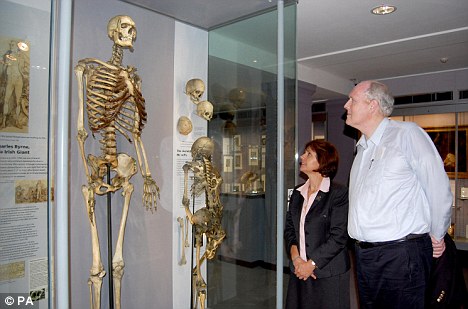
He was a giant of a man, 7 feet 7 inches tall, who left his home in Ireland when he was 19 and traveled to London to make his fortune as a freak. There Charles Byrne, known as the Irish Giant, garnered wealth and fame. But, suffering from tuberculosis and an excessive love of gin, he died a few years later, in 1783. A surgeon — John Hunter — bought Mr. Byrne’s corpse, boiled it in acid to remove the flesh, and exhibited the skeleton in his museum in London.

And there the bones remained, studied in 1909 by the renowned American surgeon Harvey Cushing, who removed the top of the skull and pronounced that Mr. Byrne had had a pituitary tumor. Other than that, Mr. Byrne remained a curiosity, a famous giant, the subject of a 1998 novel by the British writer Hilary Mantel, yet, with only a skeleton remaining, of little interest to science.

Until now: researchers in Britain and Germany have extracted DNA from Mr. Byrne’s teeth and solved the mystery of his excessive height.

It turned out to be a rare and mysterious gene mutation, discovered only in 2006. The researchers then found the mutation in four families from Northern Ireland, near where Mr. Byrne was born. Following a hunch, they decided to ask whether Mr. Byrne had had the mutation, too, and whether the mutation indicated that the four families were related to him. Their hunch was right.

The group, led by Dr. Marta Korbonits, professor of endocrinology and metabolism at Barts and the London School of Medicine and Dentistry, reports its finding in Thursday’s issue of The New England Journal of Medicine.

Symptom-producing pituitary tumors are rare, and those caused by an inherited mutated gene are rarer still. At most only 5 percent of people with pituitary tumors have them in their families.

The tumors can lead to disfigurement — patients develop bulging foreheads and large jaws, hands and feet — and chronic severe headaches. They can also cause visual problems, because the tumor presses on the optic nerve. They may even cause milk secretion, because the tumor can secrete prolactin, a hormone that is needed for fertility and to produce milk in the breasts.

Usually, tumors that secrete growth hormone start to grow in adulthood, after people have reached their full height. But when tumors start growing in children or adolescents — as they do with many patients with the mutated gene — they can result in gigantism because they make the gland churn out growth hormone, prodding bones to keep growing. Pituitary tumors are of great interest to researchers because they grow very slowly and almost never spread elsewhere in the body.

Dr. Shlomo Melmed, a pituitary tumor researcher at Cedars-Sinai Medical Center in Los Angeles, explained that the tumor cells undergo premature aging. “We think it might be protective,” Dr. Melmed said, a reason the tumor does not spread.

The involvement of the gene, known as AIP, in pituitary tumors is a surprise, researchers say. Mutations in the gene are associated with about 20 percent of inherited pituitary tumors when no other organ is involved. But it is not clear why mutations in this gene, which seems to be involved in metabolism — possibly to detoxify chemicals — can cause tumors or how these tumors form.

“There is nothing solid scientifically,” said Dr. Constantine Stratakis, a geneticist and pituitary tumor researcher who is the acting scientific director for the Division of Intramural Research at the National Institute of Child Health and Human Development.

And for unknown reasons, only about 30 percent of people with the mutated gene develop tumors.

In London, Dr. Korbonits said she had been aware of the Irish Giant because of her work on pituitary tumors. She suspected he might have had the AIP mutation when she saw a drawing of him standing with twin brothers who also were giants, who came from a nearby village, and who were said to be related to Mr. Byrne. That, she said, “suggested it was a genetic disease.”  (Me: So the Irish Giant had lived close to TWO brothers who were relative who were also giants!!)

And she had found the gene in members of four families from the same region of Ireland.

Dr. Korbonits wrote to the Hunterian Museum, where Mr. Byrne’s skeleton is still displayed, and asked to test the giant’s DNA, and then she and her colleagues removed two of his molars. She enlisted the help of an expert on ancient DNA, Joachim Burger of Gutenberg University in Mainz, Germany, to extract DNA from the giant’s teeth. She was worried that the DNA might be too degraded to analyze — after all, the giant’s corpse had been boiled in acid and then displayed in a museum for a couple of centuries.

“It was not clear at all that we would have suitable DNA,” Dr. Korbonits said. The DNA turned out to be broken in many pieces, but it could still be analyzed.

The investigators calculated that the giant and the four contemporary Irish families had a common ancestor who lived about 1,500 years ago. And, they report, there are probably 200 to 300 people living today who have inherited that same mutation.

One is Brendan Holland, a 58-year-old Irishman who sells mining equipment. Mr. Holland started growing excessively when he was 13, he said in a telephone interview.

“I kept growing and growing,” he said, eventually reaching a height of 6 feet 9 inches. As he grew, he said, he became less coordinated, developed frequent violent headaches and had sporadic episodes when he could not see. He had no idea what was wrong, but left school when he was 19, on the theory, he said, “that all that studying was giving me headaches.”

Finally, when Mr. Holland was 20 and living in London, an endocrinologist, Dr. G. Michael Besser at Barts and the London School of Medicine, figured out that Mr. Holland had a pituitary tumor. As soon as the tumor was destroyed with radiotherapy, Mr. Holland’s headaches disappeared and his growth hormone levels dropped to normal.

After the AIP gene mutation was discovered in 2006, Dr. Korbonits asked to test Mr. Holland to see if he had it. He did. Then, Mr. Holland said, she started suggesting he might be related to the giant.

“She was asking me pointed questions about where he lived and where I lived,” Mr. Holland said. “Then she said, ‘I think it is possible that you and this chap are related.’ ”

With the giant’s DNA analysis, it turned out that Dr. Korbonits was right.

Mr. Holland says he was touched thinking about the giant’s life, knowing how hard it is to be so tall and the subject of barbs and jeers.

The genetic analysis also had another effect, he said.

“I remember having a conversation with Dr. Besser when I was first diagnosed,” Mr. Holland said. “I said, ‘With eight children, why was I suffering from it and none of the others?’ He was very honest with me: He said they did not have a medical explanation.”

Now, at least, Mr. Holland has an explanation for his physical problems. And that helps, he said.

“I can only speak for myself,” he said, “but having a logical explanation helps me to come to terms with my condition.”

This article has been revised to reflect the following correction:

Correction: January 7, 2011

An article on Thursday about a genetic mutation in the case of Charles Byrne, known as the Irish Giant, misstated the year of publication of a novel by Hilary Mantel based on him. It was 1998, not 2007.


Here is the 2nd resource I wanted to use and that is from TheTallestMan.Com website. The webpage for Charles Byrne is found HERE.

This is the few parts on the page that really got me puzzled.

Charles Byrne (1761 – 1783) also known as Charles O’Brien or “The Irish Giant”, was a human curiosity or freak in London in the 1780s….His exact height is of some conjecture, but most accounts refer to him as from 8 feet 2 inches (248 cm) to 8 feet 4 inches (254 cm) tall, however true and undeniable skeletal evidence pitches him at just over 7 feet 7 inches….Although Charles Byrne was 22 years old when he died, radiographic images of his wrist bones showed that the epiphyses were not yet fused. His “bone age” was estimated to be only about 17, indicating that he was still growing at the time of death.”

The last resource I wanted to use HeightQuest.Com Again because Tyler does talk in a few articles about the fact that Gigantism and Pituitary tumors are not as simple as made to believe to the public.

FULL CRITICAL ANALYSIS: Like most of the general public my view point for gigantism was always very simple. The way gigantism and suspiciously tall humans happen is very logical and sequential.

1. The gigantism is from a benign or malignant tumor that has grown to a certain size to push against he pituitary gland at a region to cause excess release of HGH.

2. The HGH gets released to the liver which creates IGF-1 which runs gets distributed by the blood stream and causes any areas of the body like cartilage to start accelerated cell divions thus the bones and body of the person gets bigger in all dimension.

3. It does NOT matter whether the person was past puberty or before puberty. As long as the pituitary gland can release excess HGH, the human body will find a way to grow even by height. My old theory on how a post-puberty adult person with completely fused growth plates grew was that the IGF-1 reached the cartilage on the ends of their synovial joints and caused chondrocyte proliferation there. You can read up on that theory/ idea in my previous post found HERE.

4. The condition people have before growth plate closure is called Gigantism. The condition people have after growth plate closure is Acromegaly. It does not matter whether a person has Gigantism or Acromegaly, they will increase in height in both cases. So growth plate closure does not inhibit continual height increase.

5. As long as person has a pituitary gland that can release excess HGH, their body will find a way to grow in size, and height.

The issue now is that the case of Charles Byrne puts some of my deepest beliefs and ideas on how height work into question. The first problem I had with it was when the article stated the phrase “researchers have now figured out the reason for his excessive height” . For me, I didn’t think that scientist needed another reason to explain his height because his height can already be explain by the fact that he had a pituitary tumor problem, so he was just another pituitary giant. End of story. I didn’t understand why scientists and geneticists felt the need to look for another reason for his height. Didn’t the fact that an old surgeon state that he suffered a pituitary gland problem be good enough to explain his gigantism??

That was where I was coming from. Well it turns out that the situation is not that simple.

First, Bryne apparently has a genetic propensity to develop a pituitary condition causing gigantism. The researchers had found two twin brothers who were related to Byrne who also developed into giants, from gigantism. There is actually a genetic mutation in the AIP gene to cause this condition of pituitary gland disorder and gigantism to be far more prevalent. Byrne had it and his blood relatives throughout  the centuries also have it and a modern distant relative developed acromegaly. This is the first complication.

Second, on the Tallest Man’s Website, they showed that Byrne was reported in height upwards of 8′ 4″ but his skeleton measures 7′ 7″ , still a very amazing height. He died when he was 22 but X-Rays show that his growth plates were still there and that his plates indicated he was growing like a 17 year old. This excerpt challenged two beliefs I had always had.

Belief 1 – Pituitary giants even though they have the pituitary gland problem should not be able to also influence their growth plates and the rate at which the growth plates go through senescence.

Belief 2- Pituitary giants have the same age range for growth plate evolutions as the rest of us.

It seems that Byrne not only had a pituitary gland problem and had ALSO a genetic mutation to cause him to be more likely to develop gigantism, His growth plates were also very different from the average person. Could it be that either the genetic mutation or the fact that he has a putuitary condition cause his growth plates to go through with senescence a lot slower than the average human? That would be definitely something that I have never considered, but Tyler did.

I had tried to show that with cases like Sultan Kosen and Tanya Angus that they had closed growth plate because they were in their 20s or late teens when they started to go through their growth spurt. Tanya grew from 5′ 8″ to 6′ 11″ from age 18-30. Kosen grew from 8′ 1″ to 8′ 3″ from 27-29.

This just shows that Pituitary giants like Byrne , Kosen, and Angus may be even more unique than the ordinary pituitary giant (or the other possibility is that all or most pituitary giants have this skill and potential). I do note that Byrne did not start go through his massive growth spurt when he was in his adult years, but that he suffered from gigantism so he went through his massive growth spurt as a child with growth plates still open.

If this new case proves that having either the genetic mutation or pituitary condition can actually have an affect on the  rate of senescence of the growth plates in the individual, then my previous theory is wrong and that humans may NOT actually be able to grow taller naturally after puberty after all. My idea was that with enough HGH released into the body, the articular cartilage was what took over the height increase growth.

I decided not to write about the findings found by Tyler since this article post is getting quite long so I will save the discussion of the relationship between gigantism , pituitary giants, and gene mutations from the articles I found on HeightQuest for another time. That post will be very interesting so I can compare old and new theories to see which one makes more sense.