Wnt11

Overexpression of Wnt11 promotes chondrogenic differentiation of bone marrow-derived mesenchymal stem cells in synergism with TGF-β

“Wnt signaling involves regulating chondrogenesis and MSC differentiation. The aim of the present study was to investigate the role of Wnt11, a member of noncanonical Wnts, in MSCs during chondrogenic differentiation. Overexpression of Wnt11 inhibited proliferation of MSCs and caused a G0/G1 cell cycle arrest. The expression level of chondrogenic markers, aggrecan and Collagen II, was significantly increased in MSCs transduced with Wnt11 as compared with non-transduced cells or MSCs transduced with the empty lentiviral vector. Furthermore, ectopic expression of Wnt11 stimulated gene expression of chondrogenic regulators, SRY-related gene 9, Runt-related transcription factor 2, and Indian hedgehog. Finally, Wnt11 overexpression promoted chondrogenic differentiation of MSCs in synergism with TGF-β.”

“Wnt3a promotes proliferation whereas suppresses chondrogenic differentiation of MSCs. In addition, expression of either Wnt-1 or Wnt7a causes a severe block in chondrogenesis”

“Wnt11 gene is highly expressed at the late stage of chondrogenic differentiation of human MSCs in three-dimensional alginate gels”

“the inhibition rate of Wnt11 overexpression for MSC proliferation was relatively low”

“Wnt11 may enhance the chondrogenic differentiation of MSCs via repressing canonical Wnt signaling, such as Wnt1 and Wnt3a, which has been shown to inhibit chondrogenic differentiation of MSCs”

NPR2

Identification and Functional Characterization of Two Novel NPR2 Mutations in Japanese Patients with Short Stature.

“C-type natriuretic peptide (CNP) – natriuretic peptide receptor B (NPR-B) signaling is critical for endochondral ossification, which is responsible for longitudinal growth in limbs and vertebrae. Biallelic NPR2 mutations cause acromesomelic dysplasia, type Maroteaux, which is bone dysplasia characterized by severe short stature and short limbs. A monoallelic NPR2 mutation has been suggested to mildly impair long bone growth. Objective: The goal of this study was to identify and characterize NPR2 mutations among Japanese patients with short stature. SWe enrolled 101 unrelated Japanese patients with short stature. NPR2 and NPPC were sequenced, and the identified variants were characterized in vitro. In two subjects, we identified two novel heterozygous NPR2 mutations (R110C and Q417E) causing a loss of CNP-dependent cGMP generation capacities and having dominant negative effects. R110C was defective in trafficking from the endoplasmic reticulum to the Golgi apparatus. In contrast, Q417E showed clear cell surface expression.  We identified heterozygous NPR2 mutations in 2% of Japanese patients with short stature. Our in vitro findings indicate that NPR2 mutations have a dominant negative effect, and their dominant negative mechanisms vary corresponding to the molecular pathogenesis of the mutations.”

“a gain-of-function NPR2 mutation was identified in patients with tall stature and macrodactyly”

“the effect of a heterozygous NPR2 mutation on height SDS is–1.8 according to the previous report, 2.6 in 100 subjects with short stature, which is defined as height SDS less than –2.0, are expected to be heterozygous for the NPR2 mutation.”

Exercise and the articular cartilage

An increase in articular cartilage thickness will result in a slight increase in height.  There have been studies that show that mechanical loading can increase this cartilage thickness in some bones.  Whether the articular cartilage can increase in thickness could be affected by whether it is bound by the periosteum or not.  Some bones such as the fingers can grow longer and are not bound by periosteum.

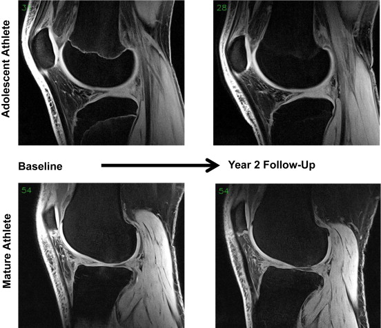
Longitudinal change in femorotibial cartilage thickness and subchondral bone plate area in male and female adolescent vs. mature athletes.

“The objective of this study was to explore longitudinal change in femorotibial cartilage thickness and tAB[Subchondral bone area] in adolescent athletes, and to compare these data with those of mature former athletes. Twenty young (baseline age 16.0±0.6 years) and 20 mature (46.3±4.7 years) volleyball athletes were studied (10 men and 10 women in each group). Magnetic resonance images were acquired at baseline and at year 2-follow-up, and longitudinal changes in cartilage thickness and tAB were determined quantitatively after segmentation. The yearly increase in total femorotibial cartilage thickness was 0.8% in young men and 1.4% in young women; the gain in tAB was 0.4% and 0.7%, respectively. The cartilage thickness increase was greatest in the medial femur, and was not significantly associated with the variability in tAB growth. Mature athletes showed smaller gains in tAB, and lost >1% of femorotibial cartilage per annum[year], with the greatest loss observed in the lateral tibia. In conclusion, we find an increase in cartilage thickness (and some in tAB) in young athletes toward the end of adolescence. Mature (former) athletes displayed high rates of (lateral) femorotibial cartilage loss, potentially due to a high prevalence of knee injuries.”

“X-rays are not capable of delineating soft tissues and cartilage”

“articular cartilage volume growth in the tibia (but not in the patella) correlated significantly with change in body height, and that – in terms of cartilage volume gain – overweight children did not differ significantly from those with normal weight. Finally, tibial cartilage volume gain was greater in those who reported an average intensity of sport above the median”

“a longitudinal gain in cartilage volume and thickness was observed post-injury[ACL rupture] and was interpreted as a potential sign of (pathological) cartilage swelling”

Microsoft PowerPoint - Figures_Eckstein_et_al__Ann_Anat.pptx
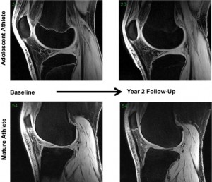
Comparison of adolescent and mature volleyball player knee by MRI.

“Adolescent men and women tended to have smaller cartilage thickness than mature participants in the medial femorotibial compartment, but greater thickness than mature adults in the lateral compartmen”

“Lateral compartment cartilage was thicker than medial compartment cartilage across all sex and age strata. Medially, the femoral cartilage was thicker compared to the tibial cartilage, while laterally, the tibial cartilage thickness was thicker compared to the medial cartilage; again, this was consistent across all sex and age strata”

“the study lacks a reference group of less physically active adults without knee injuries”

Perichondrium is key to regenerative potential of articular cartilage and unfortunately it does not cover the joint region.

Scoliosis Device and alteration of height

If you can correct scoliosis partially by stimulating growth bilaterally than you can grow taller by stimulating growth on the spine unilaterally.  Unfortunately, this study seems to only study reduction in height.

Biomechanical Simulation and Analysis of Scoliosis Correction using a Fusionless Intravertebral Epiphyseal Device.

“Computer simulations to analyze the biomechanics of a novel compression-based fusionless device (hemi-staple) that does not cross the disc for the treatment of adolescent idiopathic scoliosis.Objective. To biomechanically model, simulate and analyze the hemi-staple action using a human finite element model (FEM).Summary of Background Data. A new fusionless growth sparing instrumentation device (hemi-staple), which locally compresses the growth plate without spanning the disc, was previously developed and successively tested on different animal models.Methods. Patient specific FEMs of the spine, ribcage and pelvis were built using radiographs of 10 scoliotic adolescents (11.7±0.9yrs; Cobb thoracic:35°±7°, lumbar:24°±6°). A validated algorithm allowed simulating the growth (0.8-1.1mm/yr/vertebra){There are 33 vertebrae.  So that would 33 mm of height per year and 66 mm over a 2 year period so about 20% of an inch in height} and growth modulation process (Hueter-Volkmann principle) over a period of 2 years. Four instrumentation configurations on the convex curves were individually simulated (Config#1: 5 thoracic vertebrae with hemi-staples on superior endplates; Config#2: same as Conf#1 with hemi-staples on both endplates; Config#3: same as Config#1 +4 lumbar vertebrae; Config#4: same as Config#2 +4 lumbar vertebrae).Results. Without hemi-staples, on average the thoracic and lumbar Cobb angles respectively progressed from 35° to 56°, and 24° to 30°, while the vertebral wedging at curve apices progressed from 5° to 12°. With the hemi-staple Config#1, the Cobb angles progressed, but were limited to 42° and 26°, while the wedging ended at 8°. With Config#3, Cobb and wedging were kept nearly constant (38°, 21°, 7°). With hemi-staples on both endplates (Config#2, Config#4), the Cobb and wedging were all reduced (thoracic Cobb for Config#2 and #4: 24° and 15°; lumbar Cobb: 21° and 11°; wedging: 2° and 1°).Conclusion. This study suggests that the hemi-staple has the biomechanical potential to control the scoliosis progression and highlights the importance of the instrumentation configuration to correct the spinal deformities. It biomechanically supports the new fusionless device concept as an alternative for the early treatment of idiopathic scoliosis.”

“Simulated instrumentation of both growth plates on the convex side of the scoliotic curve
allows the optimal correction”

“The ratio of expected vertebral longitudinal growth rates (Gm: 0.8 – 1.1 mm/yr) [related] according to the difference in magnitudes between scoliotic stress in the growth plate (σ) and regular physiological stress (σm).”

“With the hemi-staple on both growth plates on each vertebra, the growth rate on the convex side was almost null while it was typically maintained to a rate of 0.48-0.66 mm/year (0.8-1.1 mm year reduced by 40% because of an increased compression of 0.2 MPa) on the concave side.”

RECQL4

RECQL4 Regulates p53 Function in vivo During Skeletogenesis.

“Rothmund-Thomson syndrome, RAPADILINO, and Baller-Gerold syndrome are rare genetic disorders caused by mutations in the RECQL4 gene. These patients have significant skeletal developmental abnormalities including radial ray, limb and craniofacial defects. To investigate the role of Recql4 in the developing skeletal system, we generated Recql4 conditional knockout mice targeting the skeletal lineage. Inactivation of Recql4 using the Prx1-Cre transgene led to limb abnormalities and craniosynostosis mimicking the major bone findings in human RECQL4 patients. These Prx1-Cre+ ;Recql4fl/fl mice as well as Col2a1-Cre+ ;Recql4fl/fl mice exhibited growth plate defects and an increased p53 response in affected tissues. Inactivation of Trp53 in these Recql4 mutants resulted in genetic rescue of the skeletal phenotypes, indicating an in vivo interaction between Recql4 and Trp53, and p53 activation as an underlying mechanism for the developmental bone abnormalities in RECQL4 disorders. Our findings show that RECQL4 is critical for skeletal development by modulating p53 activity in vivo.”

“Whole skeleton preparations of P0 mice showed that the forelimbs of Prx1-Cre+;Recql4fl/fl mice were severely foreshortened and deformed. Hindlimbs of these mutants were less affected but were also smaller and shorter compared to littermate controls”

“At three weeks of age, mutant mice that survived exhibited significantly smaller limbs, as well as abnormal ossification in the joint region of the hindlimbs. Prx1-Cre+;Recql4fl/fl mutants also displayed growth retardation”

“at E18.5, H&E staining of distal femur growth plates revealed that chondrocytes from Prx1-Cre+;Recql4fl/fl mice had significantly reduced cell density and increased cell size in the resting zone (RZ), proliferating zone (PZ) and hypertrophic zone (HZ) compared to littermate controls. At three weeks of age, mutant mice showed disorganized distal femur growth plates and abnormal chondrocyte morphology demonstrating increased cell size. It appears that inactivation of Recql4 in mesenchymal progenitor cells primarily affects the growth plate chondrocytes. These cartilage phenotypes correlate with the known expression of Recql4 in the developing cartilage and may contribute to the skeletal defects in the human RECQL4 disorders at least in the appendicular skeleton.”

Deletion of Recql4 resulted in increased apoptosis and decreased proliferation of growth plate cartilage cells.  Deletion of Recql4 results in increased levels of p53 which can result in DNA damage.

Ipamorelin

Ipamorelin, a new growth-hormone-releasing peptide, induces longitudinal bone growth in rats.

<-it was new in 1999 when this study was published.

“Ipamorelin is a potent synthetic pentapeptide which has distinct and specific growth hormone (GH)-releasing properties. With the objective of investigating the effects on longitudinal bone growth rate (LGR), body weight (BW), and GH release, ipamorelin in different doses (0, 18, 90 and 450 microg/day) was injected s.c. three times daily for 15 days to adult female rats. After intravital tetracycline labelling on days 0, 6, and 13, LGR was determined by measuring the distance between the respective fluorescent bands in the proximal tibia metaphysis. Ipamorelin dose-dependently increased LGR from 42 microm/day in the vehicle group to 44, 50, and 52 microm/day in the treatment groups{So there was no cap at the dosage so far but there was a point of dimishing returns}. There was also a pronounced and dose-dependent effect on BW gain. The treatment did not affect total IGF-I levels, IGFBPs, or serum markers of bone formation and resorption. The number of tartrate-resistant acid phosphatase-positive multinuclear cells in the metaphysis of the tibia did not change significantly with treatment. The responsiveness of the pituitary to a provocative i.v. dose of ipamorelin or GHRH showed that the plasma GH response was marginally reduced after ipamorelin, but unchanged after GHRH{So there is a negative feedback mechanism in response to ipamorelin}. The pituitary GH content was unchanged by ipamorelin treatment.”

4-month old Sprague-Dawley rats were used.  Unlike with longitudinal bone growth there were no diminishing returns with body weight gain.

This study doesn’t quite give enough evidence to be confident in Ipamorelin’s height increasing abilities such as growth plate images for example but it’s definitely worth investigating.