New Experimental LSJL routine

Here’s the old routine and I am still doing that but I’m adding something else.

I was looking through some old LSJL studies and I noticed an interesting image.


knee loading image

This image is from the study Mechanical Intervention for the Maintenance of Cartilage Bone which is not a bone lengthening study but still pertinent.  You’ll note that the loading does not appear to be strictly on the lateral side of the knee but on the diagonal so the top and bottom of the knee is getting loaded a bit as well.

Here’s a mouse and rat knee:
mouse and rat knee

Here’s the explanatory text: “MRI images of a rat (Wistar) and mouse (C57BL/6) knee joint. (A) 3D spin echo MR image (117 × 114 × 144 μm) of a rat knee ex vivo displaying the anatomical landmarks of the articular joint: a = femur condyle, b = tibia, c = patella, d = patellar ligament, e = meniscus, f = articular cartilage and g = intrapatellar fat pad. (B) Histological image of the knee joint. The MR images provided an excellent visualisation of the rat knee anatomy, with detailed observations on the subchondral bone and in the articular synovial space. (a, b, c, d) Sequential fast spin echo multi-slices images (axial views from proximal to palmar) from the proximal region of the mouse knee (512 × 512 μm). The MR images displayed the bones of the area of knee joint (1 = patella; 2 = femur; 3, 3′ = femur condyles), providing good views of the subpatellar region and the synovial cavity (see arrows). Images acquired in a 9.4-T Varian scanner (Varian, Inc., Oxford, UK) with 100 G/cm gradient coils and a Rapid bird-cage RF coil. ”  You can also see that the patella has a position relatively close to the growth plate.

Mice and rats do have patellas.

Here’s the image of the loading under Lengthening of Mouse Hindlimbs with Joint Loading which is a lengthening study:
lengthening of mousehindlimbs
On the right diagram you can see that the patella is loaded as well.  What is on either side of the growth plate?  Bone and bone.  What is on the other side of the knee joint?  Bone and bone.  Loading bone directly against bone may have a unique response.

I gained in height initially but not so much lately but I have been gaining in wingspan.  On that post I reported a wingspan of 74.5″ but lately I’ve been getting closer to 75″.

Notice something about the elbow joint:
elbow xray

Lots of potential for bone on bone contact.

Now notice the knee joint from a lateral view:

lateral knee xray

Not much potential for bone on bone contact unless you load the fibula or patella.

I found evidence that my right metacarpal grew longer than my left but so much the other parts of the bone.  My left side was longer than my right before so growth may even be more than as noted.  Growth was about 0.8% of Right metacarpal versus left.  An 0.8% increase in height would be about half an inch.

What do you notice about the palm of hand bones(which includes the metacarpal) versus the finger bones?
hand xray

Much more bone on bone action.    I clamped the palm of my hand close to the index finger metacarpal.  I’ll have to post an image of how I did it next post.  But according to my theory the index finger grew from the thumb bone pressing down on it then my middle finger bone should’ve grown from the index finger pushing down.

Also, if you look at my first method for performing LSJL when I did initially gain length:


initial LSJL

I generated force like this and this would press the fibula against the tibia.  The reason gains begin to cease is that my ability to increase force was limited with this whereas clamping can generate much more force.

Interestingly I did not any results on ankle loading producing lengthening which gives me theory credence and Hiroki Yokota stated that he did not study it.

So expect some images of how I’m going to explore this theory and some studies on bone against bone(with tissue in between) especially loading against a growth plate region like patella against the femur.

Study with insight on why Marfan’s causes tall stature

If we know why people with Marfan’s stature grow taller we can incorporate that into our quest to grow taller.

It’s interesting to note that all the things that all the things that grow taller with Marfan’s have more joint mobility especially the fingers and toes.  What doesn’t grow with Marfan’s doesn’t have as much joint mobility such as the torso.  So Marfan’s syndrome creating additional joint spacing via looser ligaments could be key to growth.

The Fibrillin Microfibril Scaffold: A Niche for Growth Factors And Mechanosensation?

“The fibrillins, large extracellular matrix molecules, polymerize to form “microfibrils.”  Fibrillin microfibril scaffold is populated by microfibril-associated proteins and by growth factors, which are likely to be latent{So more microfibrils may catch more growth factors helping people grow taller?}. The scaffold, associated proteins, and bound growth factors, together with cellular receptors that can sense the microfibril matrix, constitute the fibrillin microenvironment. Activation of TGFβ signaling is associated with the Marfan syndrome, which is caused by mutations in fibrillin-1. Mutations in fibrillin-1 cause the Marfan syndrome as well as Weill-Marchesani syndrome (and other acromelic dysplasias) and result in opposite clinical phenotypes: tall or short stature; arachnodactyly or brachydactyly; joint hypermobility or stiff joints; hypomuscularity or hypermuscularity. These different syndromes are associated with different structural abnormalities in the fibrillin microfibril scaffold and perhaps with specific cellular receptors (mechanosensors). How does the microenvironment, framed by the microfibril scaffold and populated by latent growth factors, work?  Fibrillin microfibril niche [is] a contextual environment for growth factor signaling and potentially for mechanosensation.”

“fibrillins and LTBPs may bind and sequester different members of the TGFβ superfamily of growth factors.”

“Disruption of the fibrillin microfibril scaffold was implicated in the Marfan syndrome, and mutations in the gene for fibrillin-1 (FBN1) were shown to cause Marfan syndrome. Mutations in the gene for fibrillin-2 (FBN2) were found in congenital contractural arachnodactyly (now called Distal Arthrogryposis, Type 9 or DA9)”

“fibrillin-1 deficiency [may result] in abnormal activation of TGFβ signaling”

” fibrillins interact with the BMP-7 complex”

“TGFβ propeptides bind covalently to an 8-cysteine domain in LTBPs”

“TGFβ should be regarded as a “cellular switch”, and “its true function is to provide a mechanism for coupling a cell to its environment””

“Bound to the fibrillin microfibril scaffold, the large latent TGFβ complex, as well as BMP prodomain/growth factor complexes, require activation to initiate growth factor signaling. For activation of TGFβ, integrin binding to the propeptide might change the conformation of the propeptide such that the growth factor is allowed to interact with its receptors”

“If binding to fibrillin alters the conformation of the BMP prodomain such that its growth factor is not accessible to BMP receptors, mechanisms of activation will also be required for BMP signaling.”

“With the possibility of multiple interactions between growth factors and the fibrillin microfibril scaffold, tissue-specific microenvironments can be achieved through regulation of growth factor gene expression.”

New Home Page from LSJL Scientists

HIroki Yokota and Ping Zhang are two of the scientists responsible for studying mechanical load driven bone lengthening.

What can their latest website (you must visit this site to understand the rest of the post)tell us about their plans to study this further?

On their research page, both knee loading(LSJL) and spinal loading(perhaps developing a way to lengthening the spinal column) are mentioned but only as a means of strengthening bone.

They mention that they are studying the regeneration of cartilage tissue which means that it’s a possibility that they are studying growth plate regeneration but more likely they are focused on the regeneration of articular cartilage. Their method for cartilage regeneration mentioned does not involve stimulation of mesenchymal stem cells to differentiate into chondrocytes but rather stimulation of aggreccan and type II collagen expression.

Unfortunately it says that Ping Zhang is a collaborating past lab member and Ping Zhang was the driving force behind studying lengthening effects.  It mentions that PIng Zhang is studying mechanical loading, osteoperosis, and fracture healing but nothing on bone lengthening specifically.

Interesting Review Paper on Bone Growth

Factors affecting bone growth

“Longitudinal bone growth depends on the growth plate{although some other regions may contribute in a minor way}. The growth plate has 5 different zones—each with a different functional role—and is the final target organ for longitudinal growth. Bone length is affected by several systemic, local, and mechanical factors. All these regulation systems control the final length of bones in a complicated way. ”

“Bone growth in width is also controlled by genetic factors, but mechanical loading regulates periosteal apposition”<-periosteal stem cells have the potential to differentiate into chondrocytes so it would be possible to use the mechanical loading to induce new longitudinal bone growth.

“Differences in bone size are established early in life, before puberty and perhaps even in utero. Bone begins to form when mesenchymal cells form condensations—clusters of cells that adhere through expression of adhesion molecules”

“Two models for control of bone growth in width have been suggested—the mechanostat theory (mechanical requirements regulate periosteal apposition) and the sizostat hypothesis (a master gene or set of genes regulates bone growth in width so bone reaches a preprogrammed size, independent of mechanical requirements)”

“The growth plate consists mainly of collagen fibrils, proteoglycans, and water, arranged to form a sort of sponge with very small pores. The growth plate is located between epiphyseal and metaphyseal bone at the distal end of long bones and is strain-rate–dependent, which means it is hard when squeezed rapidly but soft when deformed slowly{the means that the frequency to which stimuli is applied to the growth plate can result in a large variation in response}. The growth plate becomes ossified after puberty and epiphyseal fusion.”

“Histologically, the growth plate consists of horizontal zones of chondrocytes at different stages of differentiation. The germinal zone, at the epiphyseal end of the growth plate, contains resting chondrocytes, which seem crucial in orienting the underlying columns of chondrocytes and, therefore, in unidirectional bone growth, probably by secretion of a growth plate–orienting factor. Next is the proliferative zone, a matrix-rich zone in which flattened chondrocytes undergo longitudinal cell division and orient themselves in typical column-wise fashion. At some point, proliferating chondrocytes lose their capacity to divide; they start to differentiate and become prehypertrophic, coinciding with a size increase. Proliferating chondrocytes are located in the transition (maturation or prehypertrophic) zone. In the hypertrophic zone, round chondrocytes secrete matrix proteins in large amounts. This stage is characterized by an increase in intracellular calcium concentration, which is essential in the production of matrix vesicles. These vesicles, small membrane-enclosed particles, are released from chondrocytes and secrete calcium phosphates, hydroxyapatite, and matrix metalloproteinases, resulting in mineralization of the vesicles and their surrounding matrix. The chondrocytes in this mineralized zone eventually undergo programmed cell death (apoptosis), leaving a scaffold for new bone formation. ”

“Generally, bones increase in length as long as new material is being squeezed between the reserve zone of the growth plate and the zone of provisional calcification.”<-Can we still do this post epiphyseal fusion?

“Intracellular calcium concentration increases in the hypertrophic chondrocytes in the hypertrophic zone of growth plate cartilage; at some point, these chondrocytes begin to mineralize the longitudinal septa in the surrounding matri. At the growth cartilage junction, mononuclear cells of undetermined origin resorb the unmineralized horizontal septa of the growth cartilage. These cells are called septoclasts or chondroclasts. Blood vessels invade the area and pave the way for bone cell precursors. Eighty percent of the longitudinal septa of the growth cartilage is rapidly resorbed in the metaphyseal zone immediately behind the invading blood vessels, paving the way for bone cell precursors. About 40% of mineralized septa serves as scaffold for the formation of primary bone trabeculae; the other 60% is absorbed by chondroclasts (osteoclasts) near the vascular invasion front. ”

The scientists list factors that positively influence bone growth:

I think to or after a certain level should be applied to almost all of these like Estrogen.  This list shouldn’t be taken as gospel as there is a lot more detail in how these hormone influence longitudinal bone growth.  Innervation is to supply a body part with nerves and I think too has a more complex relationship to longitudinal bone growth than is alluded too.

“GH acts on resting zone chondrocytes and is responsible for local IGF-1 production, which stimulates clonal expansion of proliferating chondrocytes in an autocrine/paracrine manner. Infusion of GH or IGF-1 shortens stem- and proliferating-cell cycle times in the growth plate of hypophysectomized rats and decreases the duration of the hypertrophic differentiation phase, with GH being more effective. According to [an] experimental study, GH or IGF-1 treatment restores mean cell volume and height, but the growth rate is not normalized by either hormone. ”

“hyperthyroidism increases the growth rate in children but also leads to premature growth plate fusion and short stature. T3 seems to stimulate recruitment of cells from the germinal zone to the proliferating zone and facilitates differentiation of growth plate chondrocytes. Its precursor, T4, increases the number of [3H]methylthymidine-labeled chondrocyte nuclei and [35S]incorporation in Snell dwarf mice growth plates, suggesting a stimulatory role in chondrocyte proliferation and differentiation”

However, I think this view overemphasizes the importance of hormones.  You can read more on hormones in the full study which I provided a link too in the top.

“If compression always inhibited bone growth, as it was believed, growth plates would be extremely unstable, as any slight deviation from the straight alignment of the long bones of the lower extremities would induce a vicious circle of positive feedback and result in catastrophic deformities. Mild compression leads to increased, not decreased, growth. Nevertheless, when compression on one side of the growth plate exceeds a certain level, growth is indeed suppressed, and the lesion begins to worsen.”

Hypothesized theory of input of compression and tension on growth rate.

“bone cells accommodate to a customary mechanical loading environment, making them less responsive to routine loading signals”

“muscle pull affects periosteal tension and, consequently, bone form and growth in length”

“wider bones must have higher midshaft periosteal apposition rates, as this is how they
become wider.”

New info about reversing growth plate ossification: chondrocyte transdifferentiation into osteoblasts

I found earlier studies about chondrocyte transdifferentiation into osteoblast such as this.

Essentially, what’s so significant about chondrocyte transdifferentiation into osteoblasts is that it means that the growth plate genetics are maintained in the transdifferentiated-from-chondrocytes osteoblasts.  So if we can induce de-differentiation of these transgene osteoblasts then we can possibly form neo-growth plates.  This is a much more promising for height growth than the view where hypertrophic chondrocytes just undergo apoptosis and no growth plate genetic information would be maintained by apoptosis whereas if hypertrophic chondrocyes transdifferentiate into osteoblasts some of the growth plate genetic information would be maintained info the former-hypertrophic chondrocytes osteoblasts.

Chondrocytes Transdifferentiate into Osteoblasts in Endochondral Bone during Development, Postnatal Growth and Fracture Healing in Mice.

“To investigate whether cells derived from hypertrophic chondrocytes contribute to the osteoblast pool in trabecular bones, we genetically labeled either hypertrophic chondrocytes by Col10a1-Cre or chondrocytes by tamoxifen-induced Agc1-CreERT2 using EGFP, LacZ or Tomato expression. Both Cre drivers were specifically active in chondrocytic cells and not in perichondrium, in periosteum or in any of the osteoblast lineage cells. These in vivo experiments allowed us to follow the fate of cells labeled in Col10a1-Cre or Agc1-CreERT2 -expressing chondrocytes. After the labeling of chondrocytes, both during prenatal development and after birth, abundant labeled non-chondrocytic cells were present in the primary spongiosa. These cells were distributed throughout trabeculae surfaces and later were present in the endosteum, and embedded within the bone matrix. Co-expression studies using osteoblast markers indicated that a proportion of the non-chondrocytic cells derived from chondrocytes labeled by Col10a1-Cre or by Agc1-CreERT2 were functional osteoblasts{maybe we could investigate these other non-osteoblastic chondrocyte derived cells if they can be used for height increase purposes?}. Both chondrocytes prior to initial ossification and growth plate chondrocytes before or after birth have the capacity to undergo transdifferentiation to become osteoblasts. The osteoblasts derived from Col10a1-expressing hypertrophic chondrocytes represent about sixty percent of all mature osteoblasts in endochondral bones of one month old mice{this is a significant proportion which gives a lot of promise about reversing endochondral ossification}. A similar process of chondrocyte to osteoblast transdifferentiation was involved during bone fracture healing in adult mice. In addition to cells in the periosteum, chondrocytes represent a major source of osteoblasts contributing to endochondral bone formation in vivo. ”

I’d recommend visiting the study as there were some images I couldn’t get onto here.

“Endochondral bone is made of an outer compact bone (cortex) and an inner spongy bone tissue within the bone marrow cavity. The conversion from the nonvascular cartilage template to fully mineralized endochondral bones proceeds in distinct and closely coupled steps. The first step is initiated when chondrocytes in the center of the cartilage models undergo hypertrophic differentiation and cells in the perichondrium surrounding the hypertrophic zone differentiate into osteoblasts to form the interim bone cortex (bone collar). Concurrently, the initial vascular invasion occurs in the same region importing blood vessel-associated pericytes, osteoclasts and progenitor cells in the circulating blood”

“Immediately following the onset of bone collar formation, hypertrophic chondrocytes and the mineralized cartilage matrix in the center of the cartilage template are replaced by a highly vascularized trabecular bone tissue as well as bone marrow. Bone trabeculae in the primary spongiosa are formed by deposition of osteoid by osteoblasts on the surface of calcified cartilage spicules.”

“During endochondral ossification, terminally differentiated hypertrophic chondrocytes are eventually completely removed from the initial cartilage template or growth plates. Some of these chondrocytes have been shown to be eliminated through either apoptosis or autophagy (type II programmed cell death). Hypertrophic chondrocytes express osteoblast markers, such as Alkaline Phosphatase (ALPL), Osteonectin (SPARC), Osteocalcin (BGLAP), Osteopontin (SPP1) and Bone sialoprotein (IBSP), implicating potential complex functions of these cell”

in cell cultures containing ascorbic acid or in organ cultures, hypertrophic chondrocytes, instead of becoming extinct, resume cell proliferation and undergo asymmetric cell division, giving rise to cells with morphological and phenotypic characteristics of osteoblasts capable of producing a mineralized bone matrix in vitro“<-Could this be a perpetual growth plate?

“in embryonic chicks, long bone chondrocytes differentiated to bone-forming cells and deposited bone matrix inside their lacunae”

“After the growth period the Col10a1-Cre induced reporter+ cells were still present in the metaphyseal and cortical regions in 6-month-old mice”<-6-month old mice are pretty old and not really growing based on the breed of mice so this is promising that the transChondro-Osteoblasts could still be around in older humans.

“However, more of these cells were embedded within the bone matrix, and less of them were found on the bone surfaces, compared to the 2- and 3-week-old mice, implying that the number of active osteoblasts derived from chondrocytes was likely reduced after the growth period. At 8-month, there were almost no Col10a1-Cre induced reporter+ cells in the primary spongiosa and very few were on the bone surfaces.”<-This is less promising information.  TransChondro-Osteoblasts on bone surfaces are in a better position to form neo-growth plates.

“Hypertrophic chondrocytes might first dedifferentiate, then proliferate before these cells redifferentiate into osteoblasts. The existence of cells in the bone marrow that were derived from Col10a1-expressing chondrocytes. These cells displayed properties of mesenchymal progenitor cells and were able to differentiate into osteoblasts, chondrocytes and adipocytes in vitro, although we do not know whether these cells had the ability to become osteoblast cells in vivo. “hypertrophic chondrocytes to osteoblasts” transdifferentiation may indeed involve a dedifferentiation and then a redifferentiation process. In juvenile mice the chondrocyte-derived reporter+ cells, which were negative for osteoblast markers, persisted in the primary spongiosa for a considerable amount of time before becoming functional osteoblasts”<-This information doesn’t really hurt the prospects of using the genetic information of these cells to create new growth plates as even if there’s a dedifferentiation stage between chondrocytes becoming osteoblasts the genetic material will still be maintained.

According to Buried alive: How osteoblasts become osteocytes, ” the average half-life of a human osteocyte as 25 years. However, when we consider an overall bone-remodelling rate of between 4 to 10% per year, the life of many osteocytes may be shorter. Furthermore, the lifespan of osteocytes greatly exceeds that of active osteoblasts, which is estimated to be only three months in human bones”<-So we’d only have three months after growth plate fusion to induce dedifferentiation of trans-chondro osteoblasts before they don’t exist anymore.  10-20% of osteoblasts become osteocytes and osteocytes live longer but osteocytes are in a much less advantageous position to form new growth plates.

Here’s a study that mentions alternative theories:

Pubertal growth and epiphyseal fusion

“There are four theories on the cellular mechanism of epiphyseal fusion after the pubertal growth spurt. Apoptosis, autophagy, hypoxia, and transdifferentiation have been considered the causes of epiphyseal fusion”

Apoptosis: “Hypertrophic chondrocytes of the growth plate undergo death by apoptosis, leaving behind a frame of cartilage matrix for osteoblasts that invade and lay down bone.”

Autophagy: “Autophagy is another method of programmed cell death that involves a catabolic process in which the cell degrades its own components through autophagosomes. ”

Hypoxia: “a dense border of thick bone surrounding growth plate remnants [is present] in a human growth plate tissue specimen undergoing epiphyseal fusion.  The dense border might act as a physical barrier preventing oxygen and nutrients from reaching the fusing growth plate, resulting in hypoxia and eventually cell death in a nonclassical apoptotic manner.”

Transdifferentiation: “terminal hypertrophic chondrocytes transdifferentiate into osteoblasts at the chondroosseous junction of the growth plate”

Scientists find a significant increase in height due to heat application

It’s long been noticed that heat can increase longitudinal bone growth but mostly there has been a lack of concrete numbers.  It may have little impact on adult height growth due to mostly impacting the growth plate however heat may have an impact on stem cells.

Unilateral Heat Accelerates Bone Elongation and Lengthens Extremities of Growing Mice

“Current bone lengthening protocols involve invasive surgeries or drug regimens, which are only partially effective. Exposure to warm ambient temperature during growth increases limb length, suggesting that targeted heat could noninvasively enhance bone elongation. Daily heat exposure on one side of the body unilaterally increases femoral and tibial lengths. Mice (N = 20) were treated with 40C unilateral[only one side of the body] heat for 40 minutes/day for 14 days post-weaning[after no longer taking breast milk]. Non-treated mice(N = 6) served as controls. Unilateral increases in ear (8.8%), hindfoot (3.5%), femoral (1.3%) and tibial(1.5%) lengths were obtained.Tibial elongation rate was >12% greater (15 µm/day) on the heat-treated side.Extremity lengthening correlated with temperature during treatment. Body mass and humeral length{It’s interesting that heat only worked for certain bones} were unaffected. To test whether differences persisted in adults, mice were examined 7-weeks post-treatment. Ear area, hindfoot, femoral, and tibial lengths were still significantly increased ∼6%, 3.5%, 1%, and 1%, respectively, on the heat-treated side{So the heat treatment permanently increased growth and not just growth rate.  That ear area increased shows that the cartilage itself is affected}. Left-right differences were absent in non-treated controls, ruling out inherent side asymmetry. This model is important for designing noninvasive heat-based therapies to potentially combat a range of debilitating growth impediments in children.”

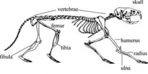
Here’s a rodent skeleton:

mouse skeleton

Let’s say your tibia and femur together are about 30 inches.  A one percent increase would be about .3inches resulting in a 3rd of an inch increase in height.  Since the heat applied is not optimized there’s the possibility for more.

“A major obstacle to successful bone lengthening by noninvasive means is difficulty in targeting therapeutics to cartilaginous growth plates, which do not have a direct blood supply{Note that LSJL is able to target the growth plates and the stem cells within the growth plates via mechanical loading}. Experimental drug delivery approaches include surgically implanted catheters and localized injections into specific growth plates”<-Note that Ping Zhang was able to stimulate length growth without injecting directly into the growth plate via localized IGF2 injections.

“Data from our lab and others demonstrate that exposure to warm ambient temperature
during growth increases bone blood supply and length in young mice.”<-The increase in bone blood supply could have an impact in adults as well if the right stimuli was in place to induce the formation of new growth plates.

“intermittent targeted heating could be accomplished with a heating pad or temperature cuff.”

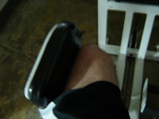
Here’s an adjustable heating pad.  I haven’t testing it myself and I’m not sure if it’s the best one but it is a sample:


“goal is to develop a low-cost, noninvasive method for lengthening bones that can translate
into practical therapy to offset linear growth impediments in children.”<-They believe that heat can work in human children to increase growth.

“Mice were anesthetized with 1.5% isoflurane and placed in lateral recumbency on a
40C heating pad for 40 minutes each day”

“The 3-5 week age interval is a time of rapid, temperature-sensitive growth in mice. By comparison, this period could be considered roughly similar to human development between toddler age and entry to middle school.”

“Limbs on the heat-treated side were wrapped in custom fitting thermal booties to ensure uniform heat distribution”

“In addition to limb length, cartilaginous ears were measured to document a treatment effect because ear size increases with ambient temperature”

“Skin temperatures of heat-treated hindfeet and ears averaged 40C during treatments. Nontreated side temperatures averaged 30C with no major fluctuations”<-So 10C of heat increase resulted in a 1% increase in leg length.  It’s unclear whether more heat would result in more lengthening.

“When nontreated and heat-treated sides were analyzed in aggregate, there were significant positive correlations between hindlimb temperature and tibial elongation rate”

“short-term (30-minute) hindlimb heating increases molecular uptake in mouse tibial growth plates”<-this doesn’t really help adults create neo-growth plates.

“Although it is unclear why humeral length did not differ, one potential explanation is the warmer starting temperature of the forelimb when compared to the hindlimb. The knee joint capsule is normally at least 3-4C lower than body core.Our treatments elevated hindlimb temperature by 10C on the heat-treated side.
However, skin temperatures in the humeral region more closely resembled body core, consistent with thermal maps for humans showing that 37C core temperature extends into the shoulder region, while extremity temperatures progressively decrease in a proximal-distal gradient.”

“Our working hypothesis is that heat-treatments do not impact temperature of the humerus
due to the proximity of this joint to the body core, and the disproportionately large volume of
warm blood delivered to the shoulder region through the large subscapular artery”

“the left-right symmetry in humeral length reflects its relatively constant temperature. This could be tested by decreasing shoulder temperature with cold, which stunts limb elongation in a dose-dependent manner”

“up to a 20% increase in bone growth rate for every one degree C increase in incubation temperature in growing chicks.”

“unilateral exposure of mild, non-damaging 40C heat for 40-minutes per day for only 14 days permanently increased ear area and hindlimb length on heat-treated sides of young mice.”

“the width of the mouse growth plate is only a fraction of that of a human growth plate. It will be important to replicate these results in a larger animal model to ensure that heat can fully penetrate a larger growth plate, so as to avoid potential angular growth deformities. This should not be problematic, however, sincewhole body heat-effects on bone length have already been demonstrated in experiments using large animals”

I already feel a bit of a temperature elevation when performing LSJL.

According to Periodic heat shock accelerated the chondrogenic differentiation of human mesenchymal stem cells in pellet culture., Heat increased chondrogenic differentiation although they were cells already committed to the chondrogenic lineage.  41C for 1 hour was the stimulus used.

 Hindlimb heating increases vascular access of large molecules to murine tibial growth plates measured by in vivo multiphoton imaging.

“Advances in understanding the molecular regulation of longitudinal growth have led to development of novel drug therapies for growth plate disorders. Despite progress, a major unmet challenge is delivering therapeutic agents to avascular-cartilage plates. Dense extracellular matrix and lack of penetrating blood vessels create a semipermeable “barrier,” which hinders molecular transport at the vascular-cartilage interface. To overcome this obstacle, we used a hindlimb heating model to manipulate bone circulation in 5-wk-old female mice (n = 22). Temperatures represented a physiological range of normal human knee joints. We used in vivo multiphoton microscopy to quantify temperature-enhanced delivery of large molecules into tibial growth plates. We tested the hypothesis that increasing hindlimb temperature from 22°C to 34°C increases vascular access of large systemic molecules{This is a limited temperature range so we don’t know if there’s a cap on how much temperature can increase height}, modeled using 10, 40, and 70 kDa dextrans that approximate sizes of physiological regulators. Vascular access was quantified by vessel diameter, velocity, and dextran leakage from subperichondrial plexus vessels and accumulation in growth plate cartilage. Growth plate entry of 10 kDa dextrans increased >150% at 34°C. Entry of 40 and 70 kDa dextrans increased <50%, suggesting a size-dependent temperature enhancement. Total dextran levels in the plexus increased at 34°C, but relative leakage out of vessels was not temperature dependent. Blood velocity and vessel diameter increased 118% and 31%, respectively, at 34°C. These results demonstrate that heat enhances vascular carrying capacity and bioavailability of large molecules around growth plates, suggesting that temperature could be a noninvasive strategy for modulating delivery of therapeutics to impaired growth plates of children.”

“transporting agents into and through the dense ECM of cartilage remains a challenge.”

“There are two components of the vascular-cartilage barrier that determine solute availability in growth plates: 1) ability of molecules to exit the vasculature and 2) ability to enter the cartilage matrix. The endpoint of interest is total uptake in the growth plate, since this is what ultimately impacts chondrocyte performance.”

“heat increases small solute (<500 Da) uptake in growth plate cartilage in vivo. In the current study, we sought to determine whether heat could increase uptake of large molecules (≥10 kDa).”

” Healthy human knee joint temperatures can range from under 20°C to over 37°C during normal and clinical activities. By modulating limb temperature within this natural range, we show that mild 34°C heating increases transport of large (≥10 kDa) molecules that are otherwise size restricted at the vascular-cartilage interface.”<-So this study doesn’t tell us whether non-physiological temperatures can help.

“The three principal vascular routes by which blood reaches the growth plate, are epiphyseal vessels, metaphyseal vessels, and a circumferential ring vessel connected to a subperichondrial plexus. The epiphyseal and metaphyseal vessels course within the bone, whereas the circumferential vessel and its plexus are located just deep to the perichondrium (i.e., subperichondrial) that surrounds the cartilage plate. Soluble factors reach the growth plate through all three vascular routes, and temperature can affect blood flow through these vessels”

“Heat increases vessel diameter and blood velocity in subperichondrial plexus vessels.”