Grant promises to use heat to increase longitudinal bone growth

This project has a decent amount of funding(over 300K).

Heat enhanced molecular delivery to growth plates for targeted bone lengthening

“Linear growth deficiencies have multiple etiologies, ranging from injury and illness to genetic bone disease. Bone elongation disorders are particularly challenging to treat because of the relative inability to regulate molecular delivery o the growing skeleton. The primary obstacle to successful clinical intervention is lack of molecular delivery to the growing skeleton. The primary obstacle to successful clinical intervention is lack of methods for targeting therapeutics to growth plate cartilage, which does not have a penetrating blood supply. Existing procedures for limb lengthening involve invasive surgery or drug regimens, which are to date only partially effective. Data generated by the applicant show that localized heating increases molecular uptake in growth plate cartilage in vivo, suggesting that heat could be a noninvasive and inexpensive alternative for augmenting delivery of bone-lengthening drugs. The long-term goal is to identify physiological mechanisms underlying temperature-enhanced bone elongation in the growth plate{is this a method just to increase drug delivery or is the temperature alone a driving factor in elongation?}. The overall objective of the problem-solving proposal is to determine whether heat augments the bone-lengthening effects of systemic growth regulators. Insulin-like growth factor (IGF)-I is a potent stimulator of linear growth and part of a drug regimen used in children. The central hypothesis, based on strong preliminary data and tested under two specific aims using dynamic in vivo multiphoton microscopy, is that heat localizes delivery of systemic IGF-I into growth plates to promote additive bone lengthening{so this implies that the heat increases delivery of IGF-1 already in the body to the growth plates}.
Specific Aim 1 uses in vivo cartilage imaging to determine dose, timing, and temperatures that maximize IGF-I uptake in 5-week-old mouse tibial growth plates. Injections of fluorescently labeled IGF-I and size proxy tracers will be given to quantify molecular uptake and clearance in the growth plate. Real time imaging will be used to determine delivery rate and total volume of labeled molecules in the growth plate and surrounding vasculature after timed injections. Permeability of the vessels and matrix will be measured at different doses and temperatures.
Specific Aim 2 uses a novel limb heating model to determine if heat-enhanced IGF-I uptake in growth plates causes unilateral limb lengthening, analyzed by quantifying growth rate and biomarkers of IGF-I activation in heat-treatable tibiae. Injections of IGF-I and its receptor antagonist will explicitly show if heat-enhanced uptake increases bone length. IGF-I deficient growth hormone receptor knockout mice will be used to test the drug-targeting efficacy of heat in a disease model. The rationale is to facilitate design of heat based drug-targeting approaches to enhance length at specific skeletal sites using noninvasive techniques. This project is innovative by using multiphoton imaging to assess growth plate physiology at the cellular level in vivo, in a dynamic way not possible with other methodologies. This contribution is significant because it can yield transformative findings that mechanistically link heat, bone lengthening, and vascular access to growth plates. Such results could fundamentally shift the approach that physicians take in treating a spectrum of growth plate disorders, leading to new therapies with better outcomes by reducing amount, toxicity and costs of high-dose systemic pharmaceuticals. ”

What’s also interesting is that when I perform LSJL I can feel the temperature of my bones increasing slightly.  Although to recreate a growth plate would require more than just IGF-1.

H3K4 may be involved in growth plate cessation

This grant by Jeffrey Baron has insights on the cessation of growth.

Regulation Of Childhood Growth

“Children grow taller because their bones get longer. This bone elongation occurs at the growth plate, a thin layer of cartilage within juvenile bones. The growth plate contains progenitor cells located within the resting zone. Children stop growing taller because the growth plate cartilage undergoes programmed senescence which involves extensive changes in gene expression, declining chonodrocyte proliferation, altered chonodrocyte differentiation, and involution of the growth plate{Note that this does not necessarily involve estrogen although estrogen may be a part of it}. Eventually growth plate senescence leads to cessation of bone elongation and epiphyseal fusion. Estrogen accelerates this developmental process, causing growth to stop earlier.  Senescence occurs because progenitor cells in the resting zone of the growth plate are depleted in number and that estrogen acts by accelerating this depletion. Body size varies enormously among mammalian species. In small mammals, body growth is typically suppressed rapidly, within weeks, whereas in large mammals, growth is suppressed slowly, over years, allowing for a greater adult size. Body growth suppression in rodents is caused in part by a juvenile genetic program that occurs in multiple tissues simultaneously and involves the downregulation of a large set of growth-promoting genes{If we can upregulate these genes via mechanical and dietary mechanisms can we reverse this body growth surpression?}. This genetic program is conserved among mammalian species but that its time course is evolutionarily modulated such that, in large mammals, it plays out more slowly, allowing for more prolonged growth and therefore greater body size. Epigenetic mechanisms may orchestrate this juvenile growth-regulating genetic program. Extensive genome-wide shifts in H3K4 and H3K27 histone methylation [occur] with age. Temporal changes in H3K4 trimethylation showed a strong, positive association with changes in gene expression whereas changes in H3K27 trimethylation showed a negative association. Genes with decreases in H3K4 trimethylation with age were strongly implicated in cell cycle and cell proliferation functions. The common core developmental program of gene expression which occurs in multiple organs during juvenile life is associated with a common core developmental program of histone methylation. In particular, declining H3K4 trimethylation is strongly associated with gene downregulation and occurs in the promoter regions of many growth-regulating genes, suggesting that this change in histone methylation may contribute to the component of the genetic program that drives juvenile body growth deceleration. In some children with subnormal linear growth, a cause can be identified, but in many the etiology remains unknown. This condition, idiopathic short stature (ISS), can sometimes be severe. We used whole-exome sequencing to study three families with autosomal dominant short stature, advanced bone age, and premature growth cessation. In these families, we identified novel heterozygous mutations in ACAN, which encodes aggrecan, a proteoglycan in the extracellular matrix of growth plate and other cartilaginous tissues. Our study demonstrated that heterozygous mutations in ACAN can cause a skeletal dysplasia which presents clinically as short stature with advanced bone age. The accelerating effect on skeletal maturation has not previously been noted in the few prior reports of human ACAN mutations. Our findings thus expand the spectrum of ACAN defects and provide a new molecular genetic etiology for the child who presents with short stature and accelerated skeletal maturation. ”

So we want to study H3K4 trimethylation and see if we can re-upregulate it during aging to find away to grow taller.  Although he only claims that H3K4 trimethylation downregulation is only associated with gene downregulation, he suggests that this H3K4 trimethylation downregulation may be a part of a larger program to cease growth.

Although it seems like it would be very difficult to manipulate H3K4 trimethylation and it would require something like a retrovirus.

Anti-Sclerostin inhibitors may be able to increase height

Sclerosteosis is characterized by tall stature.  If we understand why this occurs we can mimic it and possibly apply it to ourserves to become taller.  Sclerosteosis is typified by very low levels of sclerostin in serum.  If we also inhibit sclerostin we may be able to obtain the tall stature of those with this syndrome.

There are anti-sclerostin inhibitors that are at Stage II and III of clinical trials.  Since Sclerostin is produced by osteocytes and affects bone formation by osteoblasts it’s possible that it could have an impact on bone growth outside of the growth plate.  Although Wnt signaling, which Sclerostin inhibits also affects cartilage growth.

Sclerostin inhibition has been discussed before.

The sclerostin story: From human genetics to the development of novel anabolic treatment for osteoporosis.

Sclerosteosis and Van Buchem disease are two rare bone sclerosing disorders characterized by increased bone mineral density, tall stature and entrapment of cranial nerves due to overgrowth of a highly dense bone. Recent advances in human genetics have revealed the genetic background of these disorders by cloning the SOST gene, which is localized on chromosome region 17q12-q21 and codes for sclerostin. Sclerostin is a protein produced almost exclusively from osteocytes inhibiting bone formation by both osteoblasts and osteocytes. At the molecular level, sclerostin inhibits the Wnt signaling pathway, which plays a critical role in osteoblast development and function. Induced sclerostin deficiency in mice reproduces the bone sclerosing human diseases{so inhibiting sclerostin may increase height}, while sclerostin excess leads to bone loss and reduced bone strength. The extracellular nature of sclerostin has rendered it a promising target for the development of novel anti-osteoporotic treatment. Otherwise healthy carriers of the SOST mutation present with increased bone mass and low levels of sclerostin in serum in contrast to patients with sclerosteosis, who exhibit undetectable levels, thus pointing to the possibility of titration of sclerostin levels in the circulation. Based on these unique characteristics, human anti-sclerostin antibodies have been developed and tested in ovariectomized rats and monkeys, demonstrating very promising results in bone formation. Clinical phase II and III trials are currently underway thereby translating human genetics to drug development.”

“[Sclerostin deficiency can cause] overgrowth of skull bones, mandible, ribs, clavicles, long bones and pelvis.  Patients are usually tall and have a characteristic face because of the facial deformities of bossing of the forehead and enlargement of the mandible.”  <-There are also other problems generated but perhaps we can just lower Sclerostin levels to a certain point so they don’t get too low as to cause issues.  Although it seems only those with undetectable levels of Sclerostin have tall stature.

“Circulating sclerostin binds to the LRP5/6/4 proteins preventing the intracellular part of LRP5 from restraining Axin and free beta-catenin from the intracellular protein complex, this leading to its phosphorylation and degradation and consequently inhibition of Wnt signaling.”

“parathyroid hormone inhibits the expression of the SOST gene by osteocytes”

“Calcitonin, on the other hand, which inhibits osteoclast resorption, up-regulates sclerostin expression by osteocytes, while it decreases other osteocyte products such as MEPE and DMP.”

“Mechanical stimulation of the skeleton either through exercise or experimental loading induces bone formation, while immobilization increases the number of sclerostin positive osteocytes”

Romosozumab(A sclerostin inhibitor) is on Stage 3 clinical trials.

But it seems you need virtually total knockout of function of Sclerostin to gain the tall stature.  I couldn’t find any studies on adult individuals to see if Sclerostin could increase height post cessation of growth.

According to the study Effect of sclerostin antibody treatment in a mouse model of severe osteogenesis imperfecta., a sclerostin antibody had no effect on bone length in 20 week old adult rats(and also in 4 week old mice).  However, with an antibody elimination of sclerostin isn’t likely to be total.  In 4 week old mice there was a .3mm(about 2%) increase in bone length between the Sost-Ab and control group that was found to be nonsignificant but this could indicate that further studies could find that Sost-Ab can increase growth in the growing phase.  In the 20 week old mice there was no difference between limb lengths in control and Sost-Ab group but it’s possible that the difference could be too small to detect.

Sclerostin antibody treatment causes greater alveolar crest height and bone mass in an ovariectomized rat model of localized periodontitis

“Sixty female, 4-month-old Sprague-Dawley rats{4 month old rats have fairly senescent growth plates} underwent sham operation or bilateral OVX and were left untreated for 2 months. Experimental periodontitis (ligature) was established by placing silk sutures subgingival to the right maxillary first and second molar teeth for 4 weeks, and feeding the rats food and high-sugar drinking water during this period. Thereafter, ligatures were removed and 25mg/kg vehicle or Scl-Ab was administered subcutaneously twice weekly for 6 weeks. Rats were randomized into four groups: (1) Control (Sham+Vehicle), (2) Sham+Ligature+Vehicle, (3) OVX+Ligature+Vehicle, and (4) OVX + Ligature + Scl-Ab. Terminal blood and right maxilla specimens were collected for analyses.
Group 3 rats showed lower bone volume fraction (BVF) of alveolar bone with higher bone resorption and lower bone formation than Group 2 rats. Group 4 rats had higher alveolar crest height, as assessed by linear distance of cementoenamel junction to the alveolar bone crest and greater alveolar bone mass using Micro CT, than Group 3 rats. Significantly higher values of mineral apposition rate (MAR) and mineralizing surface/bone surface (MS/BS) were also observed in Group 4 rats by analyzing polychrome sequential labeling data. Increased serum osteocalcin and osteoprotegerin, and deceased serum tartrate-resistant acid phosphatase and CTx-1 illustrate the ability of Scl-Ab to increase alveolar bone mass by enhancing bone formation and decreasing bone resorption in an animal model of estrogen deficiency osteopenia plus periodontitis.
Scl-Ab could be a potential bone anabolic agent for improving alveolar crest height and higher alveolar bone mass in conditions where alveolar bone loss in periodontitis is compounded by estrogen deficiency osteopenia.”

Note need to analyze full study at some point.  Alveolar bone is the bone around the teeth.

This paper describes growth of the mandible.  Description of alveolar growth: “The bone of
the mandible begins to grow on each side of the tooth germ. By this growth the
tooth germs come to be in a trough or groove of bone, which also includes the
alveolar nerves and blood vessels”

“with the loss of the teeth the alveolar process is absorbed”

Cartilage pathways paper

A pathway to bone: signaling molecules and transcription factors involved in chondrocyte development and maturation.

SOX9pathway

“Mesenchymal progenitors that originate from the cranial neural crest, somites and lateral plate mesoderm contribute to the craniofacial, axial and limb skeleton, respectively. Condensations of such mesenchymal cells express the transcription factor Sox9, which is a key regulator of chondrogenesis, and give rise to cartilage primordia consisting of round immature chondrocytes that continue to express Sox9. Cells lying within the central regions of the cartilage primordia then undergo maturation. During this process, chondrocytes withdraw from the cell cycle and increase ∼20-fold in volume, giving rise to cells that are termed hypertrophic chondrocytes. As the cartilage continues to grow longitudinally, it continually deposits hypertrophic chondrocytes in its wake. These are subsequently replaced by bone in a region known as the primary ossification center. The production and maturation of chondrocytes is eventually restricted to the end of the bone (the epiphysis) in a structure termed the growth plate. A secondary ossification center then arises within the epiphysis, separating the growth plate from the distal ends of the long bones. ”

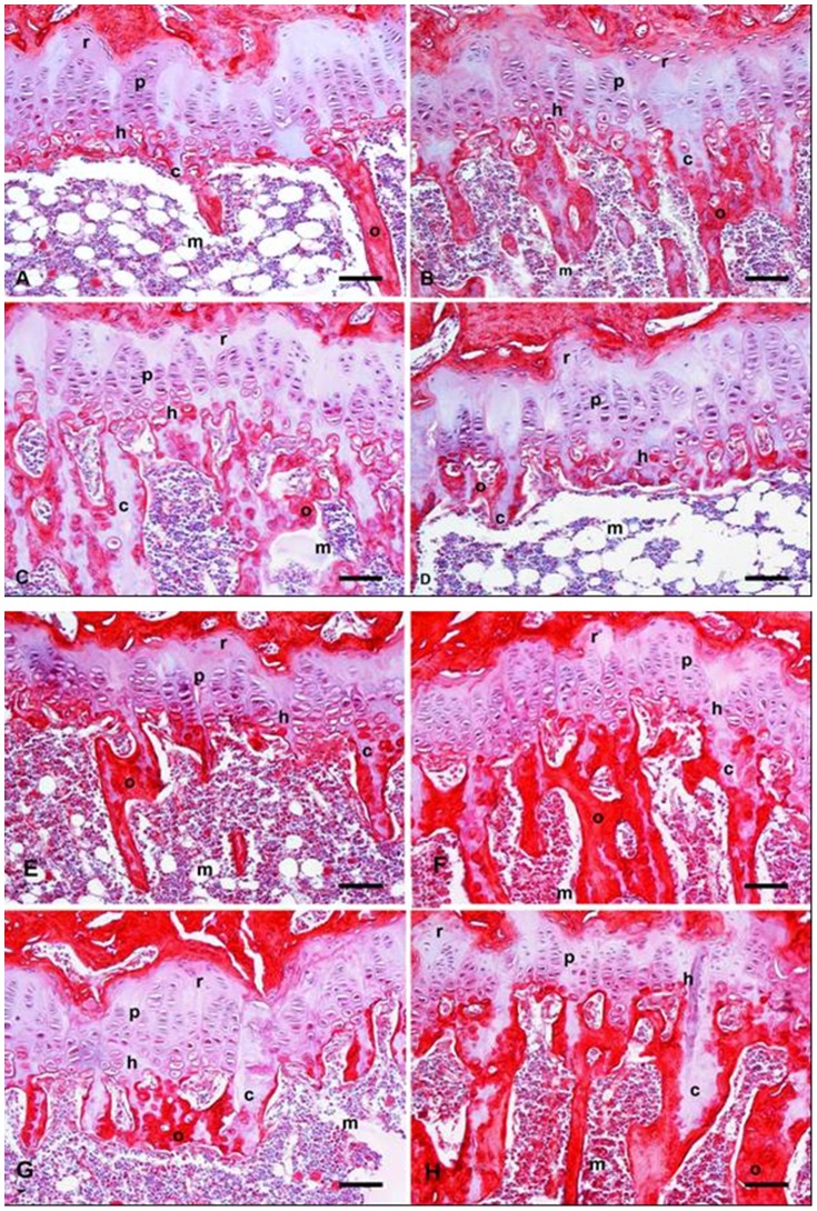
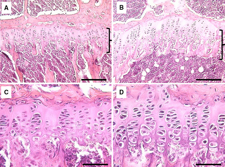
“Within the growth plate, small round distally located chondrocytes initially give rise to flattened chondrocytes that are more centrally located in the cartilage primordia{You can see the flattened and round chondrocytes in the LSJL loaded growth plates}, and which proliferate and stack into longitudinal columns. These immature chondrocytes express the transcription factors Sox5, Sox6 and Sox9, and the structural proteins collagen, type II, α1 (Col2a1) and aggrecan (Acan). The next stage of maturation into prehypertrophic chondrocytes is marked by the expression of both parathyroid hormone 1 receptor (Pth1r) and Indian hedgehog (Ihh). This is followed by maturation into early hypertrophic chondrocytes that express collagen, type X, α1 (Col10a1). Notably the induction of Pth1r, Ihh and, subsequently, Col10a1 correlates with the loss of Sox5, Sox6, Sox9, Col2a1 and Acan expression. Finally, Col10a1-expressing cells lose expression of this collagen and progress to become late hypertrophic chondrocytes, which express vascular endothelial growth factor A (VEGFA), matrix metalloproteinase 13 (Mmp13) and secreted phosphoprotein 1 (also known as, osteopontin/bone sialoprotein 1; Spp1). VEGFA and Mmp13 expression herald the invasion of the growth plate by endothelial cells, osteoclasts and osteoblast precursors. Osteblast precursors that arise from both the perichondrium and the hypertrophic chondrocytes work together with osteoclasts to remodel the growth plate matrix to form trabecular bone. ”

stagesgeneexpression

“Limb bud mesenchymal cells can undergo chondrogenesis in culture when plated at extremely high density (termed micromass culture) or at low density but only when these cells are plated in either suspension culture or collagen gels. This leads to speculation that a spherical cell shape might promote chondrogenesis”

“During development, the somite undergoes a process of differentiation. The ventral region becomes mesenchymal and forms the sclerotome, precursor to the vertebrae and to the medial part of the ribs. A signaling molecule known to be crucial for sclerotome induction is sonic hedgehog (Shh), a member of the hedgehog family of proteins that is expressed by both the notochord and the floorplate of the neural tube. Interactions between Shh and the patched transmembrane receptors Ptch1 and Ptch2 cause another transmembrane protein, smoothened (Smo), to translocate to the base of the primary cilium. This leads to activation of Gli transcription factors (Gli1, Gli2 and Gli3), which in turn regulate the expression of hedgehog-responsive genes. Shh signaling has been found to play multiple roles in sclerotome formation: promoting the survival of somitic cells, an epithelial-mesenchymal transition of the sclerotome via induction of Snail and, finally, the induction of sclerotome-specific markers such as Pax1{LSJL upregulates Pax1}, Sox9 and Nkx3-2. In addition to Shh, noggin (Nog) (which is expressed in the notochord) and gremlin 1 (Grem1) (which is expressed in the dorsal neural tube and somites) cooperate to maintain a BMP signaling-free zone that is crucial for Shh-mediated sclerotome induction. Mice engineered to lack Shh fail to form vertebrae, indicating that Shh signaling is crucial for the formation of vertebral cartilage. the subsequent differentiation of sclerotome into cartilage does not depend on maintained Shh signaling. Shh signals induce the expression of Sox9 and the transcription factor Nkx3-2, which indirectly maintains Sox9 expression in somitic cells. Bone morphogenetic protein (BMP) signals maintain the expression of these genes after their initial induction by Shh, although they cannot induce the expression of either Sox9 or Nkx3-2 in presomitic paraxial mesodermal cells that have not yet been exposed to Shh signaling. Provided that BMP signals are present, Sox9 can both regulate its own expression and induce expression of Nkx3-2. Nkx3-2 is a BMP-dependent transcriptional repressor that blocks the expression of inhibitor(s) of Sox9 transcription. Nkx3-2 blocks BMP-dependent expression of several GATA transcription factors (Gata4, Gata5 and Gata6) in explants of paraxial mesoderm, and these GATA factors can in turn block Shh-dependent induction of Sox9 gene expression ”

conditional loss of β-catenin expression in either limb or head mesenchymal progenitors both increases the expression of Sox9 in these progenitor cells and induces chondrocyte formation at the expense of osteoblasts“<-So a Beta-Catenin inhibitor may be a useful height increase tool.

“In addition to Wnts secreted by the limb bud ectoderm, FGFs secreted by the apical ectodermal ridge (AER) are necessary to maintain: (1) limb bud outgrowth; (2) the viability of a chondrogenic precursor pool that gives rise to the cartilage templates of the limb; and (3) the competence for limb bud mesenchymal cells to undergo chondrogenesis once the Wnt signals are removed. In addition, FGF signals have been demonstrated to boost the expression of Sox9 in primary chondrocytes via a mitogen-associated protein kinase (MAPK)-dependent pathway ”

“Wnt signals induce both a repressive chromatin mark (H3K27me3) and DNA methylation over the Sox9 promoter, and that Wnt-induced irreversible silencing of the Sox9 gene requires DNA methylation of this locus that is specifically countered by FGF signals. FGF blocks the recruitment of the de novo DNA methyltransferase DNMT3A to the Sox9 promoter by inducing the interaction and phosphorylation of DNMT3A by extracellular-regulated kinase (ERK) 1 and ERK2, and thereby controls whether the expression of Sox9 is irreversibly or reversibly silenced by Wnt signals in limb bud mesenchymal cells{This step may be the reason that increased FGFR3 signaling reduces height}

“Wnts secreted by the ectoderm act via a β-catenin-dependent pathway to block Sox9 expression and cartilage formation in limb bud mesenchymal cells, and that FGF signaling maintains chondrogenic competence of these cells by blocking DNA methylation of the Sox9 promoter{So neo-growth plate formation in adult bone MSCs may be partially blocked by DNA methylation of Sox9 in these cells but MSCs are definitely capable of chondrogenic differentiation as shown by distraction osteogenesis}.”

Blocking TGF-Beta signaling did not appear to block the initial stages of chondrogenesis.

“GFβ signaling inhibition has limited effects on the formation of Sox9+Scx− progenitors, TGFβ signaling is absolutely necessary for both formation of the Sox9+Scx+ progenitor population and the subsequent development of bone eminences”

” The mis-expression of BMPs or of activated BMP receptors in the limb bud results in ectopic chondrogenesis”

“Hif1α was shown to promote the expression of Sox9 and to elevate the expression of glycolytic enzymes and glucose transporters to allow chondrocyte differentiation and adaptation to hypoxic conditions. Hif1α was also shown to upregulate VEGFA expression in the growth plate as well as promote collagen hydroxylation to enable collagen secretion by hypoxic chondrocytes”

chondrogenesisstages

“Sox9 loss of function leads to premature maturation of immature chondrocytes into hypertrophic cells. Conversely, overexpression of Sox9 in either immature or hypertrophic chondrocytes of the growth plate slows the process of chondrocyte hypertrophy”

“the ectopic expression of Runx2 in immature chondrocytes drives premature cellular maturation and induces the expression of Col10a1 and other hypertrophic markers”

“Mef2c loss of function in chondrocytes results in shortening of the bones, a delay in chondrocyte hypertrophy and downregulation of Runx2 expression. Conversely, gain-of-function experiments in transgenic mice that express an activated Mef2c-VP16 fusion protein in chondrocytes results in the opposite phenotype of premature and excessive endochondral ossification


controlofproliferation

“Ihh promotes chondrocyte proliferation, at least in part by increasing the expression of cyclin D1, which regulates cell cycle progression. In addition, Ihh promotes the transition of small round chondrocytes into proliferating chondrocytes, independently of PTHrP expression (Kobayashi et al., 2005), by inhibiting the repressor activity of Gli3 (i.e. Gli3R)”

“The mechanical regulation of chondrocyte proliferation affects not only the length of the bone but also its morphology. For example, both the ‘mini’ growth plate of bone eminences and the endochondral ossification-mediated repair of bone fractures are controlled by muscle loading”

Glucosamine and Chondroitin may contribute to height by slowing down growth plate senescence

This study attempts to accelerate growth plate senescence by removing rat ovaries.  Chondroitin and Glucosamine appear to counteract the effects of senesence on growth plate fusion.  So even if say hypothetically GS+Chondroitin may do nothing normally, when growth plate is close to senesence or cessation of growth GS+Chondroitin may extend this period to be longer allowing height to grow further.  Unfortunately, they do not measure longitudinal bone growth directly.

But Glucosamine+Chondroitin is available for sale and the potential toxicity for the supplement seems to be extremely low so I consider it worth trying if growth plates appear close to fusion.

Glucosamine and chondroitin sulfate association increases tibial epiphyseal growth plate proliferation and bone formation in ovariectomized rats

“The growth plate consists of organized hyaline cartilage and serves as a scaffold for endochondral ossification, a process that mediates longitudinal bone growth. Based on evidence showing that the oral administration of glucosamine sulfate (GS) and/or chondroitin sulfate (CS) is clinically valuable for the treatment of compromised articular cartilage, the current study evaluated the effects of these molecules on the tibial epiphyseal growth plate in female rats.

The animals were divided into two control groups, including vehicle treatment for 45 days (GC45) and 60 days (GC60) and six ovariectomized (OVX) groups, including vehicle treatment for 45 days (GV45), GS for 45 days (GE45GS), GS+CS for 45 days (GE45GS+CS), vehicle for 60 days (GV60), GS for 60 days (GE60GS) and GS+CS for 60 days (GE60GS+CS). At the end of treatment, the tibias were dissected, decalcified and processed for paraffin embedding.

after 60 days of treatment, the number of proliferative chondrocytes increased two-fold, the percentage of remaining cartilage increased four-fold and the percentage of trabecular bone increased three-fold in comparison to the control animals.

GS and CS treatment drugs led to marked cellular proliferation of the growth plate and bone formation, showing that drug targeting of the tibial epiphyseal growth plate promoted longitudinal bone growth.”

An OVX female mice is a mice that has had their ovaries removed and this is used to simulate osteoperosis.  So there’s guarantee that this will be applicable to a healthy individual.  The rats in this study were 16-weeks old.

The control OVX groups in this study had disorganized growth plates and reduced growth plate size.

OVX growth plates +- CS

“Growth plate photomicrographs of the following: A – non-OVX rats treated for 45 days with vehicle (GC45); B – OVX rats treated for 45 days with vehicle (GV45); C – non-OVX rats treated for 60 days with vehicle (GC60); D – OVX rats treated for 60 days with vehicle (GV60), E – OVX rats treated for 45 days with GS (GE45GS); F – OVX rats treated for 45 days with GS+CS (GE45GS+CS); G – OVX rats treated for 60 days with GS (GE60GS); and H – OVX rats treated for 60 days with GS+CS (GE60GS+CS). Sirius red-hematoxylin staining. (r: resting cartilage; p: proliferative cartilage; h: hypertrophic cartilage, c: remaining cartilage, o: trabecular bone, m: bone marrow). Scale bar = 75 µm.”

Here’s an LSJL growth plate for reference from this LSJL study:

The mice in this study were not OVX and were 8 weeks old.  The growth plates do not appear to be more disorganized.

“Compared to GV45 and GV60, the GE45GS, GE45GS+CS, GE60GS and GE60GS+CS groups presented an organized cellular arrangement and increases in the number of resting and proliferative chondrocytes, PZ thickness, remaining cartilage and the trabecular bone area. In addition, these groups showed a decrease in the number of hypertrophic chondrocytes in the bone marrow area, as well as a decrease in RZ and HZ thickness.”

The GC45 and GC60 are the control groups were looking at since those are the control groups that are not OVX but they did not have Chondroitin and Glucosamine applied unfortunately..  They will help answer the question if Chondroitin and Glucosamine stimulated growth beyond just eliminating the deficit caused by OVX.  Compared to control, Chondroitin and Glucosamine treated OVX rats had more resting and proliferative chondrocytes and less hypertrophic chondrocyes.  So CS+GS may inhibit chondrocyte hypertrophy ie. decelerate growth plate senescence.

Cartilage thickness was about equal total but the CS+GS OVX group had more thickness in the proliferative zone.  CS+GS OVX group had more hyaluronan in the hypertrophic zone and after 60 days had more remaining cartilage but less bone marrow than the control group.

“In older rats, the growth plate structure and thickness is maintained but is not functional and longitudinal growth ceases at a certain point”

“To accelerate the senescence process in adult animals, OVX was performed in our study.”

Sermorelin, GHRH Analogue to Stimulate The Pituitary Gland Directly To Increase HGH

There seems to be a type of amino acid combination that has been developed which you can take orally to stimulate the pituitary gland to increase the amount and rate of HGH that will go through the system. From what we are seeing, it is most effective for people who are suffering from growth hormone deficiency. It is known as Sermorelin Therapy, which is a type of GHRH analogue.

It would work primarily for prepubertal children with idiopathic growth hormone deficiency. The method and dosage for use is recommended from source 1 to be “subcutaneous sermorelin 30 μg/kg bodyweight per day, given as continuous infusion or as 3 divided doses”. However, the increase in height still seems to be less than just taking somatotropin once a day.

From source 2, “Unlike recombinant hGH, which stimulates production of the bioactive hormone IGF-1 from the liver, SERMORELIN simulates the patient’s own pituitary gland by binding to specific receptors that increase production and secretion of endogenous hGH” and “Its effects are regulated at the level of the pituitary gland by negative feedback and by release of somatostatin so that the side effects of too much hGH are difficult if not impossible to achieve”. Remember that the anterior part of the pituitary releases two major chemicals, somatostatin and HGH, which have inhibitory effects on the other’s influence on the body’ physiological processes. Using sermorelin means that you are not going to develop medical conditions since any increase in HGH to a level that is bad for the body would cause the release of somatostatin as well to negate the effects of the stimulated HGH.

This is the only type of treatment that is commercially available right now.

Refer to source 1 and source 2 for more information