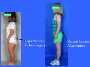
Go To The Gym – Hyperlordosis Correction Exercises Or Hyperextension Help Increase Height

HyperextensionHere is something that I recently found which will help a large percentage of people which will let them gain about half a centimeter in increase height quite quickly. Something which I have always believed is that getting exercise which can remove the load on our backs and vertebrate bone will lead to some height increases.

I was watching this Youtube video by Dr. Dror Paley when he mentioned the fact that one of the things he does when he is performing surgeon on his patients, but most especially the ones suffering from Dwarfism, or Achondroplasia, is to correct the hyperlordosis that is often most visible in people suffering from dwarfism.

HyperlordosisRefer to the Youtube Video “Achondroplasia A Guide For Parents By Dr Dror Paley.mp4”. Notice how the lower back area is curved towards the posterior direction.

When the surgery is being doing, the femur is pushed backwards during the lengthening of the lower limb bones.  Somehow it straightens out the pelvis, and by doing that, it also straightens out the spine.

The surgical part probably doesn’t seem that interesting but the surgical methods doing in the OR doesn’t always have to be. The thing is that if we can look at our own vertebral curves in an X-Ray, almost all of us have the lower back area curved a little. We can do something in our normal lives to possibly correct for those things with some serious dedication.

The study that proves that this exercise would indeed increase height, at least temporarily  – Spine Height and Disc Height Changes As the Effect of Hyperextension Using Stadiometry and MRI

So there are 2 things you can do to correct for lordosis

Part 1

Before the first video, I would like to remind people that I wrote a post maybe a year ago showing that I had found another scientific article showing that the Supine Flexion Exercise would also work to help decrease the load in the back. Refer to the post “Restore Spinal Disk Height And Increase Height Temporarily Through Land Based Supine Flexion

Part 2

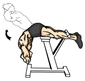
You will need to go to the local gym and use the device which is above. It is known as the Hyperextension Exercises. The machine you will need is found in almost any local Gym. It has no moving parts. You just lie down on it, stomach facing the ground. You just use your lower back muscles or the core muscles to raise the upper torso/body upwards against gravity.

The benefit of these two types of exercises will be more than just a slight increase in height. You will also notice that your mid-section will become smaller, and if you are a guy, a 6-pack of abs. Having a strong back and core from repeated using this gym machine will over time give

Proposed Theory On IGF-1 Stimulation Increases and Lengthens Muscle Mass

I think it was more than 6 months ago when I found certain publications that Dr. Dror Paley had done research to find the effects of IGF-1 on lengthening of muscle. Refer to the post How Muscle Tissue Is Lengthened – Bone Lengthening Will Not Be Limited – Breakthrough!. In that post, I had revealed a Grant that Paley along with other limb lengthening surgery surgeons had proposed to study the effects of using IGF-1 during distraction osteogenesis surgery. (IGF-1 Gene Therapy of Muscle during Distraction Osteogenesis)

In that post, I had proven that the limitation that certain posters on the online forums have set forth, on the inability of muscle tissue to stretch, has also been worked out and had the technical problems resolved. The muscle factor has now been considered and worked around.

What I would propose fro Paley’s research is a personal theory. Here it is…

It might be that IGF-1 does not really help make the growth plates grow thicker and have the chondrocytes proliferate more. What might actually be happened is that the IGF-1 is having the most affect on the muscle tissue surrounding the growth plate, while it is still active.

If we assume that the IGF-1 which is injected into the leg close to the growth plate makes the muscle tissue in that area thicker, which I believe right now could translate to mean longer, then the tension on the bone in the center would decrease.

If we remember the studies which show that “periosteal stripping” would increase the rate of bone longitudinal growth, the surgeons who did those tests more than 100 years ago had suggested that if you just remove the elements that hold the growth plates in place (ie periosteum layer) then the inhibitory factors would be removed, allowing the functional growth plate to expand much more than expected.

If the periosteum is one of those factors, acting like a tough raincoat wrapped around the body which doesn’t allow the growth plate to push outwards, then the muscles which are bound in a certain length could be thought of as the other major factor (maybe also the ligaments and tendons that are connected to the bones might have also an effect).

Since the IGF-1 would increase the muscles, and thus make them longer, as Paley suggested, maybe the growth plates would feel less inhibited by that factor, and the bone in the middle of a limb grow longer as a result of the growth plate having the chance to push outwards a little more.

Comparing “races”: Based on the idea that different “races” have different levels of IGF-1 that is stimulated during puberty, I would even make the guess that the people who are African Americans have increased IGF-1 stimulation compared to the other race groups. (Refer to “Growth Hormone and Weight Gain in African-American Girls“) Studies show that the african american community have around a 15% increased rate for developing Diabetes Type II since the African American children have higher blood insulin levels.

From the study “Relationships Between IGF-1 and IGFBP-1 and Adiposity in Obese African-American and Latino Adolescents” I refer to the following passage…

” It has been shown that Latino and Caucasian prepubertal females have lower IGF-1 levels compared to African-American females (16,20). Other studies have reported positive correlations between total body fat and IGF-1 concentrations in Caucasian children (21,22) and our laboratory demonstrated a positive relationship between IGF-1 and body fat in African-American and Caucasian children that was not explained by diet, physical activity, socioeconomic status, or adiposity”

Does that mean then African American’s would end up taller than other races since the IGF-1 level is slightly higher? Not really, since we need to realize that the natural physiological way for IGF-1 to even be developed is for it to be converted from HGH in the pancreas. HGH does make the chondrocytes in the growth plate divide faster, but the HGH also speeds up the senescence and maturity of bone. Also, IGF-1 can also be sourced from other areas of the body besides just the organ where HGH had the transformation.

So at the same time IGF-1 would cause the muscles to “loosen up”, the elevated GH levels in the system would also speed the body towards ossification. This phenomena is noticed when we see the data showing that on average, African American Females start puberty about 0.5 years earlier than their Caucasian American counterpart. However, the age at which the body stops getting taller would also be earlier as well.

Mathematically speaking, if we say that the duration of puberty is the same for black and white adolescent, from 10.5-17.5, and 11-18 respectively, then it would mean that the ‘white’ groups would on average end up slightly taller, since they had about an extra 1/2 of a year to grow on average. Then when we add in the factor that elevated IGF-1 rates might give the growth plates more growing potential during a short duration of time (from having one of the inhibitory factors removed), in the peak puberty years, then it might turn out that on average, “white” and “black” americans would end up almost exactly the same in height when we average out the entire population and truncate the outliers.

The end result is that while the adult height for the two ethnic groups eventually end up to be almost the same, the adult African American ends up having a higher BMI from a higher adult weight, which is positively correlated with the levels of IGF-1 as detected when they were still an adolescent.

3D Printing of Cartilage Tissue Is Getting Better and Easier

The more research I do on the possibility of getting the Lab grown cartilage implantation to work, the more and more it seems that the possibility is more and more true. I recently found another article showing how researchers in labs around the world have gotten the cartilage to be grown, and quite easily, using 90s printers. (source: http://www.nytimes.com/2013/08/20/science/next-out-of-the-printer-living-tissue.html?pagewanted=2&_r=4&ref=todayspaper&)

A Daryl D’Lima, who is the head of an orthopedic research lab at the Scripps Clinic, has already succeeded in getting bio-artificial cartilage from extracted bovine tissue.

First, he and his team succeeded from getting an old inkjet printer (a 1990s-era Hewlett-Packard printer, a Deskjet 500, with bigger nozzles, a thermal inkjet printer) to put on layer upon layer of a gel containing the cells that cartilages are composed of. The ideas was to replace the ink in the cartridges with their cartilage-making mixture, which consisted of a liquid called PEG-DMA and the chondrocytes. (This I am assuming is from bovine sources).

Second, he and his team have also gotten some bit of cartilage that have been extracted from people who have already gone through knee replaceent surgery.

Like any good researcher, he is cautious in his optimism, and the speed at which his research in the lab will be viable to be placed in the market for the general public to use. Technically speaking, the process might still need to be perfected a little, but it seems most of the challenges at this point is beauracratic in nature, (ie conduct clinical trials, and getting regulatory approval)

D’Lima’s hope is to have just a printer in the OR one day right next to the surgeons. It will… “custom-print new cartilage directly in the body to repair or replace tissue that is missing because of injury or arthritis…

This device that D’Lima’s team have build is not really like the 3D Printers that are conventionally used in manufacturing companies like PLA and ABS. This bioprinters print cells, but what is actually extruded through the extruder head is a gel or liquid medium. The gel medium would eventually act like the extracellular matrix of the cell, and that eventually turns into living tissue.

When we look closer at the possibility of pushing the cells in the gel medium through the 3d bioprinter’s head without killing the cells, it seems that the cells did indeed survive. The heat pulse was so rapid that most cells survived the process. (Important thing to remember: There is actually two main ways to get the tissues made. The 2nd approach on making living functional tissues involve starting with a scaffold first and then adding cells into it.)

The advantage of using a bioprinter to print layers of gel and cells on top of the previous layer is that with the bioprinter, it can control the placement of the cells to be similar to cell layout and overall cell structure and aligment of natural cell arrangements. Remember that to have a cartilage to work like a natural growth plate, the cells (chondrocytes) will need to be stacked on top of each other in a “column” like structural alignment.

What we do have in terms of bad news is that we probably will not have the bioprinters making functional hearts in a few years though. That might take more like 20-30 years. The company Organovo is mentioned again. Thomas Boland, a researcher at the University of Texas has still been able to say that the future is in regenerative medicine.

It does however say that when it comes to the cartilage tissue, it is much easier to bioprint than most other tissues. Dr D’Lima says the following “… cartilage might be the low-hanging fruit to get 3-D printing into the clinic

The reason why cartilage tissue is easier than the others is because it is simpler. The chondrocytes in the ECM is actually quite low maintenance. The cells don’t get their nutrients through blood vessels, but through diffusion through the ECM.

Here at NHGH I will be the first person to acknowledge that what Dr. D’Lima is trying to create is articular cartilage for the ends of bones, not epiphyseal cartilage types, although at the structural level, they are the same thing. Articular and Epiphyseal cartilage are both hyaline cartilage. We want to get epiphyseal type hyaline cartilage created, which will be implanted between two bone segments.

The natural growth plate or epiphyseal cartilage does seem to get its main source of nutrients from blood vessels, that run to the epiphysis and the metaphysis. That might be a technical challenge if we tried to shift the expected functon of the cartilage we are bioprinting to not just stay in cartilage form (for treating articular cartilage damage due to osteoarthritis) but to eventually grow volumetrically and push overall to become longer and make bones longer.

What is good to know however is that the body naturally produces chemical signals that would cause the local area of an osteonomy (bone cut) to start to develop vascularization. Over time, blood vessels will automatically grow into the cartilage implant. This process is however slow.

How do growth plates regenerate?

Growth plate regenaration has been covered before here and here.  Ultimately, this provides more evidence that the presence of germ immature cells are the key limiting factor in creating new growth plates.  Thus our primary focus should be on developing mechanical and supplemental methods that alter the structure of cells to be more like these growth plate chondrocyte precursor cells.

Regeneration of the Growth Plate

Pdf-growth plate regeneration

“The occurrence of growth plate regeneration has been doubted. However, in 5 different series of experiments reported between 1950 and 1986 regeneration of injured parts of growth plates in long bones of rabbits and pigs could be demonstrated. The 1st series implied partial X-ray injury of growth plates in rabbits aged 3–6 weeks.The 2nd series implied autotransplantation of the head of the fibula in rabbits aged 10–21 days. The 3rd, 4th and 5th series implied transplantation of autologous fat grafts into provoked defects of growth plates in rabbits and pigs. The findings show that regeneration of a growth plate occurs when a part of it is injured in such a manner that a bone bridge is not formed between the epiphysis and the metaphysis. Regeneration of a plate is much faster in relation to the growth in length of the bone in the rabbit than in the pig. The 1st and 2nd series suggest that regeneration takes place by interstitial proliferation of cells from the germinal layer of the uninjured parts of the plate. Signs of partial regeneration of growth plates have been seen in radiographs after operation for partial closure of growth plates in children. ”

Growth plate regeneration refers to the healing of damaged growth plates but if we learn the mechanisms by which growth plates are repaired we could potentially use those mechanisms to create new growth plates.

“Microscopic examination showed that injured portions of cartilage were pushed aside by the unirradiated carti­lage growing into the irradiated area transversely in rela­tion to the axis of the bone. In several experiments defor­mities and radiolucent foci appeared which showed similarity to those seen in dyschondroplasia{ectopic masses of cartilage}. When one  half of the distal  growth  plate  of the radius  had  been  irradiated then lagging  behind of  ossitication could  be demonstrated after 9 days.”  Part of the injured cartilage was left behind in the metaphysis as the bone was growing.  Another part adhered to the bony plate of epiphysis.  In between, there was what appeared to be normal growing regenerating cartilage.

2nd series. Another set of experiments describes growth plate regeneration via interstitial growth.  This type of growth would be very promising in terms of inducing adult height growth.  No perichondrium and no zone of ranvier ossification groove was present.  So there was no pool of typical growth plate progenitor cells.

“The regeneration seemed to take place from the viable parts of the germinal cell layer.”<-Bone and cartilage tends to come from the mesodermal layer.  But mesodermal cells implies the type of immature cells like embryonic stem cells.  But perhaps the right mechanical forces can still induce more adult mesenchymal stem cells into becoming more immature cells and into pre-chondrocyte growth plate cells.

“lt is generally accepted that interstitial latitudinal growth occurs in the very young animal, when the germinal zone cells of the growth plate are adjacent to a largely cartilaginous epiphysis as the tissues are plastic.”<-Plastic refers to irreversible deformation rather than elastic deformation where the object returns to normal.  You can get plastic deformation in adult bones it’s just much harder.

Later the study states that interstitial growth can still occur even against a more rigid epiphysis as long as the germ cells are present.

Occurding to Figure 16 area C, the germinal cells look much like resting zone cells.  We’ll have to study how much LSJL and other mechanical stimuli can induce mesenchymal stem cells to be more like mesodermal germinal cells.

A Mechanical Jack-like Mechanism Drives Spontaneous Fracture Healing in Neonatal Mice

“To study the mechanism underlying spontaneous regeneration of fractured bones, we left humeral fractures induced in newborn mice unstabilized, and rapid realignment of initially angulated bones was seen. This realignment was surprisingly not mediated by bone remodeling, but instead involved substantial movement of the two fragments prior to callus ossification. Analysis of gene expression profiles, cell proliferation, and bone growth revealed the formation of a functional, bidirectional growth plate at the concave side of the fracture. This growth plate acts like a mechanical jack, generating opposing forces that straighten the two fragments. Finally, we show that muscle force is important in this process, as blocking muscle contraction disrupts growth plate formation, leading to premature callus ossification and failed reduction.”

“The ossification of soft callus bares similarities to the process of endochondral ossification during skeletogenesis”

“another type of growth plate known as synchondrosis{these do fuse though} is located between the bones of the skull base. The synchondroses exhibit a remarkably organized structure, as each consists of two mirror-image growth plates facing opposite directions. These growth plates are fed by a shared resting zone located between them. The formation of double layers of prehypertrophic and hypertrophic chondrocytes drives growth in opposite directions, leading to expansion of the skull volume”

“the growth plate must also be able to generate considerable forces. Indeed, studies in various animal models as well as in humans suggest that the different growth plates can generate forces that range from the equivalent of 40% to 200% of body weight”

“an active bidirectional growth plate is formed at the concave side of the callus to mediate bone growth. This finding strongly supports our hypothesis that bone growth by the bidirectional growth plate generates the force required for the movement of the two fracture fragments during reduction, similar to a “mechanical jack” mechanism.”

“Expression analysis of SRY box containing gene 9 (Sox9), Col2a1, and Col10a1 revealed that the absence of muscle contraction led to symmetric chondrogenesis and loss of the bidirectional growth plate organization”

“in the absence of muscle contraction the callus fails to organize and act as a growth plate and undergoes early ossification”

“all of the characteristics of an active growth plate exist in the callus at the concave side of the fracture site, including gene expression profiles, cell proliferation, and bone growth. We therefore argue that the growth plate in the callus serves not only for intermediate stabilization, but also to actively promote bone reduction. However, unlike the epiphyseal growth plates and similar to the synchondroses that mediate cranial base expansion, the bidirectional growth plate at the fracture site drives growth in opposite directions. This generates force that moves the fragments toward straightening in a mechanical jack-like effect.”

Game Changing Breakthrough OTC Height Supplement-Meclozine

Meclozine is available without a prescription: Meclizine Chewable Tablets – 25mg – Model 85207 – Btl of 100.  Open growth plates only though but it looks like it could be very effective as it is similar to CNP which has a pretty big impact on height.

Even if you don’t understand anything below.  Please spread the word about this study.  It looks like a quite promising OTC height increase supplement.

Unfortunately, there have been no studies on Meclizine and human height and as shown by the study below there is cell toxicity to Meclizine.  Since Meclizine is a well established supplement, anyone who is currently undergoing longitudinal growth and wants to grow taller should take Meclizine at dosages recommended on the bottle and following all other directions about directed use.

Meclozine Facilitates Proliferation and Differentiation of Chondrocytes by Attenuating Abnormally Activated FGFR3 Signaling in Achondroplasia.

“Achondroplasia (ACH) is one of the most common skeletal dysplasias with short stature caused by gain-of-function mutations in FGFR3 encoding the fibroblast growth factor receptor 3. We used the drug repositioning strategy to identify an FDA-approved drug that suppresses abnormally activated FGFR3 signaling in ACH. We found that meclozine, an anti-histamine drug that has long been used for motion sickness, facilitates chondrocyte proliferation and mitigates loss of extracellular matrix in FGF2-treated rat chondrosarcoma (RCS) cells. Meclozine also ameliorated abnormally suppressed proliferation of human chondrosarcoma (HCS-2/8) cells that were infected with lentivirus expressing constitutively active mutants of FGFR3-K650E causing thanatophoric dysplasia, FGFR3-K650M causing SADDAN, and FGFR3-G380R causing ACH. Similarly, meclozine alleviated abnormally suppressed differentiation of ATDC5 chondrogenic cells expressing FGFR3-K650E and -G380R in micromass culture. We also confirmed that meclozine alleviates FGF2-mediated longitudinal growth inhibition of embryonic tibia in bone explant culture. Interestingly, meclozine enhanced growth of embryonic tibia in explant culture even in the absence of FGF2 treatment!!!!!!!. Analyses of intracellular FGFR3 signaling disclosed that meclozine downregulates phosphorylation of ERK but not of MEK in FGF2-treated RCS cells. Similarly, meclozine enhanced proliferation of RCS cells expressing constitutively active mutants of MEK and RAF but not of ERK, which suggests that meclozine downregulates the FGFR3 signaling by possibly attenuating ERK phosphorylation{Since everything . We used the C-natriuretic peptide (CNP) as a potent inhibitor of the FGFR3 signaling throughout our experiments, and found that meclozine was as efficient as CNP in attenuating the abnormal FGFR3 signaling!!!!!!!!{And CNP is a huge height increase disease}. ”

Loss of function of FGFR3 leads to tall stature and Meclozine decreases FGFR3 function even in normal cells.

“CNP has a short half-life and continuous intravenous infusion is required for in vivo experiments. The CNP analog with an extended half-life, BMN 111, has recently been developed and significant recovery of bone growth was demonstrated in ACH mice by subcutaneous administration of BMN 111″

” 0, 1, 2, 5, 10, and 20 µM of meclozine exhibited dose-dependent increases in RCS[Rat Chondrosarcoma cells] proliferation. We did not observe dose-dependency at 50 µM, which was likely due to cell toxicity. We also confirmed that 10 and 20 µM of meclozine increased the number of RCS cells”

“treating RCS cells with FGF2 for four hours induced expressions of matrix metalloproteinase 10 (Mmp10), Mmp13, and a disintegrin-like and metalloproteinase with thrombospondin type 1 motif 1 (Adamts1) transcripts{LSJL upregulates Admts1 and MMP13 but still increases height, LSJL in conjunction with Meclozine could increase height more}. We found that meclozine and CNP significantly suppressed expressions of these matrix metalloproteinases. We also quantified expressions of Col2a1 and Acan transcripts, but FGF2 treatment for 72 hours did not reduce the expression levels of these genes in RCS cells”
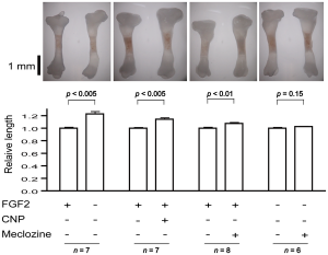
meclozine longitudinal bone growthThe rightmost figure(the one with n=6) is the Meclozine solo group.  Eyeballing it, it looks like it could be about 5% increase in longitudinal growth.  Note that a 5% increase on 5’9″ is 6’0″.
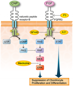
FGFR3 signalingFGFR3 signaling in chondrocytes.  LSJL does increase ERK phosphorylation.  Maybe that is a side effect of the boost of LSJL on longitudinal growth and not a cause of longitudinal growth.  And that both Meclozine and LSJL would have additive effects.

” a synthetic compound A31 is an inhibitor of the FGFR3 tyrosine kinase by in silico analysis. They demonstrated that A31 suppresses constitutive phosphorylation of FGFR3 and restores the size of embryonic femurs of Fgfr3Y367C/+ mice in organ culture. In addition, A31 potentiates chondrocyte differentiation in the Fgfr3Y367C/+ growth plate.  P3 has a high and specific binding affinity for the extracellular domain of FGFR3. They showed that P3 promotes proliferation and chondrogenic differentiation of cultured ATDC5 cells, alleviates the bone growth retardation in bone rudiments from TD mice (Fgfr3Neo-K644E/+ mice), and finally reversed the neonatal lethality of TD mice”

“These novel FGFR3 tyrosine kinase inhibitors, however, may inhibit tyrosine kinases other than FGFR3 and may exert unexpected toxic effects in humans. Meclozine may also inhibit unpredicted tyrosine kinase pathways, but we can predict that there will be no overt adverse effect, because meclozine has been safely used for more than 50 years.”

Here’s a study that will provide some insight into FGFR3 and why it inhibits growth in some cases but not in others:

The paradox of FGFR3 signaling in skeletal dysplasia: why chondrocytes growth arrest while other cells over proliferate.

Somatic mutations in receptor tyrosine kinase FGFR3 cause excessive cell proliferation, leading to cancer or skin overgrowth. Remarkably, the same mutations inhibit chondrocyte proliferation and differentiation in developing bones, resulting in skeletal dysplasias, such as hypochondroplasia, achondroplasia, SADDAN and thanatophoric dysplasia{So maybe the longitudinal growth induced by LSJL is not produced by chondrocytes but rather by the stem cells which would be promising evidence for post LSJL-inducable height growth}. A similar phenotype is observed in Noonan syndrome, Leopard syndrome, hereditary gingival fibromatosis, neurofibromatosis type 1, Costello syndrome, Legius syndrome and cardiofaciocutaneous syndrome. Collectively termed RASopathies, the latter syndromes are caused by germline mutations in components of the RAS/ERK MAP kinase signaling pathway. This article considers the evidence suggesting that FGFR3 activation in chondrocytes mimics the activation of major oncogenes signaling via the ERK pathway. Subsequent inhibition of chondrocyte proliferation in FGFR3-related skeletal dysplasias and RASopathies is proposed to result from activation of defense mechanisms that originally evolved to safeguard mammalian organisms against cancer.”

“FGFR3 [inhibits] chondrocyte proliferation. FGFR3/ERK signaling triggers disintegration of the cyclin D3-cdk6 complex in the G1 phase of the cell cycle, followed by increased association of p21WAF1 and p27Kip1 cell cycle inhibitors (CKI) with cyclin-cdk2 and cyclin-cdk4 complexes, leading to inhibition of their kinase activities. Upon FGFR3 activation, CKIs accumulate at the protein level, due to the interaction with transcriptionally induced cyclin D1. cyclin D1 upregulation also mediates the pro-mitogenic effects of FGFR/ERK signaling. Duration, magnitude and timing of cyclin D1 and p21WAF1 induction in the G1 phase of a cell cycle determines the nature of the response to an ERK signal. A strong but transient ERK activation in the early G1 induces intermittent p21WAF1 accumulation and stable cyclin D1 expression, leading to cell proliferation. In contrast, robust and persistent ERK activation leads to stable p21WAF1 accumulation and growth inhibition despite the concomitant induction of cyclin D1.

In chondrocytes, unlike most other cell types, FGFR3 activation elicits highly prolonged ERK activation lasting for up to 24 h. This phenotype is likely to stem from the maintenance of ERK pathway activation within the protein complexes interacting directly with FGFR3. ”

So FGFR3 inhibits growth in chondrocytes but not other cell types is because FGFR3 activates ERK for too long which leads to growth inhibition due to excess levels of p21WAF1.
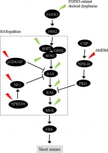
Another FGFR3 pathway“In chondrocytes, the premature senescence caused by FGFR3 activation does not involve the p53 pathway but appears CKI-dependent as induction of several CKIs (p21WAF1, p27Kip1, p16INK4a, p18INK4c, and p19INK4d) accompanies FGFR3-mediated inhibition of chondrocyte proliferation in vitro and in vivo”

NEW:

Meclozine Promotes Longitudinal Skeletal Growth in Transgenic Mice with Achondroplasia Carrying a Gain-of-Function Mutation in the FGFR3 Gene.

“Achondroplasia (ACH) is one of the most common skeletal dysplasias causing short stature owing to a gain-of-function mutation in the FGFR3 gene, which encodes the fibroblast growth factor receptor 3. We found that meclozine, an over-the-counter drug for motion sickness, inhibited elevated FGFR3 signaling in chondrocytic cells. To examine the feasibility of meclozine administration in clinical settings, we investigated the effects of meclozine on ACH model mice carrying the heterozygous Fgfr3ach transgene. We quantified the effect of meclozine in bone explant cultures employing limb rudiments isolated from developing embryonic tibiae from Fgfr3ach mice. We found that meclozine significantly increased the full-length and cartilaginous primordia of embryonic tibiae isolated from Fgfr3ach mice. We next analyzed the skeletal phenotypes of growing Fgfr3ach mice and wild-type mice with or without meclozine treatment. In Fgfr3ach mice, meclozine significantly increased the body length after two weeks of administration. At skeletal maturity, the bone lengths, including the cranium, radius, ulna, femur, tibia, and vertebrae were significantly longer in meclozine-treated Fgfr3ach mice than in untreated Fgfr3ach mice. Interestingly, meclozine also increased bone growth in wild-type mice. The plasma concentration of meclozine during treatment was within the range that has been used in clinical settings for motion sickness. Increased longitudinal bone growth in Fgfr3ach mice by oral administration of meclozine in a growth period indicates potential clinical feasibility of meclozine for the improvement of short stature in ACH.”

“A CNP analog with an extended half-life, BMN-111, has recently been developed, and significant bone growth recovery was demonstrated in amouse model of ACH by subcutaneous administration of BMN-111”

“Meclozine was administrated to 2-week-old wild-type mice for 3 weeks. As wild-type mice were weaned at 2 weeks after birth, we started meclozine treatment 1 week earlier than that for Fgfr3ach mice.  The body length of meclozinetreated mice was significantly longer than that of untreated mice after 1 week

“the plasma concentrations of meclozine used in the current study (0.2 or 0.4 g of meclozine per kilogram food).”

“Meclozine, an OTC H1 inhibitor, has been safely used for motion sickness for more than 50 years, and its optimal dose and adverse effects have already been established”

“in patients with ACH [given treatment with meclozine], the patients could be expected to increase 6.7 to 7.1 cm in height, based on the average height of adults with ACH.”
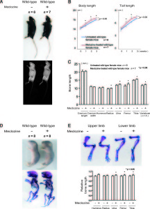
 

melcozone<-Treatment of mice with meclozine without FGFR3 deficiencies.  The meclozone treated mice are noticeably taller and lengthier.

“A-C, Wild-type mice were treated with meclozine 2 weeks after birth for 3 weeks. A, Visual images and soft X-ray images of wild-type female mice with or without meclozine. Meclozine-treated mice were larger than the untreated mice. B, Body and tail lengths of meclozinetreated
wild-type female mice were significantly longer than those of untreated wild-type female mice. Statistical significance analyzed by two-way ANOVA is shown on the right side of each graph. *P  .05 by Fisher’s LSD test for each pair. C, The lengths of the radius, ulna, femur, tibia, and vertebrae on the soft X-ray films were significantly increased by meclozine treatment by unpaired t test. D and E, Pregnant mice were treated with meclozine from embryonic day 14. D, Visual images and skeletons stained with Alizarin red and Alcian blue of wild-type mice at postnatal day 5, with or without meclozine. Meclozine-treated offspring were larger than untreated offspring. E, The lengths of the ulna, femur, and tibia measured using stained skeletons were significantly increased after meclozine treatment, as assessed by unpaired t test.”
The Calcium and Vitamin D In Milk Doesn’t Seem To Make Bones Stronger Either

I was driving home today and heard over the radio that there was news that a certain Dr. Karl Michaelsson in Sweden conducted a very large observational study to try to get the definitive answer on whether drinking milk does help make bones stronger or not. After looking at the carefully tabulated data of more than 100,000 subjects who took careful notes of their life over time, it seems that drinking more milk over a 20 year time span does NOT make the bones less likely to fracture.

This would seem to be going against what we were taught as little kids. The idea was that drinking milk was supposed to make young children taller and make the bones stronger. So far, we have proven that the correlation between young kids drinking milk and them ending up taller as adult is extremely weak. Now it seems that the claim that milk should make the bones stronger has also been sort of disproven.

Refer to the article written a month ago http://www.telegraph.co.uk/health/healthnews/11193329/Three-glasses-of-milk-a-day-can-lead-to-early-death-warn-scientists.html

Other Sources –  http://www.washingtonpost.com/news/to-your-health/wp/2014/10/31/study-milk-may-not-be-very-good-for-bones-or-the-body/

From what I could get out of the radio, drinking 3 glasses of milk a day doesn’t decrease the chances of fracture, and may in fact cause the person to die earlier. For women, the risk of fracture actually increases.

The theory proposed by Dr. Michaelsson on why this is is the following – The two types of sugar in milk, glucose and lactose, seem to cause the human body to go through even more oxidative stress. Oxidative stress is supposed to be one of the main causes for the human body to go through aging/senescence.

It turns out that when we were babies first coming out of our mothers, the first source of food was our mother’s breast milk. That breast milk had a lot of lactose in it. However, the new born baby as the lactase enzyme in the body to break down that lactose sugar. Over time, as the human develops and grows older, the level of lactase in their bodies seem to drop at a dramatic rate. In some countries like Asia, the level of the lactase enzyme is so low that people develop the condition “lactose intolerance”. The just don’t have enough of the enzyme in their body that is specifically used to break down the lactosse sugar found in milk and other dairy products.

What was shown was that instead of milk, hard cheese seemed to be able to decrease fractures. The difference between why hard cheese is effective and milk is not seems to come down to the fact that hard cheese has less levels and concentrations of lactose.

Conclusion

The first thing to realize is that drinking more than average milk does NOT decrease fracture incidences. In fact, it might have the opposite effect. This point is further validated by the PubMed Study “Milk, dietary calcium, and bone fractures in women: a 12-year prospective study.

What has been traditionally believed is that calcium is something that is desired for a developing children who is still growing taller. In the medical textbook that I have been reading, it seems that to have a developing child to be growing at their optimum level, they should be adding around 0.5 grams of extra calcium into their body everyday, and most especially during the puberty years, when they get their huge growth spurts, which should be around 1 gram of extra calcium each day

The reason milk doesn’t work in strengthening the bone is guessed by the researchers to be the negation of the effects of the calcium by the fat content in the milk.

Based on what I do know, it would still not be a good idea to stop giving the developing human child milk. Getting a reasonable amount of milk into a child is still somewhat important.

It might be that bovine derived milk is not completely compatible with the human stomach bacterial ecosystem. However, it does have some type of negligible effect.

Final TIp: Based on our research for the last two years, I am happy to tell the person who is worried about or suffers from low bone mineral density that they should instead look into a much better BMD(Bone Mineral Density) stimulant. –  Sclerostin Inhibitors (Refer to study “Sclerostin inhibition for osteoporosis–a new approach.”)