Paper states that yes stretching may be a way to grow taller

For 6 years old, who have more deformable bones. However, it is possible that strengthening the stimulus could make it worker for older and even skeletally mature individuals. If stretching works via articular cartilage endochondral ossification.

Exercise combined with lysine-inositol vitamin B12 promotes height growth in children with idiopathic short stature

Short stature is short stature of an unknown cause(not familial or genetic or unknown genetic origin). One of these causes could be inadequate nutrition including B12 deficiency. So using idiopathic short stature individuals is not ideal. The ideal test subjects would be normal stature individuals. Then ideally you’d try to find a way to reproduce the results in skeletally mature individuals by strengthening the stimulus.

“Researchers observed that many patients with ISS whose genetic target height is around the 50th percentile have no symptoms, and their bone age was only slightly lower than the age in the auxiliary examination. For these children, regular exercise of moderate intensity was employed. Stretching exercise, combined with oral lysine-inositol vitamin B12 (VB12), can effectively promote height growth. Based on
this, we systematically observed the clinical efficacy of regular stretch exercise of moderate-intensity combined with lysine and VB12 oral liquid in the intervention of ISS.”<-One possibility to make this paper work on the skeletally mature is to increase the intensity of the stretching exercise. It is possible that the exercise may work by increasing nutrient uptake in the growth plate in which case it won’t work in the skeletally mature.

“A total of 60 children with ISS who met the inclusion criteria and were treated at the traditional Chinese medicine dwarfism clinic of the Henan Children’s Hospital from June 2018 to July 2020 were selected.”

“The observation group consisted of 23 males and 7 females, aged 4.33–8.33 years. The control group consisted of 22 males and 8 females, aged 4.00–7.92 years.”

“(1) age < 3 years, or age > 10 years (male) or age > 8 years (female); (2) not meeting the ISS diagnostic criteria; (3) height growth rate ≥ 5 cm per year; (4) family history of hereditary diseases or underlying diseases of the heart, liver, kidney, etc.; (5) mental or emotional disorders or history of malnutrition.”<-it’s good that they removed malnutrition but that does not rule out vitamin deficiencies.

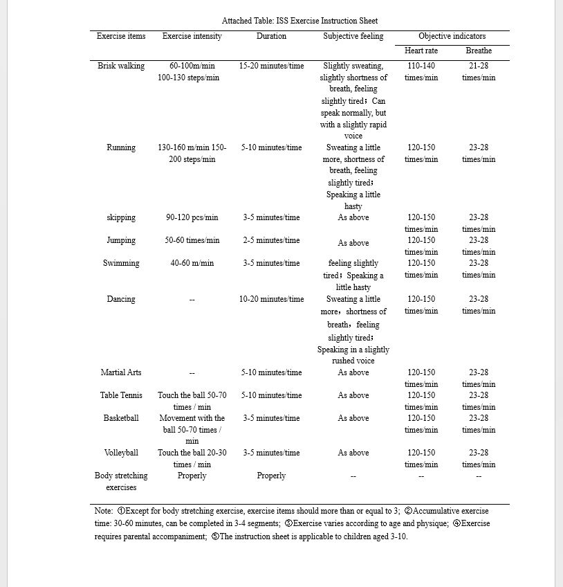
“The control group was given oral lysine-inositol VB12 oral solution (10 mL bid). Meanwhile, the observation group was given oral lysine-inositol VB12 oral solution (10 mL bid) and exercised according to the “ISS exercise guidance sheet”, which was developed in terms of exercise items, exercise intensity, exercise time, exercise days per week and so on. The children were instructed to perform aerobic exercises, such as brisk walking, running, jumping rope, etc., followed by stretching activities.”<-so really no unusual exercise activities

“The exercise could be broken down into two or three sessions per day to prevent children from
being overworked. Both groups were undergoing treatment for 12 months.”

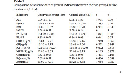
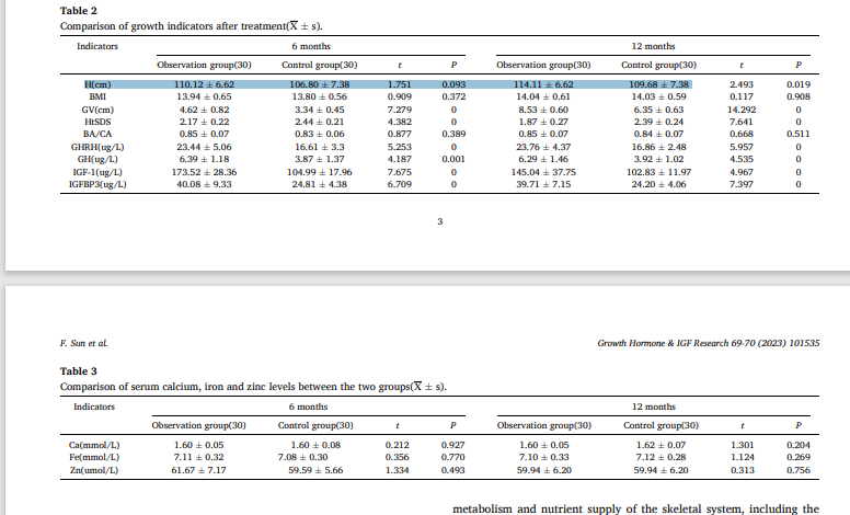
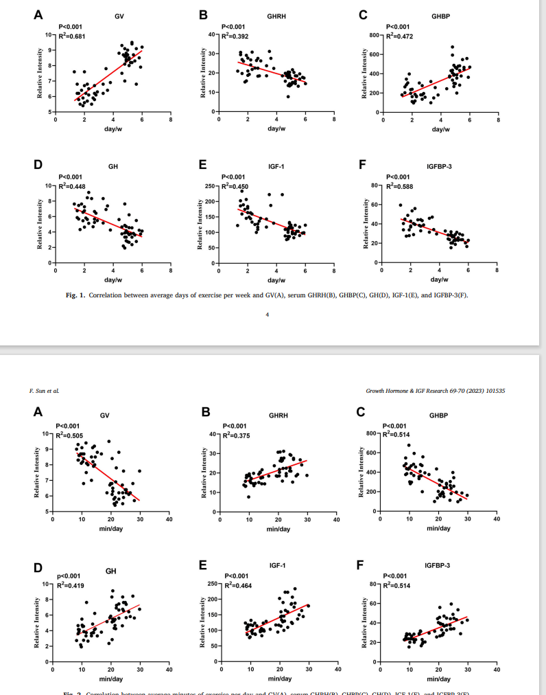
“after 6 and 12 months of treatment, GV, serum GHRH, GH, IGF-1, and IGFBP-3 levels in the observation group were significantly higher than those in the control group whereas its HtSDS[Height Standard Deviation Score] was significantly lower than that in the control group{there was less height variance in the exercised group}. The difference between the two groups was statistically significant); after 12 months of treatment, Height in the observation group was significantly higher than that in the control group, and the difference between the two groups was statistically significant

“The micronutrient Zn has been reported to promote growth rate in children with dwarfism.”

“Lysine VB12 is a complex of lysine, inositol, and VB12 that can be used to treat lysine deficiency symptoms, such as lack of appetite and poor growth. Lysine is an essential amino acid for all proteins in the human body and is a precursor substance for peptide hormones and coenzymes. In addition to maintaining metabolic balance in the body, lysine is an essential substance for children’s growth and development, since it can improve immune function, promote intellectual and physical development, improve nutritional status, and increase appetite”

So height is greater in the exercised group but so is starting height which is not ideal but still this is promising but this really needs follow up studies.

So Growth Velocity increased with days per week exercise which is greater but it decreased with the mins/days and 30 mins is not very many minutes. Maybe splitting up the exercise among more sessions would help. And growth velocity decreased with minutes per day despite positive growth stimulus hormones like GH and IGF-1 increasing. This makes it more likely that the benefit is mechanical which means there is a chance for modifying the stimulus to work on adults. Interestingly growth hormone and IGF-1 decreased with more days of exercise per week.

So interesting stuff here but we need more studies to see how it could play out. What would be interesting is spreading out the exercise more throughout the day to see if that reduce the negative correlation between minutes per day and growth velocity. Watching follow up studies will be helpful. Also we want to really optimize the optimal exercises. And find a way for any of the exercises to potentially work on adults.

HEre’s the exrcise chart. Lots of different exercises. Would be hard to know which are most beneficial as the movements are so diverse.

Breakthrough: study finds electric field can enhance jaw growth, can this be translated to height affecting bones?

There have been studies that have shown that it is possible to induce longitudinal bone growth in the jaw even potentially past skeletal maturity. Either the cells of the cartilage of the jaw are different than the cells of other joints(they could express different genes), in which case things that affect the jaw may not affect other joints, or it is not the cells themselves but rather the mechanical environment of the jaw. The jaw undergoes much more dynamic loading than other joints because the jaw is more moveable and the lateral pterygoid muscle is almost directly attached to the cartilage of the jaw providing it with a unique stimulation. Even if the cells of the jaw are different than the cells of other cartilaginous regions it is possible that these cells are different because of the unique mechanical loading that the jaw undergoes. The unique loading that the jaw cartilage undergoes maybe what alters the genetic expression that the cells of the jaw have. It is possible that if we load the joint cartilage of other cartilaginous regions in a similar way we may be able to induce growth longitudinally similar to what occurs in the jaw.

Osteogenesis Modulation: Induction of Mandibular Bone Growth in Adults by Electrical Field for Aesthetic Purposes

<-already the title is exciting

“A new technique in plastic surgery termed Osteogenesis Modulation is described. This technique uses a surgically implanted, battery-operated medical device to deliver customized electrical pulses to produce mandibular bone growth{trying to surgical implant the device to other joints may be challenging as it could potentially affect movement, we would want a way to implement the electric pulses to other joints in a way that does not require surgery}. This device was designed to be a temporary, nonpermanent implant. The purpose of this study was to review both the safety and efficacy of Osteogenesis Modulation.

This study comprises two phases. Phase I involved experimental technology development and animal experiments. Phase II included technology development for clinical use and a clinical trial. In Phase II, four patients with a diagnosis of mandibular hypoplasia and microgenia underwent surgical implantation of the novel medical device over the chin bone. Once a satisfactory change of contour of mandibular bone was achieved, the devices were removed. In all patients, the devices were left in place for 12 months, then surgically removed under local anesthesia. Preoperative and long-term postoperative cephalometric controls were done.

In all patients, symmetrical mandibular bone growth was observed with good-to-excellent aesthetic results. The overall follow-up period was 39 months. Cephalometric controls taken 3 to 6 months after the device removal showed an average increase in mandible length of 5.26mm (range, 2.83–7.60mm){this increase is not not exciting but it could improved upon}.

Preliminary clinical results suggest that Osteogenesis Modulation is a safe, minimally invasive, and effective alternative treatment for the correction of mandibular hypoplasia in selected cases.”

“he mandible thickness when low-intensity, moderate-intensity, or high-intensity negative pulses were applied was 2.70mm, 3.43mm, and 3.58mm, respectively. The thickness of the mandible on high-intensity positive pulses was 2.15mm.”<-it is very exciting that there was no decrease in results with more stimulus as it means that the stimulation may not be biphasic and it could be possible to increase the results with stronger stimulus.

Here is a before and after xray:

Before and after pic:

There are several before and after pics and x-rays in the study.

The next steps would be to try to apply this to other joints. I would try the hand and toe joints next because they are more movable compared to say the knee and ankle. Even though the stimulus to the jaw is electrical it is still possible that the enhanced jaw movement enhanced the results. Ideally we’d want only the electrical stimulation to produce the results that would make it easier to translate to other joints. That was the problem with possibly trying to mimic the bite jumping appliance on other joints. The jaw just undergoes so much more dynamic loading.

This is a very exciting study and here’s hoping it can translate to cheaper possibly greater height growth than limb lengthening surgery.

Deer antlers-could they be key to inducing new growth plates

Deer antlers are a mammalian appendage capable of regeneration. I don’t believe that eating deer antlers could induce growth plate regeneration but possibly the study of deer antlers could help enable growth plate regeneration. And perhaps studying deer antlers could enable the growth plates to be open for longer. One of the problems with limb lengthening surgery remains muscle and other soft tissue tightness. The growth plate is connected to these soft tissues via the enthesis so it is logical to think that perhaps the enthesis can help provide the cellular ingredients for those soft tissues to grow as needed via the enthesis.

A population of stem cells with strong regenerative potential discovered in deer antlers

<-perhaps for example there could be an osteotomy and an injection of these stem cells in a region near the enthesis to form new growth plates.

“The annual regrowth of deer antlers provides a valuable model for studying organ regeneration in mammals. We describe a single-cell atlas of antler regrowth. The earliest-stage antler initiators were mesenchymal cells that express the paired related homeobox 1 gene (PRRX1+ mesenchymal cells). We also identified a population of “antler blastema progenitor cells” (ABPCs) that developed from the PRRX1+ mesenchymal cells and directed the antler regeneration process.{could we use similar cells to form new growth plates?} Cross-species comparisons identified ABPCs in several mammalian blastema. In vivo and in vitro ABPCs displayed strong self-renewal ability and could generate osteochondral lineage cells. Last, we observed a spatially well-structured pattern of cellular and gene expression in antler growth center during the peak growth stage, revealing the cellular mechanisms involved in rapid antler elongation.”

“Lower vertebrates have a remarkable capacity to heal in a scar-free manner and regenerate lost appendages, even at the adult stage”

“deer antler offers a singular model to study spontaneous regeneration in mammals because its
regeneration is similar and clinically relevant to mammalian long-bone development”

“hard antlers are cast from their pedicles, then both antler bone and cartilage are regenerated from the pedicle periosteum located in the pedicle stumps”

“We further identified a population of regenerative progenitor cells, ABPCs, in the antler blastema, with impressive capacities for self-renewal, osteogenic–chondrogenic differentiation, and bone-tissue repair.”<-in the paper it goes over more what the cell characteristics of the progenitor cells might be. The question is: How do we translate what’s going on in the deer antlers to bone?

Vibration increases cartilage thickness

If vibration increases cartilage thickness. It is possible too that vibration could induce articular cartilage endochondral and stimulate growth plate growth. There are studies that show that vibration may be do this but the results have been non-overwhelming and mixed. It is possible however that it may be that the vibration stimulus needs to be improved. For example, laterally applying the vibration to the epiphysis and applying the vibration directly to the bone may improve the stimulus. But there are still several studies that show vibration is promising. It’s just the lack of a “smoking gun” study.

Vibration training intervention to maintain cartilage thickness and serum concentrations of cartilage oligometric matrix protein (COMP) during immobilization

“1) 14-days of immobilization of young healthy subjects using a 6°-“head-down-tilt-bed-rest”-model (6°-HDT) would reduce cartilage thickness in the knee and serum Cartilage oligometric matrix protein (COMP) concentration and 2) isolated whole body vibration training would counteract the bed rest effects.”

“While the control intervention resulted in an overall loss in average cartilage thickness of −8% (pre: 3.08 mm±0.6 mm post: 2.82 mm±0.6 mm) in the weight-bearing regions of the tibia, average cartilage thickness increased by 21.9% (pre: 2.66 mm±0.45 mm post: 3.24 mm±0.63 mm) with the vibration intervention. No significant differences were found in the weight-bearing regions of the femur. During both interventions, reduced serum COMP concentrations were observed (control intervention: −13.6±8.4%; vibration intervention: −9.9±3.3%).”<-it is possible that a thickness in cartilage could increase height and induce articular cartilage endochondral ossification it could also stimulate growth plate growth. The thickness of the cartilage was increased versus control which is good but COMP is reduced compared to control which is bad.

“Cartilage presumably maintains and responds to the loads placed on joints during activities of daily living. For instance, the loads generated at the knee during walking correlate with cartilage thickness in the weight-bearing regions of the knee”<-so we could potentially change the way we load to alter cartilage thickness.

“COMP plays a major role in stabilizing the extracellular matrix through its interaction with collagen fibrils and other matrix components. Serum COMP concentrations are elevated in patients with knee osteoarthritis and rheumatoid arthritis but also after a moderate walking exercise in healthy adults and after intense running exercise in athletes. Thus, serum COMP concentration appears to be sensitive to physiological loading.”

“Vibration frequencies between 15 Hz and 90 Hz have been used to achieve adaptations in muscle and bone”

“Training sessions were scheduled at least 30 min after breakfast and lunch. Subjects walked the distance between their room and the training room each session (∼25 steps). Each vibration training unit was composed of five times 60 s of isometric exercise bouts on a vibration platform (Galileo 900, Novotec Medical GmbH, Pforzheim, Germany) in an upright standing position with a knee flexion angle of 30°. Subjects carried an additional load of 15% of their body mass on a diving belt around their pelvis. Between exercise bouts subjects rested for 60 s while sitting on a chair. The vibration platform vibrated at 20 Hz with approximately 3 mm amplitude at the centre of the foot.”<-the vibration in this study could be improved.

“Average and maximum thicknesses in the tibial cartilage increased significantly by 21.9% and 26.6%, respectively[due to vibration]. The percentage change in average and maximum cartilage thicknesses did not differ between the medial and the lateral compartments of the tibia for both study phases. Cartilage thickness in the lateral and the medial compartments of the femoral cartilage did not show significant changes due to the bed rest or to the training intervention”<-maybe the way we as humans typically load does not adequate load the lateral and medial compartments of the femoral cartilage and that’s why we don’t typically gain height there. It’s possible that if all parts of the femoral cartilage was engaged we would gain height and therefore if we change our loading we could stimulate all components of the femoral cartilage and thereby gain height.

“the increased cartilage thickness after the vibration training if the mechanical stimulus increases proteoglycan content of cartilage.”

“Proteoglycans are negatively charged and thus exert a large swelling pressure that causes tensile stress on the surrounding collagen network”

Study provides evidence of microfracutre theory

There are studies that show that periosteal stripping may induce skeletal bone overgrowth pre-skeletal maturity. The below study shows evidence that fracture without periosteal damage may lead to skeletal overgrowth. Microfractures alter the fluid flow within the bone to that may be a mechanism by which microfractures can affect but that also means that it’s possible that fluid flow alone can lead to skeletal overgrowth. It’s important to note that we would need to study what exercises actually cause microfractures. There would need to be actual studies done otherwise it’s just speculation as to whether an exercise causes microfractures and don’t know if the microfractures are created to be sufficient stimulus.

Overgrowth of long bone in rabbits by growth stimulation through metaphyseal hole creation

Overgrowth of long bones was noted in pediatric patients who underwent anterior cruciate ligament reconstruction. Hyperaemia[blood vessels having more blood than normal] during creating a metaphyseal hole and the microinstability made by the drill hole may induce overgrowth{This is interesting because we could create temporary instabilities in bones and joints via exercise!}. This study aimed to determine whether metaphyseal hole creation accelerates growth and increases bone length and compare the effects of growth stimulation between metaphyseal hole creation and periosteal resection. We selected 7- to 8-week-old male New Zealand white rabbits. Periosteal resection (N = 7) and metaphyseal hole creation (N = 7) were performed on the tibiae of skeletally immature rabbits. Seven additional sham controls were included as age-matched controls. In the metaphyseal hole group, the hole was made using a Steinman pin at the same level of periosteal resection, and the cancellous bone beneath the physis was removed by curettage. The vacant space in the metaphysis below the physis was filled with bone wax. Tibiae were collected 6 weeks after surgery. The operated tibia was longer in the metaphyseal hole group (10.43 ± 0.29 cm vs. 10.65 ± 0.35 cm){about 2%, so if this happened on every bone it would take a 69 inch individual to about 70 1/3}. Overgrowth was higher in the metaphyseal hole group (3.17 ± 1.16 mm) than in the sham group (− 0.17 ± 0.39 mm. The overgrowth in the metaphyseal hole group was comparable to that in the periosteal resection group (2.23 ± 1.52 mm, P = 0.287). In rabbits, metaphyseal hole creation and interposition with bone wax can stimulate long bone overgrowth, and the amount of overgrowth is similar to that seen in periosteal resection.”

“During ACL reconstruction, there was no circumferential periosteal damage; instead, the drill hole was made from the metaphysis to the epiphysis, and the hole was filled with a tendon graft. The overgrowth was also reported in children with proximal metaphyseal fracture as Cozen’s phenomenon, and the overgrowth after proximal cortical breakage without extensive periosteal damage”

“Increased metabolic activity at the physis or bony instability has been suggested to the other possible cause of the overgrowth”

“The cortical hole made by drilling at the metaphysis could increases metabolic activity at the physis through hyperaemia and the large cortical hole would be related to the bony instability. During ACL reconstruction, a cortical hole made at the metaphysis to the phyis and interposition of the vacant space with the tendon may promote hyperaemia and bony microinstability”

The questions is if there is any instances of growth promotion in skeletally mature individuals.

Study finds that vibration can induce articular cartilage endochondral ossification

We’ve studied endochondral ossification of the articular cartilage in the condyle before. If vibration can induce endochondral ossification of the articular cartilage in the jaw it is theoretically possible that it could induce endochondral ossification in any joint unless the jaw cartilage is special. Which I don’t believe it is I believe that the jaw is more mobile than other joints and subjected to superior loading. If other joint regions were subjected to the same kind of loading as the jaw they could grow as well.

Effects of Mechanical Stimuli on Adaptive Remodeling of Condylar Cartilage

“Trabecular bone has been shown to be responsive to low-magnitude, high-frequency mechanical stimuli.
This study aimed to assess the effects of these stimuli on condylar cartilage and its endochondral
bone. Forty female 12-week-old C3H mice{this is pre skeletal maturity unfortunately, howver later in the study they say that the mice are “adult”} were divided into 3 groups: baseline control (killed at day 0), sham (killed at day 28 without exposure to mechanical stimuli), and experimental (killed following 28 days of exposure to mechanical stimuli).

The experimental group was subjected to mechanical vibration of 30 Hz, for 20 minutes per day, 5 days per week, for 28 days. The specimens were analyzed by micro-computed tomography. The experimental group demonstrated a significant decrease in the volume of condylar cartilage and also a significant increase in bone histomorphometric parameters. The results suggest that the low-magnitude, high-frequency mechanical stimuli enhance adaptive remodeling of condylar cartilage, evidenced by the advent of endochondral bone replacing the hypertrophic cartilage{vibration can induce articular cartilage endochondral ossification, if this can occur in other joints with articular cartilage then you can get longer bones by that mechanism!}.”

“there is a linear association between bone-regenerative capacity and bone density”<-So could things that increase bone density increase bone regenerative capacity?

“We achieved mechanical stimulation to the animals by placing them, while still in their cages with bedding removed, directly on a device which generated vertical ground-based vibration. This machine generated 30-Hz pulses, creating peak-to-peak accelerations of 2.9 m/sec2, referred to as a fraction of earth’s gravitational field, 0.3 g (1g = 9.8 m/sec2). Based on Rubin’s previous research, it is believed
that this produces peak strains of approximately 5 μє. Animals in the Experimental group were subjected to 20 minutes’ vibration per day for 5 days per week for a total of 28 days,”<-so the mice were subjected to vibration not specific to the jaw and it still underwent articular cartilage endochondral ossification. It would be interesting if other bones changed as well.

“the LMHF stimuli induced osteogenesis, leading to adaptive growth of the condyle in adult mice.”<interesting that they call these adult mice! But we have to be sure they’re skeletally immature

“the adaptive modeling of condylar cartilage is characterized by enhanced transition from chondrogenesis to osteogenesis. The vibrating mechanical stimulation in the present study was shown as a possible mechanism for the acceleration of the modeling.”

“A questionable aspect of this study design is the degree of transmission of the vertical ground-based oscillation, since it diminishes as the signal travels proximally through the skeleton. In a study measuring transmissibility in the hips and spines of humans standing on the oscillating plate, the authors showed
that approximately 80% of a 30-Hz ground-based signal reached the hip and spine”

This study here suggests that vibration can have benefit on growing bone:

Does Vibrational Loading Modulate the Effects of Radiotherapy on Growing Bone?

“[we evaluate] the possible beneficial effects of low-magnitude high-frequency mechanical vibration (LMHFV) stimuli on growing irradiated bone and the possibility for restoration of function of the epiphyseal plate”

ighteen 3-week old weanling male Sprague-Dawley rats were subjected to a standard radiation dose of 17.5 Gray applied to right hind limbs, with the contralateral leg serving as a non-irradiated control. Then, the animals were divided into three groups: A) rats subjected to (LMHFV) only at 45 Hz, 0.3 g for 20 minutes once per day, 7 days/week, for 3 weeks, B) rats subjected the same conditions of LMHFV plus an injection of spermine NONOate, a nitric oxide donor that that has shown weak positive results as post-irradiation recovery agent, and C) rats subjected to sham LMHFV. After euthanizing the animals, skeletal growth was measured by x-ray analysis, marrow mesenchymal stem cell osteoblastic potential was measured by CFU-F analysis, and bone morphology was measured by micro-CT analysis.

X-ray and CFU-F analyses show statistically significant differences between right and left limbs in all groups. No statistical significance was observed between vibration versus control groups, but trends suggest there could be some positive effect of vibration, although not statistically significant. Micro-CT results show a clear difference between right and left limbs in all groups. Regarding vibration versus control groups, micro-CT results are ambiguous, but do suggest that vibration may have altered local growth characteristics and stimulated local shape changes in the 20% region from the distal end of the femur, just above the growth plate.”

“Eighteen 3-week old weanling male Sprague-Dawley rats were obtained from Taconic Farms (Germantown, NY) and randomly divided into three groups: A) rats subjected to low-magnitude high-frequency vibration (LMHFV) only at 45 Hz, 0.3 g for 20 minutes once per day, 7 days/week, for 3 weeks, B) rats subjected to LMHFV with the same conditions as group A plus an injection of spermine
NONOate, a nitric oxide donor , and C) rats subjected to sham LMHFV, placed in cages
used for vibration but with no stimulus applied.”<-so we’re looking at group A versus group C as to whether vibration can increase bone length.

“there was possibly a difference, although not statistically significant, between right femora of different groups (A: 32.6±2.3 vs. B: 31.9±2.2 vs. C: 31.2±1.2, Figure 2), as well as total leg lengths of the femur
plus the tibia between different groups (A: 66.4±2.4 vs. B: 65.3±2.9 vs. C: 64.4±2.1, Figure 4).”<-So vibration group was longer than via group C.

“the ability of physical signals to influence bone morphology is strongly dependent on the signal’s magnitude, frequency and duration”