Proof that LSJL lengthened my finger

Go to this page to see the original x-rays.  Here’s a picture that shows that my right finger is longer than my left. However, what’s interesting is that the phalanx bones of the left index finger are actually longer than the right index finger.  So, I turned to the metacarpal bone of the index fingers and it turns out that the right metacarpal of the index finger is longer than the left and a longer metacarpal would explain the increased finger length in the image in the second link below.

LSJL may have lengthened all the bones of my right finger but because my left finger was always longer it may have only reduced the discrepancy.  If every single bone on the left side of body is longer than the right except for those that I performed LSJL on I submit that as proof of LSJL.  A possible reason that all my left bones are longer than my right bones but all my right bones are thicker could be related to FGFR3 and CNP.  My left side could have had less of the FGFR3 receptors or more of the CNP receptors.

I didn’t know that all my left sided bones were longer than my right until recently.  I just assumed it was due to my scoliosis or it was just an illusion because my right side was thicker.  So unfortunately, I did not account for it when designing to the experiment.  But again, if every single bone on my hands is longer on my left than my right metacarpal which is where I performed LSJL then we can take that as evidence that LSJL works.

Here’s a video with my right finger before any loading.:

Here’s the left hand before video:

My right index finger metacarpal did grow longer but it would appear that my other bones did not.  I say appear because it turns out that all my bones on the left side of my body are longer than the left.  Thus, any growth may actually just be shown by decreasing the gap between the right and left finger bones.  The right finger bones may have grown but that would not show up because the left finger bones were still longer.  If every single bone has the version on the left side be longer, but the one bone that had LSJL on it can we say that LSJL induced growth?

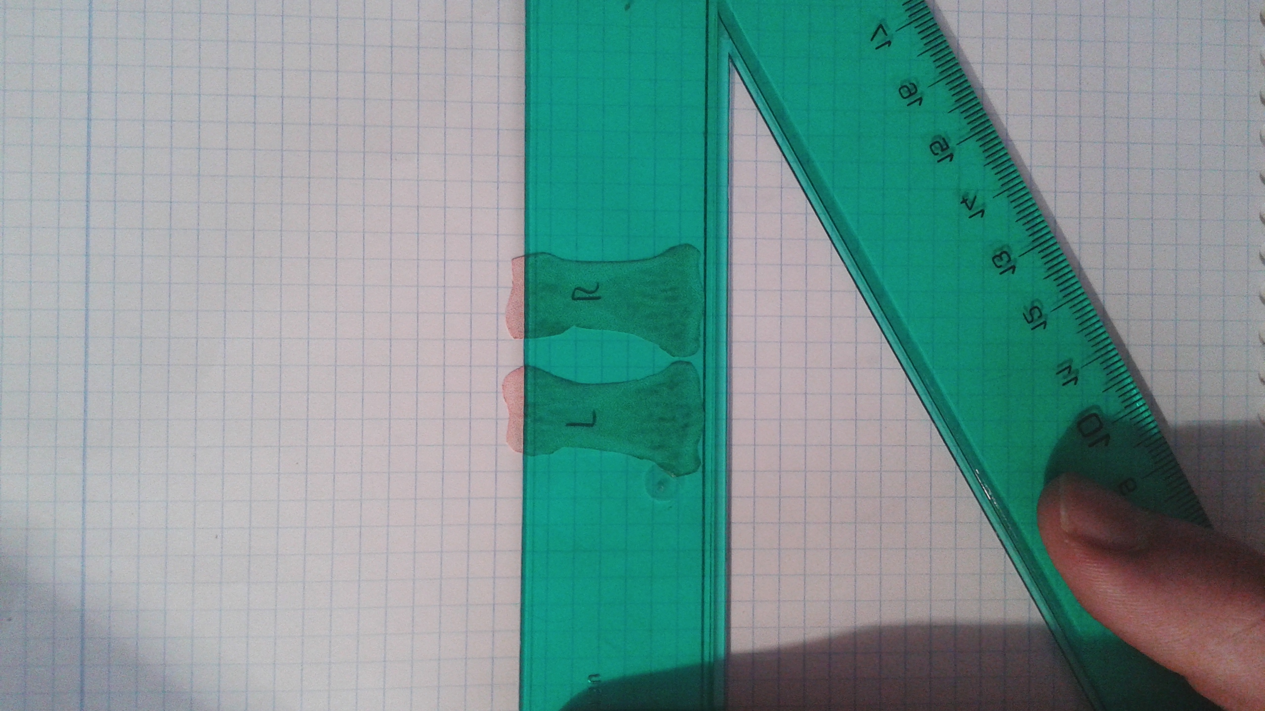
Here are the metacarpals of my right and left index fingers:

frontal index metacarpals


The lines are very straight.  I got pretty close to measuring out straight lines.  I measured the right metacarpal as being 1383.0 pixels long.  I got 1372.0 pixels long for the left metacarpal.  The left metacarpal might extend a little below the line but I zoomed in so I could get things accurate.  Right metacarpal is 0.8% longer.  Now here’s thing about the right finger being 0.8% longer.  All my left bones in my body are longer than the right bones but the right bones are thicker.  Thus, the right metacarpal may have grown to catch up to the left metacarpal and then grown further.  So actual growth may be larger than that.

I performed LSJL only on my right finger. Here’s an analysis of the other bones of the index finger:

Frontal 2 (1)


The slope of the line may be off by about 0.11 degrees or so which is very minor.  The left finger actually measures out longer.

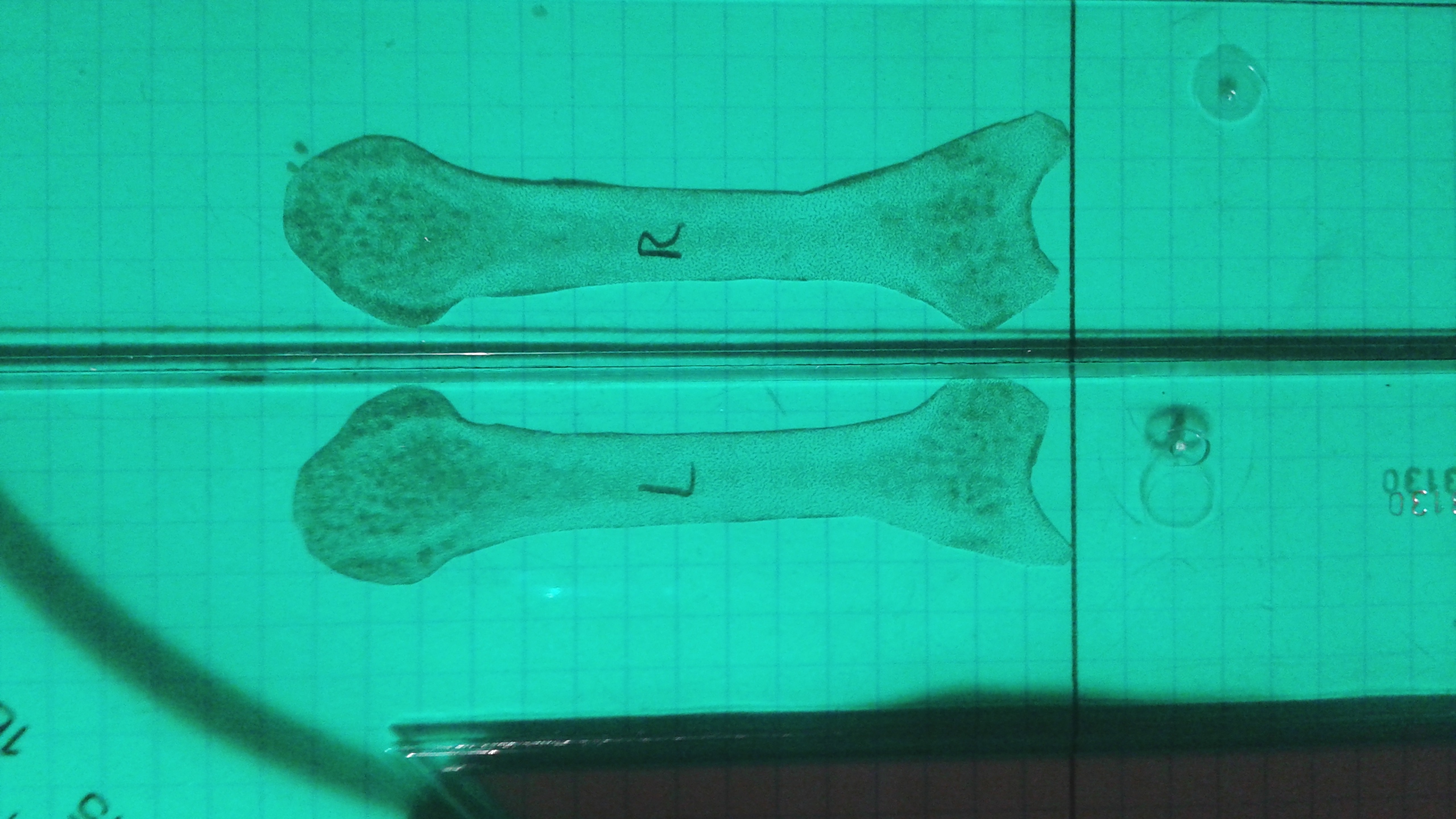
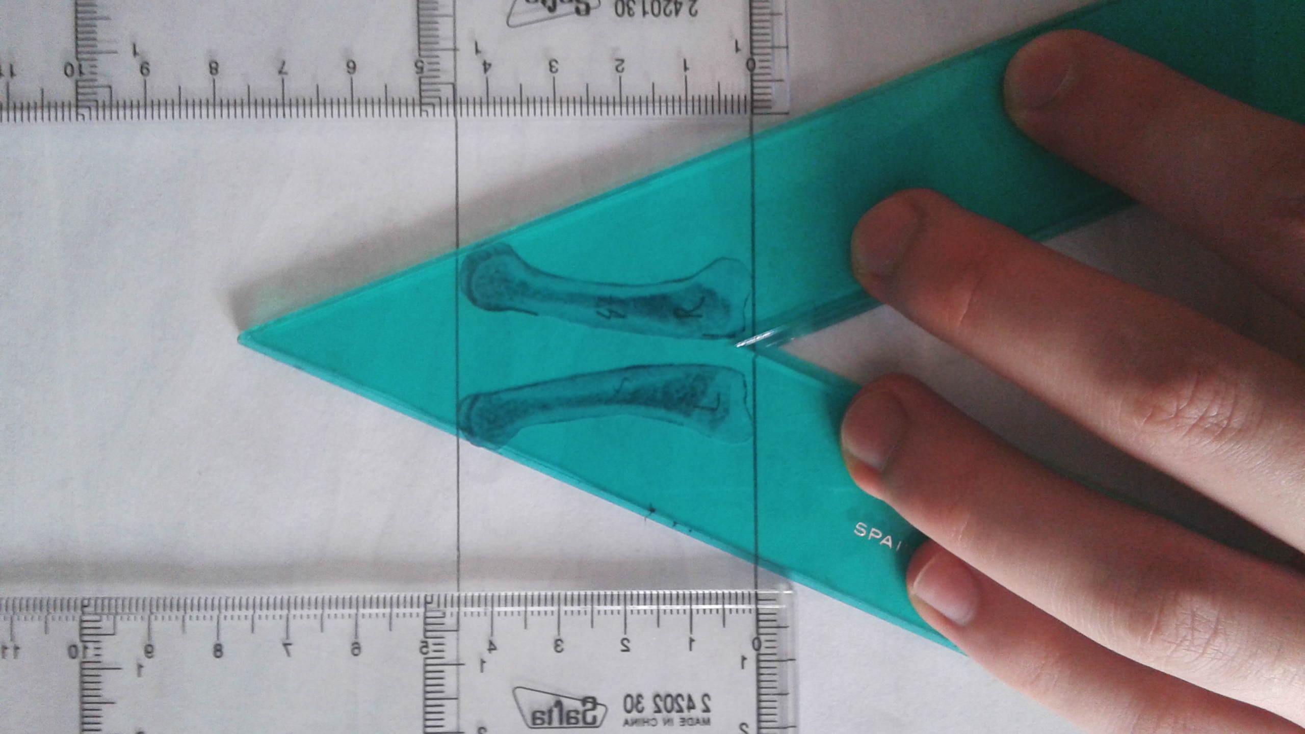
Here’s an image of my proximal index fingers:Frontal 2
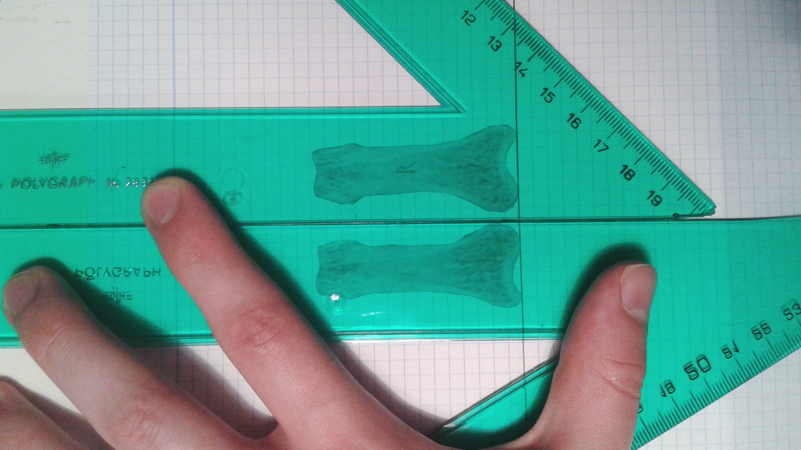
This is a cut out of the proximal index fingers of the right and left finger bones from the x-ray from the link provided above.  Click on the image as the image shows flipped for some reason.  The right bone is the one labeled with the R.  The measurements were done by adjusting the two bones until they were as long as possible.  This insures that both bones were given the same advantages and the angle at which I put my finger is not a factor.  Measurements were done by someone else.

The lines on the graph are slightly off the vertical lines sloping down at a 1.2 degree angle towards the right side of the computer screen(using the image if you click on the image not the inverted one here).  This images slope favors the right side so the right side will appear longer if you don’t correct for the one degree.

Here’s another image:

Frontal 1This one slopes to the right only at about .80 degrees(measuring the straight line downwards it is .80 off from 90 degrees.  This images slope favors the right as well but not nearly as much.  The left bone is longer.

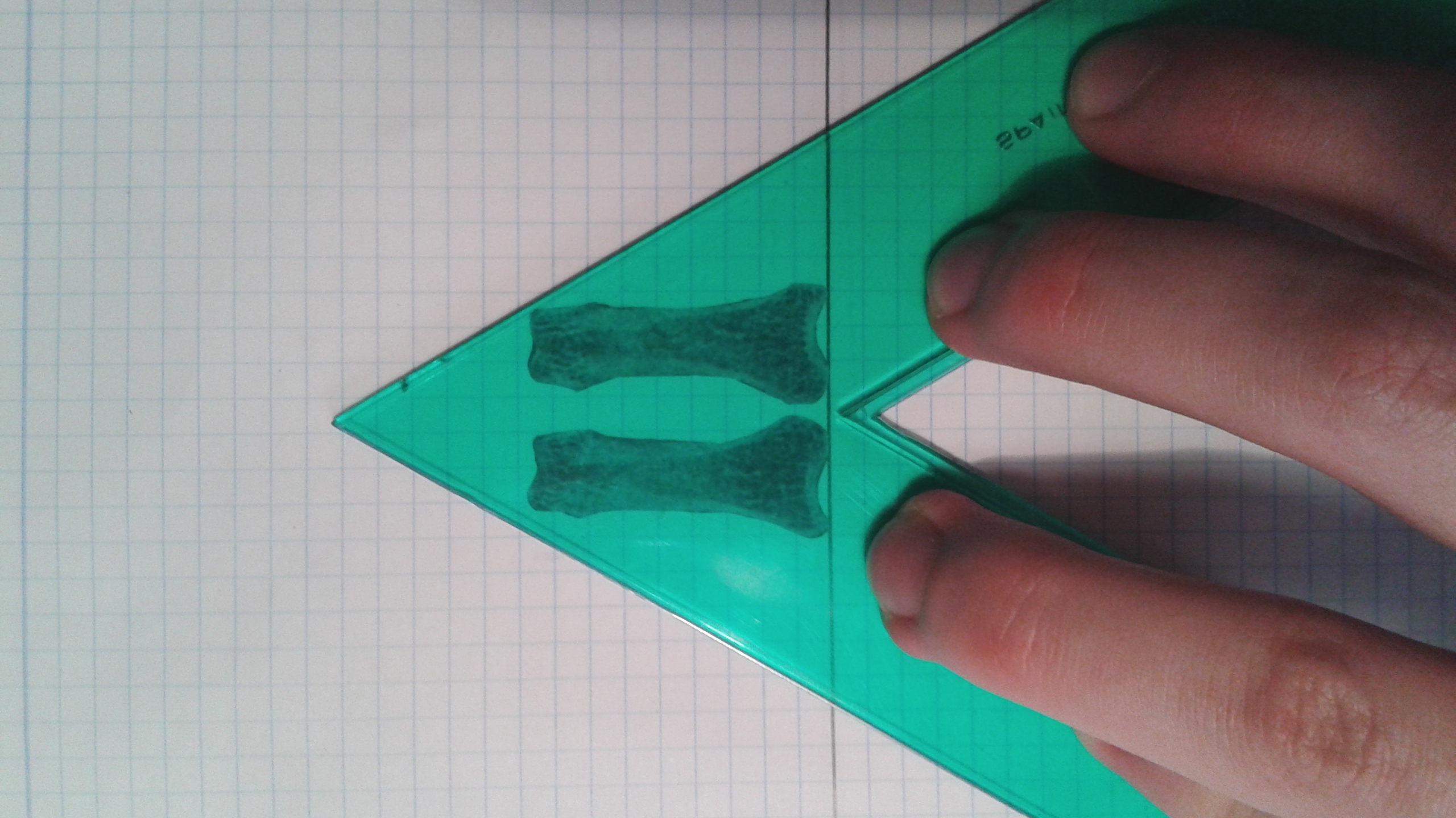
Here’s the cutout of the lateral view:

lateralYou can see here the the epiphysis of the right index finger are much larger but the left finger is longer overall.

How I’m changing my performance of LSJL now:
Next step is to confirm that all my left handed bones are longer except for that metacarpal.

Now since I have the x-rays, I’m going to be performing LSJL on both my right and left index finger.  Since I have x-ray images, I have those as a control using one of the fingers as a contralateral control is less important.

Previously, I was limiting my clamping based on my left side since my right side is stronger and thicker but since my left side is longer I’m not going to be clamping with whatever I can handle safely for my right side and whatever I can handle safely with the left side and not worrying about clamping them with the same intensity or amount of time.  Since my right side is so much stronger it should be able to withstand more clamping.  Thus, it was my goal with this to correct for the discrepancy between the right and left side in addition to increasing bone length overall.

Hoxd11 and the growth plate

This study supports that altering gene expression of Hoxa11 and Hoxd11 may be a way to grow taller as those genes can turn short bones into long bones.  Unfortunately, the genes that are known to interact with these genes are small in number and not known to be manipulatible by exercise or nutrition.  So it is more likely that in the future the Hoxa11 and Hoxd11 genes may contribute to height growth by manipulating the expression of these genes via an adenovirus.

The pisiform growth plate is lost in humans and supports a role for Hox in growth plate formation.

The human pisiform is a small, nodular, although functionally significant, bone of the wrist. In most other mammals, including apes and Australopithecus afarensis, pisiforms are elongate. the typical mammalian pisiform forms from two ossification centers. We hypothesize that: (i) the presence of a secondary ossification center in mammalian pisiforms indicates the existence of a growth plate; and (ii) human pisiform reduction results from growth plate loss. We surveyed African ape pisiform ossification and confirmed the presence of a late-forming secondary ossification center in chimpanzees and gorillas. Identification of the initial ossification center occurs substantially earlier in apes relative to humans, raising questions concerning the homology of the human pisiform and the two mammalian ossification centers. Second, we conducted histological and immunohistochemical analyses of pisiform ossification in mice. We confirm the presence of two ossification centers separated by organized columnar and hypertrophic chondrocyte zones. Flattened chondrocytes were highly mitotic, indicating the presence of a growth plate. Hox genes have been proposed to play a fundamental role in growth plate patterning. The existence of a pisiform growth plate presents an interesting test case for the association between Hox expression and growth plate formation, and could explain the severe effects on the pisiform observed in Hoxa11 and Hoxd11 knockout mice. Hoxd11 is expressed adjacent to the pisiform in late-stage embryonic mouse limbs supporting a role for Hox genes in growth plate specification.”

“Compared with humans, the pisiform of most other mammals, including primates, is substantially enlarged and elongated”

Here’s a good visual of growth plate formation:growth plate formation

“a) At birth (P0) the pisiform largely consists of undifferentiated hyaline cartilage. Note the future articular surfaces adjacent to the triquetral (right) and the transitional region near the insertion of the FCU (left). Each of these is distinct from the fibrous periosteal layers that surround the future pisiform shaft. (b) At P4 the cartilage has undergone differentiation to flattened columnar and hypertrophic chondrocytes. It is the calcified hypertrophic matrix that is staining red in Fig. 3(a). (c) By P7 the primary center of ossification begins to be replaced by bone. A broad region of flattened columnar and hypertrophic chondrocytes is preserved at the palmar end (right). (d) At P9 the preserved strip of cartilage displays all of the hallmarks of a growth plate: organized columnar and hypertrophic zones and a perichondrial ring (yellow arrowhead) adjacent to the bone collar. (e) A transverse section through the carpal tunnel demonstrates the unique ossification of the pisiform (left). Note the preserved region of red stained cartilage at the palmar end. In contrast the scapholunate (right) has ossified as a single primary center extending into the projecting tubercle. (f) At P17 the growth plate appears to be losing its activity, as there is no longer an identifiable hypertrophic zone underlying the columnar chondrocytes.”<-so if via LSJL we can induce regions of hyaline cartilage they could potentially become growth plates.

“Full deletion of Hoxa11 or Hoxd11 results in a highly penetrant phenotype with shortened pisiforms that often fuse to the triquetral (ulnare) or less commonly to the scapholunate and triquetral ”

” ‘no Hoxd land’ for short bone morphology”<-Maybe we could upregulate Hoxd in short bones to make them become long bones?

“In short bones and epiphyses, the initial process of chondrocyte differentiation is similar. However, expanded cartilaginous growth plates or active perichondrial rings do not form. Instead, the periphery of these regions largely consists of a narrow three to four cell layer of round chondrocytes that anticipate the future articular zone. In each of these respects (organized chondrocyte zones, active perichondrial ring and deposition of the bone collar), the pisiform is more similar to long bones”<-The organized chondrocyte zones, active perichondrial ring and deposition of the bone collar could be related to Hoxd.

“mice with reduced Hoxa11/Hoxd11 expression display decreased proliferation within mesenchymal condensations and dramatic shortening of the radius and ulna such that they also resemble short bones”

If we can induce Hoxd11 expression in short bones we can make those bones longer and thus ourselves taller.

Looking at the String Embl gene interaction reveals a problem in that very few genes directly interact with Hoxd11.  Although LSJL upregulates the related gene Hoxd10:

Hoxd11

Similarly with Hoxa11:

hoxa11

Stem Cell Therapy Used To Treat Short Stature – Huge Step Forward!

For a while now I have known about the existence of a type of bank known as stem cell banks, which are used to store and preserve the multipotent abilities of a person’s endogenous based cells. The billionaire Mark Cuban has called this type of “medical emergency insurance” as one of the best gifts that you can give to your children. There are probably hundreds of these banks that are around the world currently. Most people in the general public probably don’t see the long term benefits of getting something like this done. It is quite expensive to use this service, which may never be needed at all.

I keep up with many of the online sources which talk about the latest developments in biotech. One source I found linked to the 1st and largest stem cell bank in India, ReeLabs. These guys have figured out and optimized the way that stem cels can be harvested from the placenta, the amniotic sac, and the amniotic fluid. Now, technically Reelabs do not offer stem cell therapy (because they are just the bank, not a clinic), but they did show the possibility when they revealed that Stem Cell Therapy can be used in bone disorders. The only type of bone disorder that the website listed was Achondroplasia.

As we know, Achondroplasia is the most common disorder that causes severe short stature. There are dozens of types and forms of Achondroplasia. We here have studied this disorder extensively. Reelabs has categorized and defined Achondroplasia as a treatable bone disorder, using the stem cell technology. When you have this revolutionary technique compared to the conventional approach, you realize that THERE IS NO Conventional approach.

Refer to Reelab’s webpage on Achondraplasia here.

Some key things to remember

  • The term “Achondroplasia” is used to for both adults with close plates and children with open plates. This is in difference to the way Acromegaly and Gigantism are defined
  • Most likely, when Reelabs say that stem cell therapy can treat achondroplasia, they are most likely referring to children with open growth plates

Now, for some people who read our website regularly, the idea of injecting stem cells into cartilage to make it expand doesn’t seem that big of a leap, but the truth is that I personally have not found one PubMed study which have ever validated this idea. It was always guessed to be possible, but there hasn’t been a group of researchers who tried this and published positive results.

The closest any paper came to doing this was the idea of Autologous Chondrocyte Implantation ACI). An induced injury is done to the growth plates on rabbits and some part of the growth plate cartilage is severely damaged or chipped away. The implantation of some outside sources of cartilage, chondrocyte, or stem cells showed that the growth plate cartilage managed to regenerate. No study or group has come out and said that stem cell injections can cause cartilages to expand.

Since these guys say that it is possible, and maybe also been tested somewhere to get the technique to become optimized, it validates one of the last steps that we really need to get the technique to work on adults with closed growth plates. The shift from open growth plates to close growth plates involves a precursor step, the one where a step distraction is done to the bone and cartilage is implanted in between.

Of course, I had already proved the idea that growth plate cartilage transplantation was viable from the works by Teplyashin’s team and the collaboration by Ballock and Alsberg.


How much growth hormone is needed to cause gigantism?

If gigantism was only caused by excess HGH levels than why is gigantism so rare.  It is likely that extremely high levels of HGH or other factors are needed to cause gigantism.  This study will help us determine how much HGH is above the norm in cases of gigantism.

Gigantism caused by growth hormone secreting pituitary adenoma.

“Gigantism indicates excessive secretion of growth hormones (GH) during childhood when open epiphyseal growth plates allow for excessive linear growth. Case one involved a 14.7-year-old boy presented with extreme tall stature. His random serum GH level was 38.4 ng/mL, and failure of GH suppression{Resistance to GH surpression may be another factor needed for gigantism to be caused} was noted during an oral glucose tolerance test (OGTT; nadir serum GH, 22.7 ng/mL). Magnetic resonance imaging (MRI) of the brain revealed a 12-mm-sized pituitary adenoma. Transsphenoidal surgery was performed and a pituitary adenoma displaying positive immunohistochemical staining for GH was reported. Pituitary MRI scan was performed 4 months after surgery and showed recurrence/residual tumor. Medical treatment with a long-acting somatostatin analogue for six months was unsuccessful. As a result, secondary surgery was performed. Three months after reoperation, the GH level was 0.2 ng/mL{So the GH level was higher by a factor of 192 which is exceptionally high} and insulin-like growth factor 1 was 205 ng/mL. Case two involved a 14.9-year-old boy, who was referred to our department for his tall stature. His basal GH level was 9.3 ng/mL, and failure of GH suppression was reported during OGTT (nadir GH, 9.0 ng/mL). Pituitary MRI showed a 6-mm-sized pituitary adenoma. Surgery was done and histopathological examination demonstrated a pituitary adenoma with positive staining for GH. Three months after surgery, the GH level was 0.2 ng/mL{His GH was elevated by a factor of 46.5} and nadir GH during OGTT was less than 0.1 ng/mL. Pituitary MRI scans showed no residual tumor. ”

So key factors involved in Gigantism involve HGH much higher than normal and a resistance to HGH surpression.  According to this steriods forum, some experienced elevated serum HGH levels as high as 18ng/mL which is within the range of the serum levels of the two cases of gigantism.  Which means in terms of serum GH level, HGH injections may be sufficient to cause gigantism.  However, resistance of the HGH to surpression is one thing that is not conferred by the injections as the HGH injections have to be cycled.

“True gigantism is extremely rare”<-Which is why again it is not likely to be caused by elevated HGH alone.

“normal range, 0-5 ng/mL [For GH]”

In the 38.4ng/ml case for Gigantism, his IGFBP-3 was additional elevated.  IGF-1 levels were slightly above normal range.  Prolaction was within normal range.

The other boy with 9.3ng/ml case for Gigantism, had prolaction, IGFBP-3 and IGF-1 all in normal range.  But this boy was actually taller than the other case beside having about a 1/4 less HGH.

“Approximately 100 cases of children with pituitary gigantism have been reported”<-But yet gigantism was caused with levels of only about twice the maximum the normal range.

“Hyperprolactinemia is a common finding in GH excess presenting in childhood, undoubtedly related to the fact that mammosomatotrophs (GH and prolactin-secreting cells) are by far the most common type of GH secreting cells involved in childhood gigantism. However, gigantism caused by a pituitary tumor comprised of somatotropes (GH-secreting cells) show a normal prolactin level”

Why Stretching Does Work To Gain Permanent Height Increase

Usually the email to the website gets just the exact same type of messages from the same type of people. Maybe 75% of the emails we get are people asking me to upload and give them for free the E-Book PDF for the GT4I, but I have felt after dealing with too many potential legal problems that uploading that book will do more harm than good in the long term.

Yishan WongSometimes something useful actually comes along, and that happened today. An email showed me this post written on Quora by a Yishan Wong reveals that a correction of posture through stretching and maybe also yoga has let the poster increase his height by half an inch, going from 5’8″ to 5’8.5″. The person who emailed me mentioned that this Yishan Wong guy is supposed to be the CEO of reddit, but I would not know about that too much. A Quick check does reveal that this Wong guy does have a Quora account and has a similar phrase used in his profile. A Google search on this guys’s height comes up with 5′ 7.5″, which is close enough to his claims on the post about growing taller.

So in light of the new developments, it seem that even a multi-millionaire, super rich tech entrepreneur like Yishan Wong has himself noticed changes in his own height after puberty, through just the normal application of the principles of stretching and yoga.

Now, I personally don’t think there is any need for a person as rich as the CEO of Reddit to ever need to lie to others about his changes in stature. So I am going to just trust in this guy’s claims and say that somehow he increase his height by 1/2 an inch. If we go on the older claim that this guy was 5′ 7.5″ before, then his year of posture correction led to a full inch in height increase for him.

This type of change is NOT that uncommon. I remember years ago watching this televised golf tournament where the announcer noted that one of the professional golfers recently told in an television interview that he increased his height from 6′ 3″ to 6′ 4″ after spending 3 months doing stretching at a very intensive level. Why would anyone need to do that much stretching is beyond my comprehension but that professional golfer got results. Who was this golfer, or what is his name? I don’t remember, but I do remember that segment extremely well in my mind.

The point is that there are too many documented cases of people who took action to change their posture (for cosmetic or health reasons) and started doing stretching as adults and noticed height changes which never went away.

The most obvious and simple way to increase height, and not just temporary height, is to stretch, and stretch a lot. Just this last month I was at a Goodwill and found this old guide to Yoga book and it was showing Indian Hindu yogis who were like 80 years old and had amazing flexibility doing postures that teenage ballerinas can’t even do. In my opinion, stretching should be the first place we should start in our endeavors. and Join a yoga class.

Melatonin may play a role in scoliosis pathology

Scoliosis causes height reduction by causing a curvature of the spine thus it is relevant to height increase.

Abnormal Response of the Proliferation and Differentiation of Growth Plate Chondrocytes to Melatonin in Adolescent Idiopathic Scoliosis.

“Abnormalities in the melatonin signaling pathway and the involvement of melatonin receptor MT2 have been reported in patients with adolescent idiopathic scoliosis (AIS).  In this cross-sectional case-control study, growth plate chondrocytes (GPCs) were cultured from twenty AIS and ten normal control subjects. Although the MT2 receptor was identified in GPCs from both AIS and controls, its mRNA expression was significantly lower in AIS patients than the controls{Will increasing MT2 receptor levels make you taller(or shorter)?}. GPCs were cultured in the presence of either the vehicle or various concentrations of melatonin, with or without the selective MT2 melatonin receptor antagonist 4-P-PDOT (10 µM). Then the cell viability and the mRNA expression of collagen type X (COLX) and alkaline phosphatase (ALP) were assessed by MTT and qPCR, respectively. In the control GPCs, melatonin at the concentrations of 1, 100 nM and 10 µM significantly reduced the population of viable cells, and the mRNA level of COLX and ALP compared to the vehicle{Whether this would increase or decrease height is unclear}. Similar changes were not observed in the presence of 4-P-PDOT. Further, neither proliferation nor differentiation of GPCs from AIS patients was affected by the melatonin treatment.

“[The] abnormality [that] is manifested during the peripubertal period in patients with AIS in that they tend to be taller, leaner and have a longer arm span than their healthy peers”<-So perhaps MT2 receptor makes you shorter?  So an MT2 receptor inhibitor would make you taller or something that inhibits melatonin?

“Melatonin failed to inhibit the increase of 3′,5′-cyclic adenosine monophosphate (cAMP) induced by forskolin in osteoblasts from AIS patients when compared with cells from normal control subjects”

“melatonin inhibited both proliferation and differentiation of rat vertebral body growth plate (VBGP) chondrocytes with the involvement of MT1 and MT2 receptors”

“After incubation for 24 h in medium containing melatonin, the cell proliferation, gene expression of collagen type II and aggrecan, as well as protein expression of proliferating cell nuclear antigen (PCNA), Sox9 and Smad4 were significantly reduced. Moreover, it was found that the effects of melatonin could be reversed by the melatonin receptor antagonist luzindole, indicating the involvement of membrane melatonin receptors in these functions”

“AIS patients with a low level of expression of MT2 receptor in osteoblasts showed a longer arm span than those with a normal expression level of MT2 receptor”

Thus Luzindole may be a way to inhibit melatonin and grow taller at the expense of possibly inducing scoliosis.