Ecommia Leaf Extract for height

This supplement is available for sale and it seems to be promising for those who have existing growth plates:

Effects of Eucommia ulmoides Extract on Longitudinal Bone Growth Rate in Adolescent Female Rats.

Full study -> eucommia

“[We] investigate the effects of E. ulmoides extract on longitudinal bone growth rate, growth plate height, and the expressions of bone morphogenetic protein 2 (BMP-2) and insulin-like growth factor 1 (IGF-1) in adolescent female rats. In two groups, we administered a twice-daily dosage of E. ulmoides extract (at 30 and 100 mg/kg, respectively) per os over 4 days, and in a control group, we administered vehicle only under the same conditions. Longitudinal bone growth rate in newly synthesized bone was observed using tetracycline labeling. Chondrocyte proliferation in the growth plate was observed using cresyl violet dye. In addition, we analyzed the expressions of BMP-2 and IGF-1 using immunohistochemistry. Eucommia ulmoides extract significantly increased longitudinal bone growth rate and growth plate height in adolescent female rats. In the immunohistochemical study, E. ulmoides markedly increased BMP-2 and IGF-1 expressions in the proliferative and hypertrophic zones. In conclusion, E. ulmoides increased longitudinal bone growth rate by promoting chondrogenesis in the growth plate and the levels of BMP-2 and IGF-1. Eucommia ulmoides could be helpful for increasing bone growth in children who have growth retardation.”

“Because components in E. ulmoides extract activate osteoblast differentiation, we hypothesized that treatment with E. ulmoides extract would increase longitudinal bone growth rate.”<-Interesting considering it’s the growth plaate that increases height.

If you look at figure 2 in the full study, the growth plate looks bigger but it doesn’t have the dramatic differences that other chemicals or methods induce in the growth plate.  Which means that the risk is that this chemical only increases growth rate and not adult height.  According to Table 1, this compound increased levels of BMP-2 and IGF-1 by up to 50% in the resting, proliferative, and hypertrophic zone.

“At the dose of 100 mg/kg, E. ulmoides caused a significant acceleration of longitudinal bone growth rate, which was 373.1 ± 24.4 µm/day (6.4%) compared with the control group, which was 350.8 ± 18.5 µm/day. At the dose of 30 mg/kg, E. ulmoides caused an acceleration of longitudinal bone growth rate of 360.5 ± 23.5 µm/day (2.8%) compared with the control group.”

Cilia’s involvement to load on the growth plate

I wrote more about Cilia and the growth plate here.  By understanding the growth plate response of load we can understand how LSJL influences growth plate development and how crucial it is to have a growth plate in place for LSJL to work.  This study provides evidence that the adaptation to LSJL is atypical to normal load on the growth plate.

The growth plate’s response to load is partially mediated by mechano-sensing via the chondrocytic primary cilium.

Growth Plate Cilia<-link to pdf

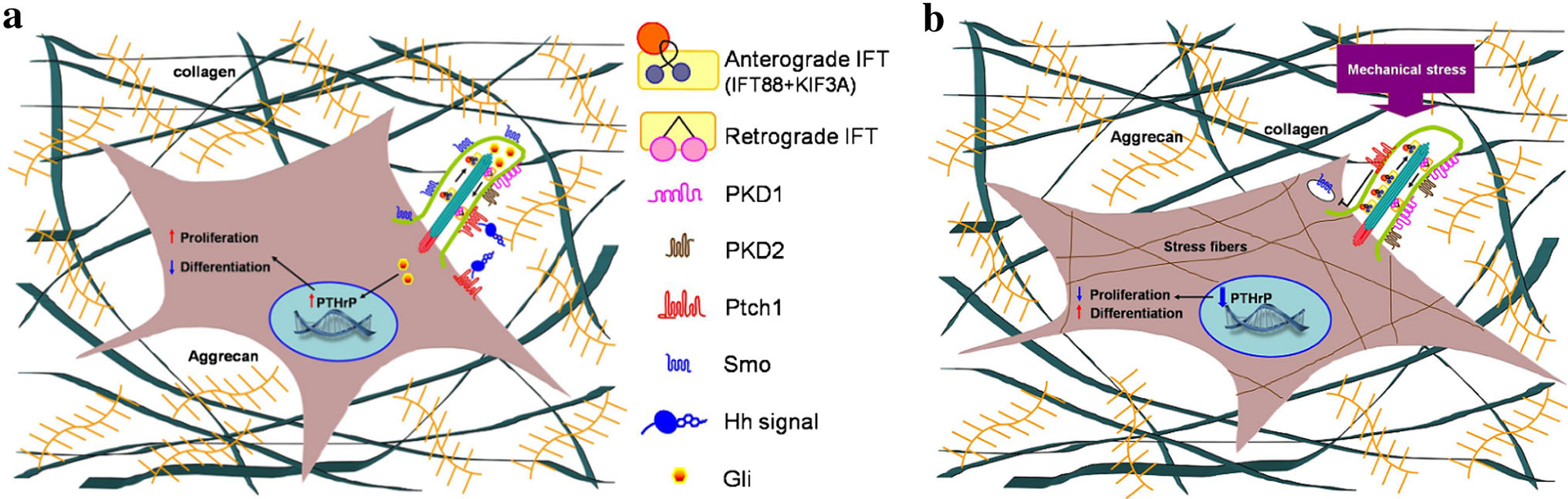
“Chondrocytes sense and respond to mechanical stimulation. The primary cilium has been identified as a mechano-sensor in several cell types, including renal epithelial cells and endothelium, and accumulating evidence connects it to mechano-transduction in chondrocytes. In the growth plate, the primary cilium is involved in several regulatory pathways, such as the non-canonical Wnt and Indian Hedgehog.  It mediates cell shape, orientation, growth, and differentiation in the growth plate. Mechanical load enhances ciliogenesis in the growth plate. This leads to alterations in the expression and localization of key members of the Ihh-PTHrP loop resulting in decreased proliferation and an abnormal switch from proliferation to differentiation, together with abnormal chondrocyte morphology and organization. We use the chondrogenic cell line ATDC5, a model for growth-plate chondrocytes, to understand the mechanisms mediating the participation of the primary cilium, and in particular KIF3A, in the cell’s response to mechanical stimulation. This key component of the cilium mediates gene expression in response to mechanical stimulation.”

“The primary cilium is critical to skeletal development; the embryonic cilium plays a role in the earliest cellular determinative events establishing left–right axis asymmetry and primary cilia in the early mesenchyme is necessary for proper anterior-posterior limb patterning”

“the primary cilium is required for bone cell response (increase in the expression of osteopontin) to dynamic fluid flow”

“The primary cilium mediates cell shape, orientation, growth, and differentiation in the growth plate as deletion of KIF3A, a subunit of the motor protein kinesin-II, results in defects in the columnar organization of the growth plate together with reduced cell division, accelerated hypertrophic differentiation, and disruption of cell shape and orientation relative to the long axis of the bone”

“Ihh, directly through its receptor Patched-1 (ptc1), increases chondrocyte proliferation and inhibits its hypertrophic differentiation through induction of Parathyroid hormonerelated
protein (PTHrP) expression”

For mechanical stimulation, cells were stetched at 1HZ by 20% elongation.

“the transition between the proliferative zone (positive for collagen II) and hypertrophic zone (positive for collagen X) was more homogeneous in the growth plates that were subjected to loading, suggesting that not only proliferation is altered by the load, but also the switch between proliferation and differentiation is altered.”<-Thus suggesting that PTH and IHH was involved.  However, this was not the case with LSJL growth plates. In that study, the hypertrophic and proliferative zone was less homogeneous.

“[In loaded growth plates], cells in the proliferative zone deviated less from the center of the column compared with the control growth plates in which more cells deviated from the column line”

“Morphometric analysis showed a significant increase in the number of cells per defined area and a decrease in the average cell area”<-Both number of cells and cell area increased during LSJL loading.

“the cells in the proliferative zone were more spread out, whereas those in the hypertrophic zone were more spherical, suggesting that mechanical load affects chondrocyte morphology and organization within the growth plate.”

A diagram depecting a chondrocytes response to stress:

chondrocyte stress

“a Unstimulated GP chondrocyte: Ihh, directly through its receptor Patched-1 (ptc1), located in the cilium, increases chondrocyte proliferation and inhibits its hypertrophic differentiation through induction of Parathyroid hormone-related protein (PTHrP) expression.  b Mechanical stimulated GP chondrocyte: morphological change of the cell together with up-regulation of cilia related genes (IFT88, KIF3A, PKD1 and PKD2) and formation of stress fibers.  Decrease in the expression of Ihh and ptc1 results in major decrease of PTHrP expression following reduced proliferation and switch for differentiation”

“we did not observe any significant difference in the cilia length caused by the mechanical stimulation.  In all checked samples, around 80 % of the cells presented cilia (counted according to acetylated-tubulin staining in comparison to nucleus DAPI staining), and cilia length was 2.4 lM,”

“primary articular chondrocytes [were subjected] to cyclic tensile strain up to 20 % for 1 h at 0.33 Hz and primary cilia prevalence was not altered in response to this stimulation”  However other studies found that cilia subjected to higher strain were reduced by 15.1%.

In this study C-Fos and egr1 were upregulated two genes that were also upregulated by LSJL.

“tensile load is induced during stretching while compression load is induced during release from stretch. Both acts induce fluid flow, thus creating shear load.”

“in control cells, mechanical stimulation induced Ihh expression, and activation of the pathway by SAG stimulation increased the expression of ptc1 and its accumulation in the cilium. Knockdown of KIF3A abolished these responses.”

Osteoclast inhibition increases growth plate height?

osteosclerosis <-the pdf.  Osteosclerosis is increased bone density.

Osteosclerosis induced by denosumab

“A 10-year-old boy, 144 cm tall, was referred to our hospital in October, 2012, with a 2 month history of persistent pain in the buttocks. He had altered gait due to pain, but no disturbance of bladder or bowels. Radiographs, CT, and MRI showed a large osteolytic lesion in the sacrum, and examination of a bone biopsy sample confirmed a giant cell tumour of bone, which we considered to be unresectable because of the potential risk of neurological deficit and massive bleeding. We obtained informed consent from the patient and his parents and the review board for off-label use of denosumab, a potent inhibitor of osteoclastic bone resorption, to reduce the tumour mass. We gave subcutaneous denosumab 120 mg every 4 weeks, with loading doses on days 8 and 15 of the first cycle. Due to the excellent clinical response and the obvious sclerotic changes along the growth plates (figure) we stopped treatment after five cycles (seven injections). The sclerosing bands were seen in almost all the radiographs of metaphyses, most prominently in the distal radius and ulna, and also in the proximal humerus, proximal femur, and phalanges of the fingers. During the 5 months off treatment the tumour grew again, so we restarted treatment with denosumab for 4 months until the tumour had reduced enough in size for surgery to be safely carried out. Before surgery repeat radiographs showed double-layered sclerotic bands at the metaphysis (figure), reflecting the longitudinal bone growth during the periods on and off denosumab. At last follow-up in March, 2014, the patient showed no signs of growth retardation (151 cm tall), was able to participate in sports without pain, and showed no evidence of tumour recurrence.”

“(A) Plain radiograph of the right wrist before denosumab. (B) Metaphyseal sclerotic bands in the distal radius and ulna after five cycles of treatment. (C) Double-layered sclerotic bands (arrows) after two courses of denosumab with an interval between the treatment.”


growth plate images

The growth plates seem to be longer after osteoclasts have been inhibited but note that taller growth plates doesn’t always lead to increased height.

Growth retardation assessment was done after two years so permanent alterations will have to be longer.

Cysteine for Height Growth?

ijmm_36_2_571_PDFCysteine induces longitudinal bone growth in mice by upregulating IGF-I.

“Cysteine (Cys) is known to exert various effects, such as antioxidant, antipancreatitic and antidiabetic effects. However, the effects of Cys on longitudinal bone growth have not been elucidate to date. Thus, the aim of the present study was to evaluate the effects of Cys on bone growth. Growth‑plate thickness and bone parameters, such as bone volume/tissue volume (BV/TV), trabecular thickness (Tb.Th), trabecular number (Tb.N), connectivity density (Conn.D) and total porosity were analyzed by means of micro-computed tomography (µCT). The levels of serum insulin‑like growth factor‑I (IGF‑I) were measured by enzyme‑linked immunosorbent assay (ELISA). Hepatic IGF‑I mRNA expression was analyzed by quantitative polymerase chain reaction (qPCR). The phosphorylation of Janus kinase 2 (JAK2) and signal transducer and activator of transcription 5 (STAT5) was investigated by western blot analysis. Our results revealed that Cys increased IGF‑I mRNA expression in HepG2 cells. The thickness of the growth plates was increased following treatment with Cys. Moreover, BV/TV, Tb.Th, TbN, Conn.D and total porosity were improved following treatment with Cys. Hepatic IGF‑I mRNA expression and serum IGF‑I levels were increased by Cys. The levels of phosphorylated JAK2 and STAT5 were elevated by Cys. The findings of our study indicate that Cys increases the thickness of growth plates through the upregulation of IGF‑I, which results from the phosphorylation of JAK2-STAT5. Thus, our data suggest that Cys may have potential for use as a growth-promoting agent.”

Cysteine is found in dietary sources but it may be possible to be deficient.

“Treatment with 50 mg/ml of Cys had the most prominent effect, and thus we evaluated the effects of treatment with 50 mg/kg of Cys in the next set of experiments, i.e., in vivo mouse models.”

“The thickness of the growth plates in the proximal tibias in the CON and PEM{protein deficient group} groups were 124.0±2.9 and 90.1±3.1, respectively; the growth-plate thickness in the Cys group was 117.0±4.3″<-Cysteine did not increase growth plate thickness versus control group.

“the levels of p-JAK2 and p-STAT5 in the liver were decreased due to malnutrition. However, the decrease in the levels of p-JAK2 and p-STAT5 was reversed by treatment with Cys”

“In general, endochondral cell proliferation in the growth plate results in bone growth. Thus, growth-plate thickness is a direct indicator of linear bone growth”

LSJL Studies 4: Some LSJL effects summarized

Unfortunately, not a lot of insight for LSJL for height growth as most of the effects are those related to the ability of joint loading to inhibit catabolism.

Mechanical intervention for maintenance of cartilage and bone.

Moderate loads to the synovial joint suppress the expression levels of matrix metallproteinases (MMPs), while loads above a threshold tend to increase their destructive activities{although some catabolic effects of MMPs may be good for height growth, as MMPs may degrade bone allowing for cartilage growth}.

“Moderate shear stress(2–5 dyn/cm2) reduced MMP expression levels, while high shear stress (10–20 dyn/cm2) increased them. Moderate hydrostatic pressure (1–5 MPa) suppressed MMP-1 expression, while higher loads (10 MPa) elevated it.”<-Since I have gotten more results with higher clamping force it could indicate that increased MMP expression is crucial to induce new length growth.

“The required magnitude of loads for joint loading is in general smaller than that for axial loading (e.g. 0.5 N for elbow loading and 2–3 N for ulna axial loading in mice). Bone is less stiff in a lateral direction than an axial direction.”<-Note that more than 0.5N(100N is mentioned) is likely required for humans. 0.5N is what was used in the mouse arm lengthening study.

“Joint loading periodically alters the pressure in the medullary cavity and activates molecular transport in a lacunocanalicular network in cortical bone.”<-It is our hypothesis that this increase in pressure in the medullary cavity induces chondrogenic differentiation.  The medullary cavity is continuous into the spaces of the spongy bone of the epiphysis.  It is these spaces where we aim to induce chondrogenic differentiation and thus induce endochondral ossification to grow taller.

“A pressure gradient in the medullary cavity generates oscillatory fluid flow in the porous bone cortex. Induced fluid flow then enhances molecular transport in the lacunocanalicular network and applies shear stress to osteocytes residing in lacunae”

“A pressure gradient in the medullary cavity generates oscillatory fluid flow in the porous bone cortex.”<-and fluid flow into the spongy bone spaces of the epiphysis and the increased MMP expression could allow the neo-growth plates to spread to other parts of the bone but this is highly speculative.

“Modulation of the intramedullary pressure with knee loading is exerted throughout the length of the tibia and the femur.”<-the epiphysis is part of the entire length thus knee loading like by LSJL alters pressure in the epiphysis.

“Proinflammatory cytokines such as IL-1β upregulate the expression and activity of MMP-1 and MMP-13. [In] cultured chondrocytes mechanical stimulation, given in a form of fluid flow shear stress, can suppress the IL-1β-induced upregulation of MMP-1 and MMP-13. In accordance with those in vitro results, joint motion in vivo is able to reduce inflammatory responses in a murine collagen-induced arthritis model. Additionally, in an antigen-induced arthritis model in rabbits, continuous passive motion suppressed transcription of IL-1β and synthesis of inflammatory mediator COX-2 and MMP-1. These mechanical signals also induced IL-10 synthesis, suggesting that moderate joint loading can generate anti-inflammatory signals.”<-MMP’s have complicated effects on height growth.  MMP-1 and MMP-13 are vital for the endochondral ossificatiion process.

“When knee loading was applied to one leg, the loaded tibia and femur were reported to be longer than the non-loaded contralateral bones. In response to knee loading, the number of cells in the growth plate of the proximal tibia increased and their cellular shape was altered.“<-If LSJL increases the number of cells in the growth plate by differentiation of stem cells into chondrocytes than LSJL will work in adults as well.  It’s possible that during knee loading only chondrocyte proliferation was increased but chondrocytes have a finite proliferative capacity and an increase in chondrocyte proliferation without increasing stem cell differentiation into chondrocytes should accelerate the the transition of proliferating chondrocytes into hypertrophic chondrocytes and not the number of cells in the growth plate.

Homeostasis of the articular cartilage is affected through interactions with the subchondral bone underneath the cartilage.  Both MMPs and ADAMTS need to be post-translationally activated, and this activation process is regulated by many factors including MMPs themselves and many proteoglycans.”<-Thus loading of the articular cartilage may itself play a role in the height gain by triggering a response in the subchondral bone in response to the stimulation of the articular cartilage.  Thus underlying the importance of loading the synovial joint.  The activation of MMPS and ADAMTS in the subchondral bone may play a role in neo growth plate formation.

“flexion of the joint in the presence of axial loads (5 N) increased the level of MMP-13 mRNA and its activity.”<-LSJL height growth can not be due to higher levels of MMP13 alone or axial loading would increase height!

“Whether mechanical loading can suppress or induce the integrated stress response is largely dependent on the loading intensity. This stress response leads to translational de-activation by a mechanism involving phosphorylation of eIF2α, with preferential translational activation of a particular set of proteins linked to cellular survival or apoptosis. In cultured chondrocytes, administration of thapsigargin and tunicamycin induces stress to the endoplasmic reticulum, which triggers an integrated stress response. In this response, the level of phosphorylated eIF2α was elevated together with the expression of MMP-13. Joint loading reduced the level of phosphorylated eIF2α by suppressing activity of Perk, one of the four known eIF2α kinases”

Substance P impact on endochondral ossification

All this may have an impact on LSJL but I’m not sure what it is yet.  However, nerves are something that you can feel.  However, different nerves have different effects so it’d be hard to gauge what the effect is.  But as alluded to in the study reduced pain sensitivity is a sign of reduced substance P and SNF.  So if you have reduced pain sensitivity due to LSJL you may have less SP and SNF.  The reduced mechanical stability may be beneficial to neo-growth plates also.  However, mesenchymal stem cells and chondrocytes stained heavily for SP so enhanced pain sensation may be more indicative of the success of LSJL.

Absence of substance P and the sympathetic nervous system impact on bone structure and chondrocyte differentiation in an adult model of endochondral ossification.

substance P pdf<-Read the full study.

“Sensory and sympathetic nerve fibers innervate bone and epiphyseal growth plate. The role of neuronal signals for proper endochondral ossification during skeletal growth is mostly unknown. Here, we investigated the impact of absence of sensory neurotransmitter substance P (SP) and removal of sympathetic nerve fibers (SNF) on callus differentiation, a model for endochondral ossification in adult animals, and on bone formation.
In order to generate callus, tibia fractures were set in the left hind leg of wild type (WT), tachykinin 1-deficient (Tac1-/-) mice (no SP) and animals without sympathetic nerve fibers. Locomotion was tested in healthy animals and touch sensibility was determined early after fracture. Callus tissue was prepared for immunofluorescence staining for SP, neurokinin1-receptor (NK1R), tyrosine-hydroxylase (TH) and adrenergic receptors α1, α2 and β2. At the fracture site, osteoclasts were stained for TRAP, osteoblasts were stained for RUNX2 and histomorphometric analysis of callus tissue composition was performed. Primary murine bone marrow derived macrophages (BMM), osteoclasts and osteoblasts were tested for differentiation, activity, proliferation and apoptosis in vitro. Femoral fractures were set in the left hind leg of all three groups for mechanical testing and μCT-analysis.
Callus cells stained positive for SP, NK1R, α1d- and α2b adrenoceptors and remained β2- adrenoceptor and TH-negative. Absence of SP and SNF did not change general locomotion but reduces touch sensitivity after fracture. In mice without SNF, we detected more mesenchymal callus tissue and less cartilaginous tissue 5days after fracture{so more sympathetic nerve fibers are pro-differentiation?}. At day 13 past fracture, we observed a decrease of the area covered by hypertrophic chondrocytes in Tac1-/- mice and mice without SNF, a lower number of osteoblasts in Tac1-/- mice and an increase of osteoclasts in mineralized callus tissue in mice without SNF. Apoptosis rate and activity of BMM, osteoclasts and osteoblasts isolated from Tac1-/- and sympathectomized mice were partly altered in vitro. Mechanical testing of fractured- and contralateral legs 21days after fracture, revealed an overall reduced mechanical bone quality in Tac1-/- mice and mice without SNF. μCT-analysis revealed clear structural alteration in contralateral and fractured legs proximal of the fracture site with respect to trabecular parameters, bone mass and connectivity density. Notably, structural parameters are altered in fractured legs when related to unfractured legs in WT but not in mice without SP and SNF.
The absence of SP and SNF reduces pain sensitivity and mechanical stability of bone in general. The micro-architecture of bone is profoundly impaired in the absence of intact SNF with a less drastic effect in SP-deficient mice. Both sympathetic and sensory neurotransmitters are indispensable for proper callus differentiation. Importantly, absence of SP reduces bone formation rate whereas absence of SNF induces bone resorption rate{maybe this could be beneficial as bone resorption would leave room for neo-growth plates}. Notably, fracture chondrocytes produce SP and its receptor NK1 and are positive for α-adrenoceptors indicating an endogenous callus signaling loop. We propose that sensory and sympathetic neurotransmitters have crucial trophic effects which are essential for proper bone formation in addition to their classical neurological actions.”

“Under rigid, stable fixation regimen, bone regenerates with no or only minor callus formation”

“When applying more flexible fixation regimens, bone healing occurs in consecutive stages which involve intense callus formation. Firstly, an acute inflammatory response and recruitment of mesenchymal stem cells (mesenchymal callus) occur in order to subsequently generate a primary cartilaginous callus populated mostly with chondrocytes (soft callus). Later, this cartilaginous callus undergoes revascularization and calcification (calcified hard callus) and is finally remodeled to fully restore a normal bony structure and architecture”<-The idea with LSJL is that perhaps the accute inflammatory respose and recruitment of mesenchymal stem cells can occur without a fracture.

“SP plays a role in pain transmission; tibial fractures cause an early and strong induction of sensory nerve regeneration and growth into the site of injury (sensory sprouting). The presence of NK1 receptors was demonstrated on bone cells”  SP can also affect proliferation in mesenchymal stem cells.

“skeletal growth or activity of bone tissue might be regulated by SNF”

“At day 5 after fracture, when chondrogenic differentiation starts, a substantial number of mesenchymal and chondrocyte-like cells stained positive for NK1R and some cells double-stained for SP. At day 9 after fracture, when most of the callus matrix has adopted a cartilaginous phenotype (soft callus), nearly all of the callus chondrocytes were SP- and NK1R-positive. At 13 days after fracture, when remodeling of the callus progressed toward tissue mineralization and the bony, hard callus was about to be formed, number of SP-positive callus cells appeared to be reduced compared to day 9 but NK1R staining seems to be unaltered in hypertrophic chondrocytes. SP- and NK1R staining pattern in sympathectomized mice was similar to WT”

“mesenchymal callus cells and periosteum stained positive for α1d adrenergic receptor 5 days after fracture whereas only few chondrocyte-like cells were α1d-positive”