Regenerate Articular Cartilage Using Silver Electrode and Direct Current Electrical Stimuli

Regenerate Articular Cartilage Using Silver Electrode and Direct Current Electrical Stimuli

This post will be a followup post to one of the most important posts I have ever written for this website “Electromagnetic Stimuli Will Increase Ossification and Make Bones Longer, Big Breakthrough!“. This is an idea which will definitely lead to at least 1-2 mm of extra increases in height, but it would probably be most applicable for people who are already suffering from some type of cartilage tissue degeneration.

As some of us might know, certain types of arthritis that older people suffer from is from the loss of thickness in the articular cartilage at the end of their long bones, at the joint area. The decrease in thickness means that bones will be rubbing against bone. There is no lubricant. That means that it is extremely painful to walk if your knees have all the cartilage gone.

Apparently if you take an electrode of at least a silver–platinum combination soldered with a resistor in the middle (to lower the current) and then apply a DC current, you can stimulate an area of articular defect to regenerate the hyaline cartilage back.

Regenerate Articular Cartilage

Dr. Becker with Bruce Baker showed in a standard experimental study using lab rabbits which they performed and then sacrificed, that you can regenerate hyaline cartilage back in using this method. Of course their approach was again similar to what they have been promoting for decades.

Here are the key points that they stated which we should remember

1. Use Silver

It turns out that silver is extremely effective in killing bacteria. The way that the silver ions diffuse, somehow they can disrupt the protein layers that form the outer membrane of bacterias (and even some viruses). Somehow Silver is also not poisonous to the human body and its cells.

2. Using a low level Current

You don’t need a very high current to stimulate the cells to do much. It is more likely that high currents would do more damage than good, since the current could cause the cells to go into rapid mitosis and turn malignant. Brighton and Friedenberg used high levels of current (10,000-20,000 nanoamps) but that might not be needed. Becker’s group found that you can use just around 100-200 nanoamps per centimeter of electrode and it would have some effect.

That is also the reason you see in the schematic above (taken from page 189 of his book) there is a resistor. The resistor is used to lower the current to a very, VERY low rate. The rating on the resister is at 10 MEG!!. We are talking about 10 MegaOhms. The voltage you need is less than 1 Volt. Using V=IR, and find for the current, we should understand why just a high level resistor is used. You want just a tiny bit of current, and that is enough apparently.

3. Stimulate close to either the periosteum and/or the marrow, but not the actual bone

Notice how there is a defect made on the joint area purposely on the lab rabbit. One of the electrode tips gets embedded into the bone layer underneath, which has the bone marrow in the epiphysis/ joint head.

Previously I had talked about the virtues of micro-fracture surgery “A Proposed Height Increase Method Using Microfracture Surgery Techniques With Fibrocartilage Formation“. However at the end of that post, I had lamented that it would be fibrocartilage we have to work with, instead of the hyaline cartilage. If you drill holes around the entire perimeter of the bone, you would theoretically for maybe a few days turn the defect into cartilage, which we can actually manipulate. If instead, we can use silver electrodes and small DC electrical stimuli to create our preferred hyaline cartilage, then we would be much, much closer in created a full plan of cartilage which we have work with.

We want the cells in the adult bone marrow (adipocyte derived stem cells) to turn into the type which we want to work with, which is either chondrocytes or cartilage which is stable.

The experiment that they did back in the 1970s-1980s showed that with such a small battery and simple device, you can regenerate hyaline articular cartilage almost completely back.

Something to remember: Bone tissue don’t actually grow.

It is the periosteum and the bone marrow which has the cells that can actually make the bone tissue expand in any way.

Low-Intensity Pulsed Ultrasound LIPUS Does Not Increase Longitudinal Growth Of Bone

Low-Intensity Pulsed Ultrasound LIPUS Does Not Increase Longitudinal Growth Of Bone

Update March 6th 2014: Tyler did leave a response to this post originally but there is now even more evidence found which tips the question of the effect of Low-Intensity Pulsed Ultrasound on bone longitudinal growth to the side of having no affect at all.

Study #1: Effects of therapeutic ultrasound on longitudinal growth of the femur and tibia in rats

The conclusion for this particular study was that using the three different intensities of LIPUS at 0.5, 1.0 W/cm2, and 1.5 W/cm^2 it was found that the emissions had no effect. There was no inhibitory effect and there was no stimulatory effect. If we were to be extremely accurate, there was a very slight stimulatory effect in the first two groups and an inhibitory effect in the third group, but the slight deviations were too small to be considered significant.

There have been some studies like the Chapter 5 “Low-intensity ultrasound stimulates endochondral ossification in vitro” which shows that using LIPUS is critical when going through the beginning stages of endochondral ossification though.

This recent news is slightly sad to hear but this is one study I did find by Joseph Spadaro, who collaborated with Dr. Robert Becker about 30 years ago, which shows that the LIPUS technology which we had previously believed to work in increasing bone growth (at least density wise) seems to do very little towards the longitudinal growth of bones “Application of low-intensity ultrasound to growing bone in rats.

Abstract Below

Low-intensity pulsed ultrasound recently has been shown to accelerate long bone fracture healing, but its effect on bone growth and development is unknown. The longitudinal growth and bone density of the femur and tibia in young rats was measured after application of an ultrasound transducer emitting 1.5-MHz pulsed ultrasound (30 mW/cm2, SATA) for 20 min/day. After 28 days, no length difference was detected (< or = 2%) compared to the sham-treated leg or to unexposed controls. Also, no significant difference in bone mineral density (BMD) of the femur or tibia was found (< or = 6%). In a repeated experiment in which a periosteal trauma stimulus was created in the femoral diaphysis, the ultrasound also had no effect on growth or BMD. This results suggests that physeal bone growth is far less sensitive to this level of ultrasound application than is fracture repair. This may be related to the cascade of cellular events and regulatory factors that are present after a fracture.

So what does this mean for us?

Recently I have been looking into the piezoelectric properties of the bone, as described by the book The Body Electric. Dr. Becker wrote that back in the 1975-1980 year range, he had found that from the arrangement of how the collagen fibers and apatite crystals are aligned next to each other, they created a PN Junction as well as being piezoelectric. The bones in our body are actually semi-conductors.

While the research group originally thought that the apatite, being a crystal, would be piezoelectric, it turned out that the large collagen fibers seemed to be the real compound.

The author of this paper was one of his colleagues, whom he collaborated extensively with. I’ve searched through the PubMed database to see what he has written in the decades that followed. This particular study done by the current professor emeritus of at SUNY suggest that even using young lab rats (which means that they have healthy growth plates available), the LIPUS technology of placing a transducer emitting Ultrasound at levels of 1.5-MHz pulsed ultrasound (30 mW/cm2, SATA) for 20 minutes every day.

If the transducer has no effect on young rats with clearly available cartilage to possibly stimulate, then it would most likely not have any effect on humans with no cartilage at all.

Note the following points made

  • There was no effect on longitudinal growth of the leg bones (femur and tibia) after 30 days
  • There was no effect on the bone mineral density of the same bones after 30 days.
  • When a load was tried on the diaphysis of the femur, there was no effect on the factors again

It seems that while LIPUS might work in somehow increasing the rate of bone non-union healing, it has almost no effect on bone physeal growth. It doesn’t even work on young lab rats.

At this point, I propose that we STOP going down this line of research, on the possibility of using the Low-Intensity Pulsed Ultrasound as a possible way to increase height, especially for adults with no cartilage to work with.

Tyler-I looked over this full study

“Four-week-old Sprague–Dawley male rats (approximately 90 g) were treated for 20 min/day with ultrasound applied to the left leg during four weeks, while the right leg served as internal control”

leg differenceExperiment 2 is with periosteal abrasion.

Exposure time (days) Treatment group Experiment 1 (ultrasound only)


Experiment 2 (ultrasound + periosteal trauma)


n Left femur (treated) Right femur (control) Left tibia (treated) Right tibia (control) n Left femur (treated) Right femur (control) Left tibia (treated) Right tibia (control)
0 Active 6 22.00 ± 0.50 21.82 ± 0.39 25.80 ± 0.87 25.90 ± 0.79 6 19.24 ± 0.80 19.11 ± 0.81 26.75 ± 0.70 26.89 ± 0.79
Sham 6 21.47 ± 0.45 21.93 ± 0.60 25.65 ± 0.47 26.13 ± 0.90 6 19.64 ± 0.23 19.89 ± 0.75 27.25 ± 0.79 27.20 ± 0.93
Normal 4 22.32 ± 0.25 21.55 ± 0.89 24.29 ± 0.78 26.07 ± 0.92
14 Active 6 28.39 ± 0.71 28.55 ± 0.65 32.17 ± 0.47 32.35 ± 0.87 6 25.89 ± 0.70 26.36 ± 0.62 33.32 ± 0.62 33.49 ± 0.71
Sham 6 27.91 ± 0.97 28.00 ± 0.60 31.64 ± 0.45 31.53 ± 0.62 6 26.77 ± 0.80 26.93 ± 1.09 34.14 ± 0.80 34.20 ± 0.17
Normal 4 28.69 ± 0.47 28.52 ± 0.77 33.42 ± 1.53 32.84 ± 0.28
28 Active 6 31.25 ± 0.64 31.07 ± 0.64 35.36 ± 0.41 35.34 ± 0.69 6 27.58 ± 1.43 28.72 ± 1.33 36.82 ± 0.59 36.83 ± 0.58
Sham 6 31.02 ± 0.67 31.18 ± 0.97 34.49 ± 0.72 34.45 ± 0.81 6 28.13 ± 1.68 27.87 ± 1.35 37.20 ± 1.11 37.17 ± 1.01
Normal 4 32.16 ± 0.73 32.03 ± 0.41 36.03 ± 0.62 35.89 ± 0.40

In one study I mentioned on this post on ultrasound, Pulsed Wave Ultrasound on the metatarsal was able to increase longiudinal bone growth rate.

Looking at this study:

Effects of growth hormone and ultrasound on mandibular growth in rats: MicroCT and toxicity analyses.

“Mandibular growth can be enhanced by the systemic administration of recombinant growth hormone (rGH) and/or local application of therapeutic low intensity pulsed ultrasound (LIPUS). The purpose of this study was to determine if local injection of rGH and application of LIPUS to the temporomandibular joint (TMJ) would synergistically enhance mandibular growth. In an animal study, the effect of rGH, LIPUS, and combination of rGH and LIPUS on male Sprague-Dawley rats was observed. Mandibular growth was evaluated by measuring total hemimandibular and condylar bone volume and bone surface area as well as condylar bone mineral density (BMD) after 21 days on dissected rats’ mandibles using micro-computed tomography (MicroCT). The expression of c-jun mRNA extracted from the liver of each of these rats was also quantified by real-time polymerase chain reaction to evaluate possible systemic effect of local rGH administration. Significant growth stimulation was observed in the mandibular and condylar bone of the animals treated with rGH, LIPUS, and rGH/LIPUS combined when compared with the control group. Bone volume, surface area, condylar bone mineral density, and c-jun expression were also compared between the treatment groups and the control in the liver. The results suggest that mandibular growth may be enhanced by injection of rGH or LIPUS application. The current study although showed synergetic effect of rGH and LIPUS application in increasing mandibular condylar head length, there was no significant changes in mandibular bone volume using both treatments together when compared to the two individual treatments. Moreover, combined rGH and LIPUS decreased condylar bone mineral density than each treatment separately. Future research could be directed to investigate the effects of different rGH doses and/or different LIPUS exposures parameters on lower jaw growth. ”

Both LIPUS and HGH increased c-Jun expression but HGH did it to a far greater degree.

” 8-week-old male Sprague–Dawley rats weighing 200 g”

Mandibular Condyle seems to respond to different things than other bone types(like femur and tibia) respond to stimuli though.  It’s been shown that LIPUS can affect stimuli involved in height growth like the growth plate directly and mesenchymal stem cells.  Maybe just specific application of LIPUS is needed.  Different frequencies for example could be required to induce longitudinal bone growth.

Update #11 – Decalcification of Bone Layer – February 1, 2014

Update #11 – Decalcification of Bone Layer – February 1, 2014

Decalcification of Bone LayerIn a seminal post I wrote about half a year ago (Why Growing Taller With Closed Growth Plates Is So Difficult To Figure Out And Impossible To Almost All People), I was talking about why it is close to impossible to stretch out bones to make them longer. Bones are not designed to be easily stretched out. From billions and billions of years of natural evolution, the way that the individual units inside the bones are aligned next to each other means that it would be rather difficult to mold the bones into a certain size or shape. (and yes, we are perfectly aware of Wolff’s Law). The problem is not just one problem, but multiple problems. For a long time, I was not sure how to tackle this problem, since it was a nested layer of problems we have to resolve. The way that the bones are actually structured, with the osteons, lamellar layers, and lacunae of osteoblasts, suggest that it would be very hard with today’s technology to say lengthen the bones or regrow growth plates in any way.

In recent months, we have been inundated with quite a few people who commented and said that we need to search for real solutions to the problem. Move away from talking about the problem and actually search for a real solution. Well, I can only say that we are trying our best with the resources that we have at our disposal at this time.

If you add in the fact that we all have our own lives to live, with other responsibilities, it means that the research is going at a snail’s pace. However, that doesn’t mean that we can’t get and find breakthroughs. So far, I would say that we have found at least half a dozen discoveries which can open more doors and paths for us to look into.

How to reopen growth platesA lot of people have been finding this website through searching for terms like “How to reopen growth plates” on Google. Well, we are not sure that you can using some type of normal pill or chemical reopen those growth plates. A common argument we hear is that there is still a thin line called the epiphyseal growth plate line which stays around and never goes away. Maybe we can do something to turn that line back. Well, from the research Ive done, I suspect that even that epiphyseal line would eventually go away after maybe 10 years after full one maturity. After I had to get an X-Ray on my knee about 4 years ago to check for bone spurs from knee pain, I looked very closed at the X-Ray of my tibia/fibula bone couplet and did not see any trace of even the epiphyseal line.

What I would be willing to propose at this point is a two pronged approach, if not a 3 pronged approach.

1. We first have to use something to change the adult adipocyte stem cells in the yellow bone marrow to start to differentiate into the chondrogenic lineage.

Two PubMed studies I haven’t read over yet suggest that it might be possible, if we keep up with the research for another 3-5 years and then create our own lab to test on ourselves.

2. We than have to figure out how to remove the crystals in the cortical layer of the bone which is the main reason we can’t stretch out the bone. Yes, we realize perfectly that the hydroxyapatite is the main component that gives the long bone the ultra-high compressive strength and the collagen is the main component that gives the long bone the nearly as high tensile strength. I however believe that if we can remove enough calcium crystals, but still keep the collagen, the range of a externally loaded Pressure (force/area) at which the bones can stretch, without causing a major fracture would be lowered. Not only that, the range at which you can use the pressure  (as drawn on a stress-strain curve) would be widened. You would be less likely to get a major fracture.

3. The third option I propose is to use a type of high intensity vibration amplifier to get the resonance frequency of the bone, place a sharp edge to the bone, and make micro-fracture cracks on it. The marrow seeps through the cortical bone layer, and turns into cartilage tissue. Of course we have not considered the trabecular bone area at all though, and that could be a big issue.

It might be worth looking over the University of California, Berkeley’s paper The effect of matrix stiffness on the differentiation of mesenchymal stem cells in response to TGF-Beta

The Main Issue

Thee main problem (in my opinion) have always been the fact that the bones are just too strong in material strength. They are as strong as stainless steel. I personally think that while the first strategy can be figured out in say 3-5 years, using the right type of electrical stimuli or chemical compound, the 2nd strategy is something that will perplex even the highly trained orthopedic surgeon.

There is the idea of using a series of needles with some type of weak acid (like Apple Cider Vinegar) to slowly diffuse through the skin and react with the calcium crystals of the bone. In theory the calcium crystals are bases and the acid mixed with the bases, mixed with whatever enzyme and catalyst is in the human body, might be able to dissolve the crystals into the blood stream, making a band on the lone bone just weak enough to allow for pulling of the bone.

The last month has been focused on me trying to look up on Google Scholar and Google Patents ideas that people have proposed on how to decalcify or reverse the calcification of bone. I’ve only found mostly quack ideas by people on how to decalcify the pineal gland related to ancient indian medicine, so the search has been elusive.

What I would love to figure out now is what compound is safe enough for us to diffuse into the bone to remove the crystals (aka decalcification of bone layer) and make the bones weaker. Of course, based on how high the material strength is, this idea would not be practical. If a small band on your long bones are weak enough to be stretched out, your body weight pushing down on it due to gravity would shorten it as well. That would be the other problem.

As for My Own Height Increase Progression

I recently spent the half of January in Osaka, Japan for a business trip so there was no way for me to check my height and see whether it has increased. I have been taking the glucosamine sulfate 1000 mg, Vitamin K3, and started to do about 10 minutes of daily stretching so that is fine for now.

I realize now that instead of keeping people on track on the progress of my height increase and whether anything really works, these monthly posts are a good way to tell people thoughts, ideas, and research that I have been getting into. This is good enough for me.

Can MSCs form new growth plate?

One of the issues behind neo-growth plate formation in adults is that adult MSCs are different from embryonic stem cells.  So can adult MSCs form new growth plates despite being more heterogeneous than embryonic stem cells and having less replicative potential?  Are the stem cells that become the epiphyseal growth plate special and have effects that cannot be replicated by adult mesenchymal stem cells?

Common Skeletal Growth Retardation Disorders Resulting from Abnormalities within the Mesenchymal Stem Cells Reservoirs in the Epiphyseal Organs Pertaining to the Long Bones

“the key role of the cartilage-bone regions is their responsibility to replenish the physis with committed chondrocytes, during the developmental, maturation and puberty periods.”

“The concentration gradients of substances such as decreased nutrients, decreased oxygen and decreased specific agents, such as NADNicotinamide-adenine-dinucleotide and poly ADP (Adenosine diphophate) ribose, most probably resulting due to the devascularization [affect morphogenic development].”

“The common metabolic features of a cartilaginous tissue are as follows: A tissue poor in cell numbers, a lack of vascularity, lymph and neurits. Also, slow, glycolytic metabolism, low oxygen tension, flagmatish nature and behaviour expressed in cell proliferation, with special capabilities and routes for the synthesis and secretion of extracellular substances. The spatial three-dimensional structure of the tissue is responsible for the tissue function{so maybe another structure could replicate growth plate function?}, rather than the cells themselves. They possess poor capability for wound healing and regeneration; cells present their final differentiation and maturation stages”

“The nutrition of the cartilaginous tissues is mainly by diffusion of small molecular weight
substances as well as limited diffusion of charged molecules. The transport barrier consists of the negative sulfate and carboxylic groups. The collagens and the proteoglycans carry the main mechanical and biomechanical responsibilities, the hydratation-imbibition of the proteoglycans
contribute to the resistance to compressive strains and their distribution, while the collagen fibers are responsible for the tensile strength.”

“cells arrive from a peripheral site of the physis – the so-called La Croix’s ring and Ranvier’s groove, at the interface of the epiphysis – metaphysis –periosteum – perichondrium, as the sanctum of the developmental activities of the precartilaginous stem cells. At these centers of cell populations and along the routes of their migration towards the germ cell zone of the
physis, chemoattractants, receptors and ligands operate in concert, inducing specific recruitment mechanisms”

“Migration assays of cells in vitro by chemoattractants and in vivo migration testing by
an operational approach, installing a flap of tissue as a barrier on the way of cell migrating route, events that lead to a change in their migration direction, almost by 90°, in pendicular to the physis”

“The epiphyses of the long bones behave as organs with specific functions during the developmental processes, responsible for normal skeletal growth, while pathological growth
retardation maladies are expressed as mutations (EXT1&2 and FGFR-3) within the very same cell populations. Among the pathologies the current review will be focused on two of the main
disorders: Osteochondromas, Hereditary Multiple Exostoses (HME) and solitary exostosis, and
Achondroplastic dwarfism; both ailments are initiated and originally expressed in the
mesenchymal stem cell reservoir populations of the epiphyses”

“At the margins of the periphery of the growth plate, at the metaphysis, on the border between
the epiphyses and diaphysis, exist a population of unique cells with the morphology of fibroblasts, present at the early stages of life, populating the structures of the so-called La Croix’s ring and Ranvier’s groove. These cell centers, also named metaphyseal perichondrium, are believed to be the storage sites of the mesenchymal stem cells,
prone to undergo several differentiation steps within the chondrogenic anlage direction. Starting with prechondroblasts and heading towards mature chondrocytes, simultaneously with the migration of the cells from the physeal periphery to the top of the growth plate columns, they replenish the physis with new differentiated – mature chondrocytes.{We have to mimic these events in order to grow taller} These events take place and last all along the developmental stages and ended post puberty, with the closure and the elimination of the physis. The above described events relate to the healthy state of matters. However under
disturbed, pathological circumstances, the cellular differentiative fate and the cellular migration are deviated, leading to extreme clinical manifestations.
a) Formation of bone protuberances perpendicular to the growth plate, the so-called exostoses
as a result of mutations in the EXT tumor suppressing genes coding for glycosyltransferases, responsible for the synthesis of heparan sulfate molecules.  Heparan sulfate molecules in the pericellular sites are known to serve as activators of FGF receptors. These receptors in turn control the metabolic activity of the cells. In states of shortage, or complete lack of heparan sulfate molecules, the direction of cell migration seems to be altered from upward, along the
periphery of the physis, to perpendicular to the growth plate. The final outcome is the
formation of bone protuberances and clinical difficulties including pain, short stature, as
well as a potential to undergo malignant transformation to chondro and osteosarcoma,
since the healthy EXT genes are considered as malignant tumor suppressing genes. Further
details on HME-osteochondromas clinical entities are outlined and differentiated from
the solitary osteochondroma in the next paragraph on neoplastic disorders.
b) Failure of proper function of FGFR3 (fibroblast growth factor receptor 3) (FGF receptor3) with a negative control in response to its ligand FGF-9, expressed as a deviation of the migration of cells. Instead of migrating to the top of the physis columns and elongating the bone height, cells remain in their original vicinity at the physis periphery and cause the typical widening of the epiphyseal head diameter”{how do we induce this migration?}

“In Achondroplasia there is widening of the long bone heads at the metaphyseal regions which is most probably due to the accumulation of cells that fail to migrate due to the mutation in
FGFR3. The responsible cells for both phenomena are the mesenchymal stem cells – the
prechondrocytes in the reservoirs, that fail to migrate along the proper route due to improper
signalling molecules.”

“Improper concentrations of HA, e.g. overproduction induced by Has 2 in the mesoderm, leads to severely malformed –shortened limbs, lacking one or more skeletal elements, with abnormal morphology and inappropriate positioning of the limb elements.
The sustained production of HA perturbs limb development, affecting precartilaginous condensations, chondrogenesis, growth patterning and cartilage differentiation, normal progression and chondrocyte maturation”

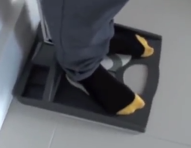
“Cell migration tests have been developed by our team in tissue culture plates. Human mesenchymal-precartilagonous cells were seeded to confluence on a half of the plates’ surface,
while an examined agent (chemoattractant) was placed – embedded in a gel of agarose within a
glass ring at the other pole of the plate. Cells from the confluent half migrated towards the other pole pending on the gradient of the treatment. Among the examined agents were TNF γ; IL-LTB-4; c- AMP; MCP-1 (monocytes chemoattractantprotein-1); IL-1; GM-CSF (granulocyte macrophage colony stimulating factor); IL-2; leptin. The last two agents (IL-2 and leptin) accelerated migration of the cultured cells towards the ring containing the chemoattractant.”

“The last two agents (IL-2 and leptin) accelerated migration of the cultured cells towards the ring containing the chemoattractant.”<-Leptin has been established as having a role in endochondral ossification before.  IL-2 will have to be investigated later.

“Leptin affects the chondrogenic axis lineage, especially at the early phase of chondrogenic differentiation, chondrogenic proliferation and differentiation in the epiphyseal growth plate.”

“Leptin is believed to accelerate skeletal growth via the expression of IGF-I receptors.”

“Osteochondromas are usually classified as benign bone tumors, but they are not neoplastic in nature{not growing new tissue}.
They appear to result from aberrant epiphyseal development with displacement of physeal
cartilage through the perichondrial fibrous ring and subsequent growth at right angles to the long axis of the bone.  The lesion is composed of a mature bony stalk that is covered by a cartilaginous cap”

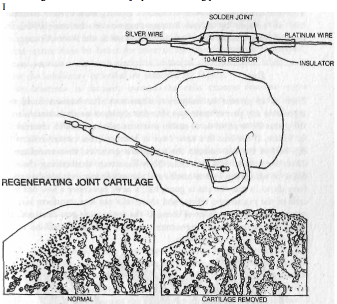
“removal of the ring of La Croix by surgery causes a complete growth arrest of the limbs”<-Would installation of a new ring of La Croix result in a growth restoration?

“Exposure of the periost to TGF-β1 enhances periosteal chondrogenesis”

“Periosteal chondroma is a benign cartilage-forming lesion, located on the surface of the bone, under the periosteum. It is also referred to as juxtacortical chondroma. Priosteal osteosarcoma is a low- to intermediate grade bone-forming sarcoma with predominantly chondroblastic differentiation that develops on the surfaces of long bones in children”

“[These compounds have a migration effect on chondrocytes]: PDGF, IGF-I, TGF-beta, and Hepatocyte growth factor, BMP-2, IL-1 and 2, and calcitonin”

“adhesive molecules and integrins, HA and its cell surface receptors affect cell mobility and migration”

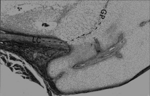
Image of Ring of La-Croix(LC):
jpem.2010

Fibroblast growth factor receptor-3 as a marker for precartilaginous stem cells.(Full Study not available)

“The epiphyseal organ contains two kinds of cartilage, articular and growth plate. Both enlarge during the growth phase of life. However, mitosis is not apparent in these tissues. In the current study, a search to trace the reservoirs of stem cells needed for the growth of these cartilages is done. A disorder in which the stem cells responsible for bone growth are mutated is achondroplasia; the mutation resides in the fibroblast growth factor receptor-3. Epiphyses stained with antifibroblast growth factor 3 antibodies reveal clusters of positively stained cells residing in the perichondrial mesenchyme, known as the ring of La Croix. Removal of the ring of La Croix causes a drastic growth arrest in the limbs of rat neonates. Cell cultures derived of the ring of La Croix biopsy specimens show high rates of cell proliferation and cell migration in vitro, in contrast to articular or growth plate derived chondrocytes. These cells stain intensely by antifibroblast growth factor receptor-3 antibodies and antiproliferative cells nuclear antigen, in contrast with articular and epiphyseal chondrocytes. Transfection of cells from the ring La Croix by an adenovirus vector containing the gene encoding for Escherichia coli beta-galactosidase (lacZ), allows tracing of these cells in tissues. Local injections were performed either to the ring of La Croix or to the joint cavity in a guinea pig model. A characteristic distribution was seen after injection. The transfected cells migrated to areas of bone and cartilage formation in the subchondral bone plate and on either side of the growth plate. This labeling and distribution is maintained for as many as 3 months after injection. The cells from the ring of La Croix appear to be responsible for bone growth. Furthermore, perichondrial cells and other precartilaginous cells expressing fibroblast growth factor-3 have been shown to be good cells for implantation to correct defects of articular cartilage.”

CMF608-a novel mechanical strain-induced bone-specific protein expressed in early osteochondroprogenitor cells.

“Microarray gene expression analysis was utilized to identify genes upregulated in primary rat calvaria cultures in response to mechanical force. One of the identified genes designated CMF608 appeared to be novel. The corresponding full-length cDNA was cloned and characterized in more details. It encodes a putative 2597 amino acid protein containing N-terminal signal peptide, six leucine-rich repeats (LRRs), and 12 immunoglobulin-like repeats, 10 of which are clustered within the C-terminus. Expression of CMF608 is bone-specific and the main type of CMF608-positive cells is mesenchymal osteochondroprogenitors with fibroblast-like morphology. These cells reside in the perichondral fibrous ring of La Croix, periosteum, endosteum of normal bone as well as in the activated periosteum and early fibrous callus generated postfracture. Expression of CMF608 is notably absent from the regions of endochondral ossification. Mature bone cell types do not produce CMF608 with the exception of chondrocytes of the tangential layer of the articular cartilage, which are thought to be under constant mechanical loading. Ectopic expression of CMF608 in HEK293T cells shows that the protein is subjected to post-translational processing and its N-terminal approximately 90 kDa polypeptide can be found in the conditioned medium. Ectopic expression of either the full-length cDNA of CMF608 or of its N-terminal region in CMF608-negative ROS17/2.8 rat osteosarcoma cells results in transfected clones displaying increased proliferation rate and the characteristics of less-differentiated osteoblasts compared to the control cells. Our data indicate that CMF608 is a unique marker of early osteochondroprogenitor cells. We propose that it could be functionally involved in maintenance of the osteochondroprogenitor cells pool and its down-regulation precedes terminal differentiation.”

“Mechanical triggering of rat primary calvaria cells was achieved by stretching following irreversible deformation of the cell attachment surface of the culture dish. For the facilitated identification of de novo transcribed genes, nuclear RNA was extracted following 20 min of stretching.”

“genes transactivated following mechanical strain [include] tenascin C, collagen type XII{up in LSJL}, fibronectin, and connective tissue growth factor”

“genes like ADAMTS-1{up}, thrombospondin 1, endothelin-1{up in LSJL} (its activating enzyme was found upregulated), and desmoykin (AHNAK) [are] previously associated with osteoblastic differentiations.”

No other genes from Table 1 were found to be shared between this and LSJL.

“CMF608 expression was significantly increased in response to all the bone formation promoting stimuli applied, the maximal effect being caused by estrogen administration. In contrast, sciatic neurotomy led to down-regulation of CMF608 mRNA levels”

“In situ hybridization analysis revealed peculiar topography and cell specificity of CMF608 expression in the skeletal tissue. The hybridization signal delineated accumulations of fibroblast-like cells in several locations throughout the long bone: perichondral fibrous ring of LaCroix, periosteum, Volkmann’s canals and endosteum”

CMF608 expression

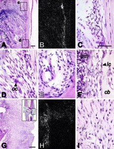
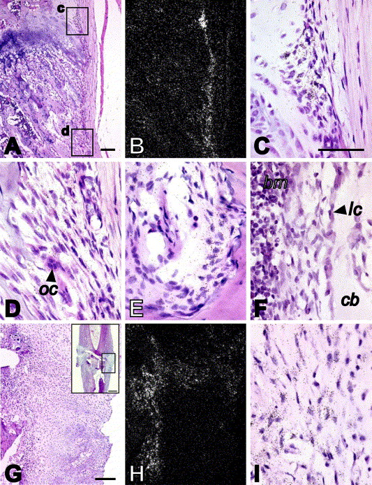
“n situ hybridization analysis of CMF608 expression in normal and fractured rat bones. Hybridization signal is evident as dark dots in the higher magnification brightfield images (C, D, E, F, I), or as white dots in the lower magnification darkfield images (B, H). Expression of CMF608 mRNA in normal rat tibia is shown in panels A–F. Expression of CMF608 in the healing fracture callus 1 week after the fracture is shown in panels G–I. Panels A and B are respectively low power brightfield and corresponding darkfield microphotographs of the metaphysis. Boxed areas, c and d, shown in panel A, are presented at higher magnification in panels C and D, respectively. Hybridization signal for CMF608 concentrates over fibroblast-like cells in the areas of perichondral fibrous ring (C) and metaphyseal periosteum (D). No hybridization signal is detected above progenitor cells in primary and secondary spongiosa. Panel E demonstrates expression of CMF608 in mesenchymal cells within Volkmann’s canal. Panel F shows expression of CMF608 in endosteal fibroblast-like cells in tibial diaphysis. No hybridization signal is detected above osteoblasts. Panels G and H are, respectively, low-power brightfield and corresponding darkfield microphotographs of the area of fracture callus, which is boxed in the inserted image appearing in the right upper corner of panel G. Hybridization signal concentrates over the fibrous part of the callus but not above the cartilaginous or woven bone areas. Panel I is a high power image of the fibrous part of the callus. Hybridization signal is associated with fibroblast-like cells. oc—osteoclast, bm—bone marrow, lc—lining cells, cb—compact bone. Scale bars: A—100 μm; G—200 μm; insert in G—1 mm; C, D, E, F, and I—50 μm.”

” At 3 and 4 weeks postoperation[induction of a fracture], the mature callus was composed mainly of a cancellous bone undergoing transformation into the compact bone with little if any cartilage or woven bone present. Vascularized periosteal tissue was reduced and covered the center of the callus and was entrapped within the newly formed bone. Multiple undifferentiated cells within this tissue continued to show CMF608-specific hybridization signal

“in situ hybridization results suggest that in adult rat bones, activity of the CMF608 gene is confined to a certain subset of skeletal progenitor cells.”

“The presence of a signal peptide, the absence of a transmembrane domain(s), and the presence of structural domains typical for extracellular proteins (leucine-rich and Ig-like repeats) predict that the CMF608 protein product should be secreted out of the cell.”

“[An] important structural feature, present in all the CMF608 orthologous proteins, is conserved integrin recognition RGD motif found, however, outside the 663 amino acid N-terminal fragment. No RGD sequence was identified in adlican. Integrins are signaling receptors that connect the cytoskeleton to the extracellular matrix, and are involved in mechanotransduction. Mechanical stimulation of bone cells by fluid flow induces recruitment of integrins to focal adhesions. Since CMF608 may theoretically serve as a secreted integrin ligand and its expression is responsive to mechanical stimulation, participation of CMF608 in integrin-mediated bone-specific mechanotransduction may be anticipated.”

“Progenitor cells expressing CMF608 have a fibroblast-like morphology and are found at several locations throughout the bone sections. The lack of CMF608 expression in pluripotent C3H10T1/2 mouse mesenchymal progenitor cells allows us to suggest that CMF608 expressing fibroblast-like cells represent already committed skeletal progenitors, whereas their specific localization hints to their involvement in bone modeling by taking part in circumferential growth of the physis (cells within perichondral ring) and bone shaft (endosteal and periosteal cells of diaphysis) as well as the involvement in the reshaping of metaphysis into bone shaft.”<-so less differentiated progenitor cells can be induced to express CMF608 thus becoming growth plate progenitor cells.

In the study Superior Osteogenic Capacity of Human Embryonic Stem Cells Adapted to Matrix-Free Growth Compared to Human Mesenchymal Stem Cells,  CMF608 is expressed in higher levels in embryonic stem cells than human mesenchymal stem cells.

The growth plates does seem to need a specialized stem cell from the Ring of La Croix as no other growth occurs if the ring of la croix is disrupted from delivering cells to the growth plate.  However, there is a specific gene known as CMF608 that can be activated by mechanical loading or fracture.  So it is possible to create these stem cells and it is possible that a LSJL type loading regime can induce the CMF608 gene.

Also the presence of CMF608 in regions other than the ring of LaCroix such as the periosteum, Volkmann’s canal and endosteum suggests that other cells can be used to form new growth plates other than specific zone of LaCroix cells.

Also, a potential height increase method could be to inject CMF608 positive stem cells underneath the periosteum to form newo-growth plates.

If you look at this image of the groove of Ranvier(A) and zone of La Croix(b) you can see that the growth plate chondrocytes seem to spill out of the groove of ranvier.

02F11

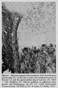
According to one site, “On the sides of the growth plate (physis) the Ossification groove of Ranvier provides cells for growth in width. The Fibrous Ring of La Croix lies outside the physis and keeps the cells from oozing out under axial loading”

Decreasing BMC for Height Growth?

Demineralized Bone Matrix is used as a scaffold to induce chondrogenesis and mineralized bone is an inhibition of interstitial growth which is how growth plates make you taller.  Demineralized bone matrix binds fibronectin which may be necessary for chondroinduction.  DBM also changes Wnt pathway signaling which may also affect chondroinduction.  In fact, one paper suggests that bone growth on DBM occurs endochondral means which suggests that there is always a cartilage intermediate.  Demineralized bone matrix also induced de novo endochondral ossification when implanted into muscle tissue.

If we could cause the demineralization of bone matrix just in certain spots that may enable the creation of growth plates in those particular areas.

Here’s what demineralized bone looks like:

Divergent Significance of Bone Mineral Density Changes in Aging Depending on Sites and Sex Revealed through Separate Analyses of Bone Mineral Content and Area.

Bone mineral density (aBMD) is equivalent to bone mineral content (BMC) divided by area. We rechecked the significance of aBMD changes in aging by examining BMC and area separately. Subjects were 1167 community-dwelling Japanese men and women, aged 40-79 years. ABMDs of femoral neck and lumbar spine were assessed by DXA twice, at 6-year intervals. The change rates of BMC and area, as well as aBMD, were calculated and described separately by the age stratum and by sex. In the femoral neck region, aBMDs were significantly decreased in all age strata by an increase in area as well as BMC loss in the same pattern in both sexes. In the lumbar spine region, aBMDs decreased until the age of 60 in women, caused by the significant BMC decrease accompanying the small area change. Very differently in men, aBMDs increased after their 50s due to BMC increase, accompanied by an area increase. Separate analyses of BMC and area change revealed that the significance of aBMD changes in aging was very divergent among sites and between sexes. This may explain in part the dissociation of aBMD change and bone strength, suggesting that we should be more cautious when interpreting the meaning of aBMD change.”

So in men Bone Mineral Content increases with age.  A lower bone density means more potential sites of demineralized bone in the bone.  However, since BMC is only measured at certain sites it may be hard to extrapolate to the entire bone.

However, in some cases in aging bone area increases while BMC decreases which would be ideal for creating sites for de novo growth plate formation.

According to Sex-specific relationships between insulin resistance and bone mineral content in Korean adolescents., insulin resistance is linked to decreased bone mineral content. In one study, insulin resistance was linked to increased longitudinal bone growth.  Maybe this is due to insulin resistance causing decreased BMC?

According to Cortical bone is more sensitive to alcohol dose effects than trabecular bone in the rat., alcohol decreases BMC but also decreases other bone parameters.  Ideally, we’d only want a BMC decrease.

In some cases changes in bone architecture may decrease length:

Anti-osteoporotic activity of methanolic extract of an Indian herbal formula NR/CAL/06 in ovariectomized rats.

“Bilateral ovariectomy was performed in female Sprague-Dawley rats under aseptic conditions and the rats were divided into five groups (n=10). Two different doses of methanolic extract of NR/CAL/06 (200 and 400 mg/kg, per oral (p.o.)) were evaluated for anti-osteoporotic activity and raloxifene (5.4 mg/kg, p.o.) was used as a reference standard. Treatment was given for 90 d.

The bilateral ovariectomy in rats resulted in decreased bone strength, bone mineral content and bone weight. The SEM images showed porous, perforated and disintegrated femur bone architecture and decreases in bone length and thickness in OVX rats. These changes were associated with elevated serum levels of calcium, phosphorus and ALP. Increases in body weight and adipose weight and a decrease in uterine weight were also observed and the changes were highly significant when compared with the sham-control group. Treatment with methanolic extract of NR/CAL/06 (200 and 400 mg/kg, p.o.) for 90 d dose-dependently restored the ovariectomy-induced alterations in bone weight, bone mineral content, bone strength, serum calcium, phosphorus and ALP, body weight and adipose weight nearly to normal levels. Furthermore, the SEM images of the femur bones of NR/CAL/06 (200 and 400 mg/kg, p.o.)-treated rats showed reduced pore formation and improved bone compactness compared with the OVX-control group.”

“The NR/CAL/06 is an herbal formulation consists of mixture of three different plant parts namely, Hibiscus rosasinensis (flowers), Cestrum diurnum (leaves){helps Vitamin D synthesis} and Glycyrrhiza glabra (whole plant){possess estrogen-like activity} in the ratio of 1∶1∶1. ”

The rats were 8 to 10 weeks old when the study began.

Femur length was actually slightly higher in OVX-control group than the sham control.  NR/CAL/06 400mg group had longer femur length than both.  The supplemented groups had a much larger variance of femur length than the sham control meaning that how much OVX or the supplements helps with height depends on the individual rat.

Effect of prior treatment with resveratrol on density and structure of rat long bones under tail-suspension.

“effects of resveratrol on bone mineral density (BMD), bone mineral content, and bone structure were examined in the femora and tibiae of tail-suspended and unsuspended rats using X-ray micro-computed tomography (micro-CT). Rats were treated with 400 mg/kg/day of resveratrol for 45 days and half of them were suspended during the last 2 weeks of treatment. Suspension caused a decrease in tibial and femoral BMD and deterioration of trabecular and cortical bone. Bone deterioration during suspension was paralleled by increased bone marrow area, which could be caused by an increase in stromal cells with osteoclastogenic potential or in adipocytes. Resveratrol had a preventive effect against bone loss induced by hindlimb immobilization. In particular, trabecular bone in the proximal tibial metaphysis was totally preserved in rats treated with resveratrol before tail-suspension.”

Resveratrol can delay fusion.

“[Resveratrol] increases epiphysial bone mineral density (BMD) and inhibits the decrease of femur calcium content in ovariectomized rats”<-interesting that this increasing in bone mineral density is in the epiphysis.

Tibia length in mm

Control

Nonsuspended: 41.58 ± 1.14

Suspended: 42.63 ± 0.68

Resveratrol

Nonsuspended: 40.90 ± 0.33

Suspended: 40.60 ± 0.57

Resveratrol decreased bone length and eliminated the bone length advantage from the suspended group.  Note that the suspended group had longer bones than the non-suspended group.  Also, remember that growth rate does not necessarily affect total growth.  So this change in growth rate would not necessarily be reflected in adult height.

Cortical BMD was lower in the suspended group thus possibly suggesting an inverse relationship between BMD and height.  The two resveratrol groups had the highest BMD.

Femur length

Control:

Nonsuspended: 37.73 ± 0.80

Suspended: 38.38 ± 1.06

Resveratrol

Nonsuspended: 38.75 ± 0.56

Suspended: 37.80 ± 0.32

In the femur the resveratrol nonsuspended femur growth was the highest and it was tied for having the highest bone density.

“In the tibial middiaphysis, measurement of marrow area showed an 1.3-fold increase after 2 weeks of tail-suspension compared to control animals. Resveratrol treatment prevented this increase. In the femoral middiaphysis, the increase in marrow area in suspended rats was more pronounced (1.5-fold compared to unsuspended rats) and could not be totally prevented by resveratrol pretreatment (resveratrol-treated suspended rats showed an 1.2-fold increase in marrow area compared to control rats).”

suspended non-suspended bones

“Micro-computed tomography (micro-CT) slices showing femoral distal (a–d) and tibial proximal (e–h) metaphysis. Suspended rats (b, f) present less trabecular bone than control (a, e), resveratrol-treated (c, g), and suspended + resveratrol-treated rats (d, h)”

“There are mainly two sites that regulate bone structure and bone mass by responding to mechanical stress: one is the osteocytes, surrounded by bone tissue and the osteoblasts next to the osteocytes, and the other is bone marrow composed of mesenchymal and hematopoietic cells. Osteoblasts differentiate from mesenchymal cells and osteoclasts from hematopoietic cells”<-our goal with LSJL is to cause chondrogenic differentiation from mesenchymal stem cells.

According to [Relationship between bone mineral content and growth disorders in children with juvenile idiopathic arthritis]., growth inhibition was associated with lower bone mineral density.

Effects of Khaya grandifoliola on red blood cells and bone mineral content in rats.

“The therapeutic efficacy of a crude water extract of Khaya grandifoliola has been established in mice. This study was designed to assess the effect of the extract on the red blood cells and bone for 7 days, 3 weeks and a recovery period of 3 weeks. Daily administration of the extract showed no related adverse effects on the mortality rate, physical appearance or behaviour of the rats. A general pattern of significant increases in the red blood cell (RBC) count, PCV, haemoglobin and plasma iron content was shown by groups administered with extract after 7 and 21 days when compared with control rats. There was a general trend of reduction in the bone minerals determined (Ca, P, Mg and Cu) in the extract administered groups. Significant decreases were observed at the 500 mg/kg concentration. The bone potassium and iron content was significantly increased in rats administered with extract in a dose-dependent manner. There was an observed significant decrease in alkaline phosphatase (ALP) activity in the rats administered with the extract when compared with the control animals. During the recovery period, the haematological indices regressed to values which were still significantly higher than those of the control values. These results indicate that K. grandifoliola has a positive effect on erythropoeisis, but no significant effect on bone mineral contents at therapeutic doses. At extremely high doses and during prolonged administration, it may have an adverse effect on bone minerals.

The red blood cell count of the 500mg/kg is 23% than the control group.  500 mg/kg also reduced phosphorus, calcium, zinc, and magnesium levels but not iron levels nor potassium levels(which were higher than control).  Perhaps this could decrease the tensile strength of the bone making it more susceptible to stretching forces.

Khaya grandifoliola also decreased alkaline phosphate levels.

There haven’t been a lot of studies on khaya grandifoliola but if reducing bone mineral content does help in a height increase routine that involves stretching the bones than compounds derived from khaya grandifoliola will be extremely helpful.

One study suggested that BMC is linked to height and stated that height loss is predictive of osteoarthritis.

The study also stated that “Most of us are the tallest at around 30-35 years old because of the peak bone mass at that time.”

However, BMC is useful when fighting against the forces of gravity in instances such as suspension then BMC content could be a detriment to the stretching of bones.

New GTPS System or Grow Taller Pyramid Secrets Review

New GTPS System or Grow Taller Pyramid Secrets Review

On Jan 28 we received a message by someone who asked us the question “When are you going to publish your article about GTPS?” – It was by a person who calls themselves calisthenics – There was also another person named Sergi who said the same thing. We are guessing that the two people who sent almost the exact same message is probably the same person. The truth is that we have already in the past reviewed the Grow Taller Guru (Review Here) (aka Lance Ward) and the new system he created called the Grow Taller Pyramid Secrets (The 1st Review is Here). What we sort of figured out about this one person who calls himself the GTG aka the Grow Taller Guru is that the main way he has been marketing and advertising himself is through YouTube, which has become a very good platform to create a unique brand and profile. (You can find his profile link here – https://www.youtube.com/user/TheGrowTallerGuru)

His recent series of video posts have been the new program called Grow Taller Pyramid Secrets. It is also  called the VLog or Video Blog or Video Proof Series. He uses the Slogan “GTG (aka Grow Taller Guru) of the UK to the NYC

Here is the listing of measurements he had done to his students over a 8 week course of exercising, stretching, and diet/nutritional coaching.

Week 1 of Harry & James

Grow Taller Pyramid Secrets 1Time of Reading – Roughly half past five pm (5:00 PM). First Reading during the daytime – 12:00 Noon

  • Harry – Shaggy hair, thin, Caucasian. Took off shoes and stood relaxed in front of the stadiometer. Is wearing black & yellow socks. First Reading – Almost exactly 68 inches (5′ 8”) or around 172.6 cms tall.
  • James – Short clean cut hair w/ Glasses, slightly taller, very pale, thin. Also in socks. Measured reading – Exactly 182 cm in height

Week 11 Measurements

Local Time- 4:49 UK Time

  • Harry Measured Height – 174 -174.1 cm – Posture is slightly more rigid, with hands out to the side
  • James Measurement – 183.9 cm – Posture is much more upright this time, puffed up chest

Week 20 Measurements

Time – 12:40 on Sept 8th, (assuming year is 2013) – (also assuming local UK time)

  • Harry Measured Height – 174.6 cm
  • James Measured Height – 185.4-185.5 – posture is more relaxed with chest not puffed out as much.

Week 30 Measurements

Time – Sunday 12:42 on Nov 17th, 2013

  • Harry Measured Height – 175. 8 cm
  • James Measured Height – 185.6 cm

Week 40 Measurement

Grow Taller Pyramid Secrets 2Time is 12:15 for Saturday, 25th of January

  • Harry Measured height – 178.4-178.5 cm
  • James Measured Height – 185.4-185.5 cm

Other things to mention about the videos that were sent.

The guy is selling Ayurvedic Urea which is supposed to be $550/bottle for free. He is giving away a free height chart. In the background is a picture of Sultan Kosen who was at the time still around 8′ 1″.

Our Analysis On The Height Increase Measured

We had previously tried to explain why one of his clients managed to increase his height by upwards of around 3 cm over a 1 year time span. (Read that review Here). The client was named Michael from Singapore. He originally measured at 8 AM at 161.6 cm on Day 1 to 164.3 & 164.4 on Days 260 and Day 300 respectively. In that past post we had only speculated that the height gain from Lance Ward’s exercise program was real, but only for a certain specific range of person. We had guessed that due to Michael’s previous lifestyle of poor eating habits and lack of exercise, his posture was slouching. Once Michael learned to improve his behavior, his posture improved. His overall increase in 3 cm over a 1 year time is completely reasonable, which could be completely explained from vertebral decompression.

There was a guy on YouTube last year called TightSkinFlash who also through daily stretches gained about 4 cms in height. We did a review on him on the post Reviewing A Height Increase Success Story By SkinTightFlash From Youtube, A Lance Ward Supporter

However that reasoning can’t be used for the cases here, because of Harry.

Where James, who is the taller of the students increased by 3.5 cms, Harry, the shorter of the two, really increased his height beyond would should be possible. I think that the primary focus on our analysis should be on Harry, who grew past the range of what could be considered just stretching and disc or spinal decompression.

Grow Taller Pyramid Secrets 3If we analyzed the videos of Harry of when the videos were started and around the end at 40 weeks, if we looked really closely, we should see that there has been a definite increase in height and change. If we took the height of Lance as the standard, assuming that he did not change in height, (also assuming that his footwear did not change in thickness) then we can see that the top of Harry’s head has increased relative to the position of Lance’s head.

Notice how in the first pictures, which we clipped from the 1st video (for Day #1) the top of Harry’s head is around the level of Lance’s mouth/lips level. By the end of the video series, the top of his head has reached the position of Lance’s Nose position. Of course all of the pictures we clipped is from the video series from the GTG Youtube Channel. The pictures are indeed grainy and the angles have changed, but I have personally tried my best to get the two pictures of the two measurements and positions to be as similar as possible. Harry has definitely increased in height from one picture to the other, at least by our judgement.

Grow Taller Pyramid Secrets ReviewWe can only propose some ideas which can never be validated or discredited, at least without real contact with Lance himself.

Here is a list of our explanations.

The most scientific reason is from jut looking at the body shape of Harry. Notice how skinny both of the students are compared to Lance.

Something that is not well known except among people who study bone growth and auxology is that adolescents and kids who still have growth plates grow vertically first, than followed by horizontally. The long bones in a person’s body first gets longer. Once the growth plates disappear, the bones then get thicker, due to periosteal bone cell growth.

If anyone would look at the relative wrist size of say their father, compared to his son, they would notice that almost always the wrist of the father, who might be shorter than his son, is thicker.

Bones get thicker and wider due to bone growth in the radial direction, as opposed to he axial direction when the hyaline cartilage was still separation the primary ossification center and the secondary ossification center. Once a male reaches full bone maturity, they actually start to grow wider, which is what NBA scouts would describe as to very long & skinny NBA prospects body to “fill out”. The difference between a male body in high school and adulthood is very different, even though they might be the same height.

The adult male body increases in muscle density and content. In fact, the muscle density on a male is highest when he is around the 25-35 year range.

From understanding the linear mechanics on how the growth progression of bones go, we can say that due to how skinny the male students are, they have not ‘filled out” yet, suggesting that they have not reached the end of their bone maturity.

Remember also the fact that there is more than just growth plates in the long bones. The irregular vertebrate bones also have a few layers of hyaline cartilage. While the legs and arms may not be getting longer & contributing to the overall height, we should not forget that the torso makes a major contribution to. If we assume that even 5 of the 33 vertebrate bones have a slight layer of cartilage left, they would lead to some extra height.

Based on our previous research to see how do growth plates actually disappear, we’ve found that the growth plates don’t ossify at the same time. While the hyaline cartilage in the distal tibia fuses the earliest, around 16-18, the cartilage in the vertebrate bones might not fuse until 20-22.

That is what I propose are the list of explanations for Harry’s height increase.

This is all of course assuming that the students are not wearing any type of height increasing socks (we did a report on these unique products which give you a full 1 inch of extra height here), have not reduced the stadiometer, or done any other type of trick as a setup beforehand.

As a person who used to perform sleight of hand tricks and practiced to be an illusionist for a few years, who understands how to trick (and/or misdirect) people through stage setups, preparations, and even using shills, I understand the need to be very sceptical and suspicious of people who make very extreme and extravagant claims. I am willing to believe that Lance might have helped these two British lads increase their height, but would their bodies have gone through the same amount of growth, without his help? That would of course require something very experimental unbiased.

I am not sure where we would be able to do some type of double-blind non-biased test using identical twins for real testing. At this point, the only thing we can do is to take his word on faith while be slightly reserved at the same time.